

Development and Utilization of 3D Printed Material for Thoracotomy Simulation
- PMID: 30581626
- PMCID: PMC6276476
- DOI: 10.1155/2018/9712647
Development and Utilization of 3D Printed Material for Thoracotomy Simulation
Abstract
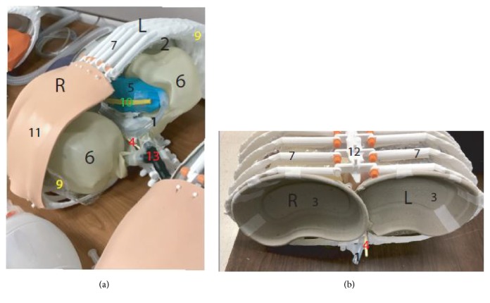
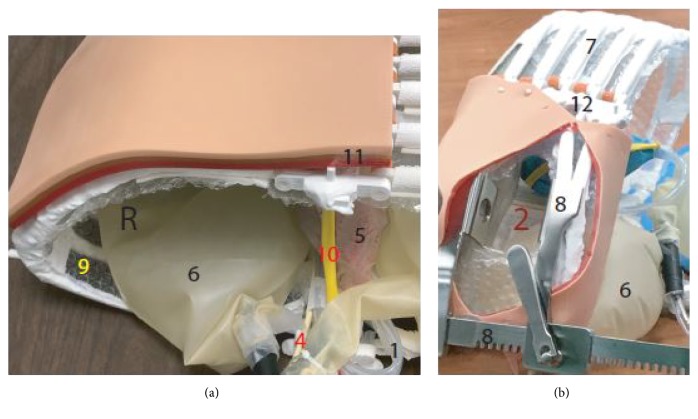
Medical simulation is a widely used training modality that is particularly useful for procedures that are technically difficult or rare. The use of simulations for educational purposes has increased dramatically over the years, with most emergency medicine (EM) programs primarily using mannequin-based simulations to teach medical students and residents. As an alternative to using mannequin, we built a 3D printed models for practicing invasive procedures. Repeated simulations may help further increase comfort levels in performing an emergency department (ED) thoracotomy in particular, and perhaps this can be extrapolated to all invasive procedures. Using this model, a simulation training conducted with EM residents at an inner city teaching hospital showed improved confidence. A total of 21 residents participated in each of the three surveys [(1) initially, (2) after watching the educational video, and (3) after participating in the simulation]. Their comfort levels increased from baseline after watching the educational video (9.5%). The comfort level further improved from baseline after performing the hands on simulation (71.4%).
Figures
Similar articles
-
Emergency department burr hole simulation using 3D-printed model.Am J Emerg Med. 2023 Sep;71:104-108. doi: 10.1016/j.ajem.2023.06.032. Epub 2023 Jun 19. Am J Emerg Med. 2023. PMID: 37356338
-
Effectiveness of High-Fidelity Simulation in Training Emergency Medicine Physicians in Point of Care Ultrasonography in Pakistan: A Quasi-Experimental Study.Cureus. 2020 Jun 17;12(6):e8659. doi: 10.7759/cureus.8659. Cureus. 2020. PMID: 32699659 Free PMC article.
-
Emergency Department Thoracotomy: A Cost-Effective Model for Simulation Training.J Emerg Med. 2019 Sep;57(3):375-379. doi: 10.1016/j.jemermed.2019.06.022. Epub 2019 Aug 1. J Emerg Med. 2019. PMID: 31378446
-
Cadaver-based training is superior to simulation training for cricothyrotomy and tube thoracostomy.Intern Emerg Med. 2017 Feb;12(1):99-102. doi: 10.1007/s11739-016-1439-1. Epub 2016 Mar 28. Intern Emerg Med. 2017. PMID: 27021389
-
See one, do one, teach one: advanced technology in medical education.Acad Emerg Med. 2004 Nov;11(11):1149-54. doi: 10.1197/j.aem.2004.08.003. Acad Emerg Med. 2004. PMID: 15528578 Review.
Cited by
-
3D printing in critical care: a narrative review.3D Print Med. 2020 Sep 30;6(1):28. doi: 10.1186/s41205-020-00081-6. 3D Print Med. 2020. PMID: 32997313 Free PMC article. Review.
-
Creation and Implementation of a Mastery Learning Curriculum for Emergency Department Thoracotomy.West J Emerg Med. 2020 Aug 24;21(5):1258-1265. doi: 10.5811/westjem.2020.5.46207. West J Emerg Med. 2020. PMID: 32970583 Free PMC article.
-
Development of a Gastight Thoracotomy Model for Investigation of Carbon Dioxide Field-Flooding Efficacy.Cureus. 2022 Jan 11;14(1):e21099. doi: 10.7759/cureus.21099. eCollection 2022 Jan. Cureus. 2022. PMID: 35165558 Free PMC article.
References
-
- Horn G. T. Manikin Human-Patient Simulator Training. Journal of Special Operations Medicine. 2017;17:89–95. - PubMed
-
- Ilgen J. S., Sherbino J., Cook D. A. Technology-enhanced simulation in emergency medicine: a systematic review and meta-analysis. Academic emergency medicine : official journal of the Society for Academic Emergency Medicine. 2013;20:117–127. - PubMed
LinkOut - more resources
Full Text Sources
Research Materials
