. 2018 Nov 19:2018:6362716.
doi: 10.1155/2018/6362716. eCollection 2018.

Distinct Nasopharyngeal and Oropharyngeal Microbiota of Children with Influenza A Virus Compared with Healthy Children

Affiliations

Distinct Nasopharyngeal and Oropharyngeal Microbiota of Children with Influenza A Virus Compared with Healthy Children

Zhixin Wen et al. Biomed Res Int. .

Abstract

Background: Influenza A virus (IAV) has had the highest morbidity globally over the past decade. A growing number of studies indicate that the upper respiratory tract (URT) microbiota plays a key role for respiratory health and that a dysfunctional respiratory microbiota is associated with disease; but the impact of microbiota during influenza is understudied.

Methods: We recruited 180 children, including 121 IAV patients and 59 age-matched healthy children. Nasopharyngeal (NP) and oropharyngeal (OP) swabs were collected to conduct 16S rDNA sequencing and compare microbiota structures in different individuals.

Results: Both NP and OP microbiota in IAV patients differed from those in healthy individuals. The NP dominated genera in IVA patients, such as Moraxella, Staphylococcus, Corynebacterium, and Dolosigranulum, showed lower abundance than in healthy children. The Streptococcus significantly enriched in patients' NP and Phyllobacterium could be generally detected in patients' NP microbiota. The most abundant genera in OP microbiota showed a decline tendency in patients, including Streptococcus, Neisseria, and Haemophilus. The URT's bacterial concurrence network changed dramatically in patients. NP and OP samples were clustered into subgroups by different dominant genera; and NP and OP microbiota provided the precise indicators to distinguish IAV patients from healthy children.

Conclusion: This is the first respiratory microbiome analysis on pediatric IAV infection which reveals distinct NP and OP microbiota in influenza patients. It provides a new insight into IAV research from the microecology aspect and promotes the understanding of IAV pathogenesis.


Figures

Figure 1
Figure 1
NP/OP microbiota structure in IAV patients and healthy children. (a) Shannon index of NP and OP microbiota in patients and healthy children. (b, c) Principal components analysis (PCA) of NP/OP samples. (d, e) Comparison of dominated genera of NP/OP microbiota between patients with IAV and healthy children. The vertical axis represents genus name, and the horizontal axis shows the log⁡10 value of relative abundance. , ∗∗, and ∗∗∗ represent q-values ⩽ 0.05, ⩽ 0.01, and ⩽ 0.001, respectively. Objects painted green or red represent healthy or disease samples.
Figure 2
Figure 2
Co-occurrence network of NP/OP microbiota in patients with IAV and healthy ones. The circle size represents relative abundance, and the density of the dashed line represents the Spearman coefficient.
Figure 3
Figure 3
Hierarchical clustering analysis of microbiota in the NP and OP. The circle dendrograms were constructed based on the dissimilarity of microbiota composition between samples. Adjacent to the dendrogram branch ends, stacked bar charts show the relative abundance of the dominant genera in the NP and OP. Subclusters (defined as more than three samples) are designated by the dotted red lines originating from the center of the dendrogram.
Figure 4
Figure 4
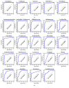
Prediction of biomarkers in the NP/OP microbiome. Receiver-operating characteristic (ROC) plots were used to estimate the efficiency of five key genera in NP (a) and 19 key genera in OP (b). The area under curve (AUC) of each genus shows the high accuracy to distinguish patients with IAV from healthy controls.

References

    1. Bradley J. S., Byington C. L., Shah S. S., et al. The management of community-acquired pneumonia in infants and children older than 3 months of age: Clinical practice guidelines by the pediatric infectious diseases society and the infectious diseases society of America. Clinical Infectious Diseases. 2011;53(7):e25–e76. doi: 10.1093/cid/cir531. - DOI - PMC - PubMed
    1. Artois J., Jiang H., Wang X., et al. Changing geographic patterns and risk factors for avian influenza A(H7N9) infections in humans, China. Emerging Infectious Diseases. 2018;24(1):87–94. doi: 10.3201/eid2401.171393. - DOI - PMC - PubMed
    1. Recommendations for Prevention and Control of Influenza in Children, 2017 – 2018. Pediatrics. 2017;140(4):p. e20172550. doi: 10.1542/peds.2017-2550. - DOI - PubMed
    1. Gong X., Yin H., Shi Y., et al. Evaluation of the immunogenicity and protective effects of a trivalent chimeric norovirus P particle immunogen displaying influenza HA2 from subtypes H1, H3 and B. Emerging microbes & infections. 2016;5:p. e51. doi: 10.1038/emi.2016.51. - DOI - PMC - PubMed
    1. Zou S., Gao R., Zhang Y., et al. Molecular characterization of H6 subtype influenza viruses in southern China from 2009 to 2011. Emerging microbes & infections. 2016;5(7):p. e73. doi: 10.1038/emi.2016.71. - DOI - PMC - PubMed