Comparative Study
. 2018 Nov 18:2018:4692197.
doi: 10.1155/2018/4692197. eCollection 2018.

Comparative Biochemical and Histopathological Studies on the Efficacy of Metformin and Virgin Olive Oil against Streptozotocin-Induced Diabetes in Sprague-Dawley Rats

Affiliations
Comparative Study

Comparative Biochemical and Histopathological Studies on the Efficacy of Metformin and Virgin Olive Oil against Streptozotocin-Induced Diabetes in Sprague-Dawley Rats

Khadijah Saeed Balamash et al. J Diabetes Res. .

Abstract

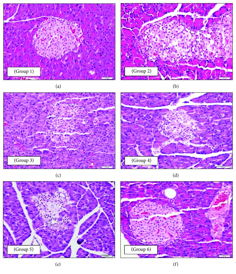
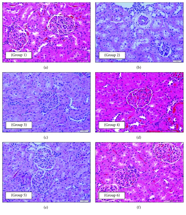
Treatment of diabetic patients with antioxidant, such as extra virgin olive oil (EVOO), may be beneficial in numerous debilitating complexities. This study was aimed at assessing the protective role of virgin olive oil in reducing hyperglycemia in streptozotocin- (STZ-) induced diabetic rats. Thirty-six healthy male Sprague-Dawley rats were divided into six groups (6 rats per group) including nondiabetic control (NC), diabetic control (DC), and animals treated with metformin, olive oil, and a combination of olive oil and metformin, respectively. The protective effect of olive oil was evaluated by determining the biochemical parameters (lipid profile, liver, and kidney) and by studying the histopathological alterations in pancreas, liver, and kidney tissues. The results showed a significant increase in alanine aminotransferase (ALT) and alkaline phosphatase (ALP) levels in diabetic rats. ALP levels remained significantly elevated in the diabetic rats that were treated with metformin and/or olive oil, and the highest level was noted in the group treated with olive oil (568.33 U/L). Contrarily, pretreatment with olive oil significantly decreased ALT (67.64 U/L) and ALP (226.17 U/L) levels. Histopathological data revealed that all the disorganized islets of Langerhans along with the clusters of inflammatory cells were absent in the group pretreated with the combination of virgin olive oil and metformin, which shows that prophylactic administration of this combination reduces the diabetic complications in a much better way. Therefore, pretreatment with olive oil with or without metformin is an encouraging approach for diabetes therapy with immense potential.


Figures

Figure 1
Figure 1
Photomicrographs of a section of the endocrine pancreatic tissue of rats: (a—group 1: NC) normal pancreatic structure and exocrine acini surrounding the islets of Langerhans (400x magnification); (b—group 2: DC) disorganized islets of Langerhans and clusters of inflammatory cells (β-cells show an unstained vacuolated cytoplasm and dark-stained degenerated nuclei) (400x magnification); (c—group 3: DM) normal islets of Langerhans and blood sinusoids (100x magnification); (d—group 4: DO) islets of Langerhans with a normal-looking population of β-cells (100x magnification); (e—group 5: DOM) normal islets of Langerhans with a normal population of β-cells and absence of any degenerative change (400x magnification); (f—group 6: DOP) normal islets of Langerhans with a normal β-cell population similar to that of group 1 (NC) indicating protection from the damaging effect of STZ (400x magnification). All sections were stained with H and E stain and viewed with a light microscope.
Figure 2
Figure 2
Photomicrographs of a section of the liver tissue of rats: (a—group 1: NC) almost normal hepatic structure (hepatocytes being radially arranged around the central vein and presence of von Kupffer cells lining the sinusoidal spaces) (200x magnification); (b—group 2: DC) numerous apoptotic dark-stained shrunken hepatocytes with small degenerated nuclei (400x magnification); (c—group 3: DM) nearly normal radially arranged hepatocytes around the central vein (it also shows that blood sinusoidal spaces and their von Kupffer cells are like those of the NC group) (400x magnification); (d—group 4: DO) almost normal hepatic structure with radially arranged hepatocytes around the central vein with the absence of any degenerated apoptotic cells (it also shows that hepatic sinusoidal spaces and von Kupffer cells are like those of the NC group) (400x magnification); (e—group 5: DOM) almost normal hepatic structure with radially arranged hepatocytes around the central vein (it also shows the presence of von Kupffer cells lining the sinusoidal spaces) (400x magnification); (f—group 6: DOP) almost normal hepatic structure with radially arranged hepatocytes around the central vein (it also shows active and healthy hepatocytes with active vesicular nuclei like that of the NC group) (400x magnification). All sections were stained with H and E stain and viewed with a light microscope.
Figure 3
Figure 3
Photomicrographs of a section of the kidney tissue of rats: (a—group 1: NC) normal renal corpuscles with normal glomerular capillaries and renal tubules (40x magnification); (b—group 2: DC) deformity of renal corpuscle with atrophy of glomerular capillaries and unstained degenerated cytoplasmic regions in the renal tubular epithelium and arterioles showing diffused muscular media thickening with perivascular edema, fibrosis, and minimal interstitium lymphocytic infiltrate (400x magnification); (c—group 3: DM) normal renal corpuscles and glomeruli (it also shows normal renal tubules) (400x magnification); (d—group 4: DO) marked protection from diabetic changes with renal corpuscle, glomeruli, and tubules looking like those of the NC group (200x magnification); (e—group 5: DOM) marked protection against diabetic changes (it also shows normal and healthy renal corpuscles, glomeruli, and tubules) (400x magnification); (f—group 6: DOP) marked protection against diabetic changes (it also shows normal and healthy renal corpuscles, glomeruli, and tubules like those of the NC group) (200x magnification). All sections were stained with H and E stain and viewed with a light microscope.

References

    1. Wu C. T., Tsai Y. T., Lin J. G., Fu S. L., Lai J. N. Chinese herbal products and the reduction of risk of breast cancer among females with type 2 diabetes in Taiwan: a case-control study. Medicine (Baltimore) 2018;97(31, article e11600) doi: 10.1097/MD.0000000000011600. - DOI - PMC - PubMed
    1. NCD Risk Factor Collaboration (NCD-RisC) Effects of diabetes definition on global surveillance of diabetes prevalence and diagnosis: a pooled analysis of 96 population-based studies with 331 288 participants. The Lancet Diabetes & Endocrinology. 2015;3(8):624–637. doi: 10.1016/s2213-8587(15)00129-1. - DOI - PMC - PubMed
    1. Lau L. H., Lew J., Borschmann K., Thijs V., Ekinci E. I. Prevalence of diabetes and its effects on stroke outcomes: a meta-analysis and literature review. Journal of Diabetes Investigation. 2018 doi: 10.1111/jdi.12932. - DOI - PMC - PubMed
    1. American Diabetes Association. Diagnosis and classification of diabetes mellitus. Diabetes Care. 2011;28(Supplement 1):s37–s42. doi: 10.2337/diacare.28.suppl_1.S37. - DOI - PubMed
    1. Emerging Risk Factors Collaboration, Sarwar N., Gao P., et al. Diabetes mellitus, fasting blood glucose concentration, and risk of vascular disease: a collaborative meta-analysis of 102 prospective studies. The Lancet. 2010;375(9733):2215–2222. doi: 10.1016/s0140-6736(10)60484-9. - DOI - PMC - PubMed

Publication types

MeSH terms

LinkOut - more resources