. 2018 Nov 20:13:1-13.
doi: 10.1016/j.omtm.2018.11.003. eCollection 2019 Jun 14.

Gene Transfer with AAV9-PHP.B Rescues Hearing in a Mouse Model of Usher Syndrome 3A and Transduces Hair Cells in a Non-human Primate

Affiliations

Gene Transfer with AAV9-PHP.B Rescues Hearing in a Mouse Model of Usher Syndrome 3A and Transduces Hair Cells in a Non-human Primate

Bence György et al. Mol Ther Methods Clin Dev. .

Abstract

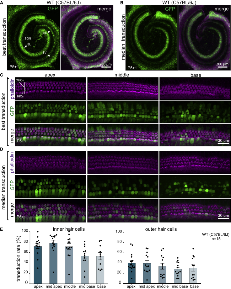
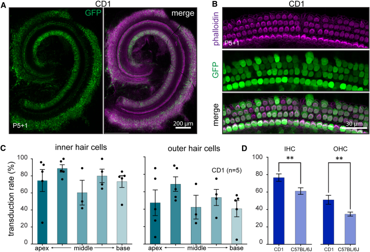
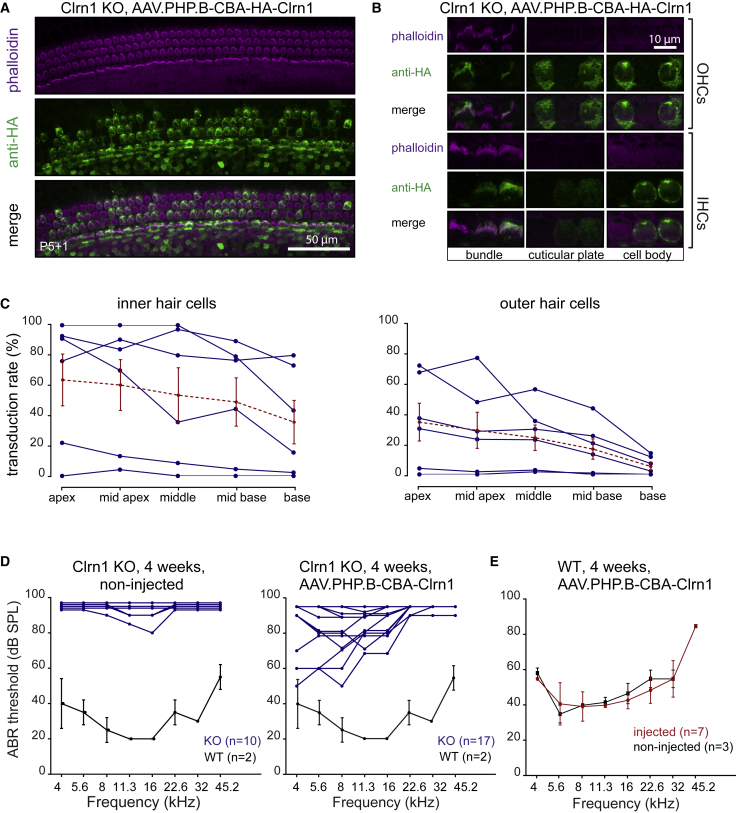
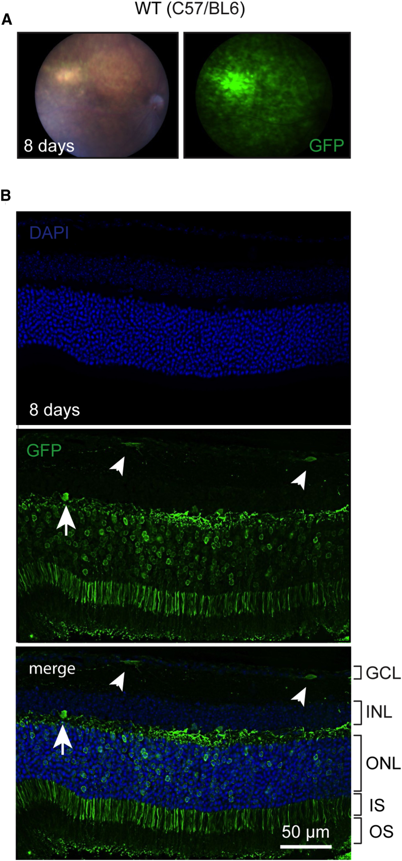
Hereditary hearing loss often results from mutation of genes expressed by cochlear hair cells. Gene addition using AAV vectors has shown some efficacy in mouse models, but clinical application requires two additional advances. First, new AAV capsids must mediate efficient transgene expression in both inner and outer hair cells of the cochlea. Second, to have the best chance of clinical translation, these new vectors must also transduce hair cells in non-human primates. Here, we show that an AAV9 capsid variant, PHP.B, produces efficient transgene expression of a GFP reporter in both inner and outer hair cells of neonatal mice. We show also that AAV9-PHP.B mediates almost complete transduction of inner and outer HCs in a non-human primate. In a mouse model of Usher syndrome type 3A deafness (gene CLRN1), we use AAV9-PHP.B encoding Clrn1 to partially rescue hearing. Thus, we have identified a vector with promise for clinical treatment of hereditary hearing disorders, and we demonstrate, for the first time, viral transduction of the inner ear of a primate with an AAV vector.

Keywords: AAV; adeno-associated virus vector; cochlea; gene delivery; hair cells; hereditary deafness; inner ear; non-human primate.


Figures

Figure 1
Figure 1
Transduction Efficiency in C57BL/6J Mice of AAV9-PHP.B-CBA-GFP after Neonatal RWM Injection (A) Low-magnification images of the cochlea showing the highest transduction level. WT, wild-type. (B) The median transduction (n = 15). Animals were injected at P1 with 5 × 1010 vector genomes (VGs), and the cochlea was explanted at P5 and cultured for another day (P5+1). The left panel shows GFP staining only; the right panel shows GFP overlaid with phalloidin staining. SGN, spiral ganglion neurons; SE, sensory epithelium; SV, stria vascularis; SL, spiral limbus. (C) High-magnification images of different regions of the cochlea with the highest transduction. (D) High-magnification images of the cochlea with median transduction. (E) Quantification of transduction efficiency in IHCs (left panel) and OHCs (right). Bars indicate mean ± SEM. To determine the specified regions, we measured the distance from the apex and used the same value for all cochleas (for details, see Figure S2). Not all the cochleas had a preserved basal region, so there are fewer data points for the base.
Figure 2
Figure 2
Transduction Efficiency in CD1 Mice of AAV9-PHP.B-CBA-GFP after Neonatal RWM Injection (A) Low-magnification image of a representative injection in CD1 mouse. Animals (n = 5) were injected at P1 with 5 × 1010 VGs, and cochleas were explanted at P5 and cultured for another day (P5+1). Left panel shows GFP staining only; right panel shows GFP overlaid with phalloidin staining. (B) High-magnification image of the sensory epithelium of mid-apex region from the same cochlea as in (A). (C) Quantification of transduction efficiency in IHCs and OHCs. Error bars indicate mean ± SEM. (D) Transduction efficiency in CD1 compared to C57BL/6J mice (unpaired t test, p < 0.001 for IHCs, and p < 0.009 for OHCs; data are from all regions analyzed).
Figure 3
Figure 3
AAV-Mediated Clrn1 Delivery Restores Protein Expression in IHCs and OHCs and Rescues Hearing in Clrn1 KO Animals (A) Anti-HA immunostaining of injected C57BL/6J mice. Animals were injected at P1 through the RWM with 7.4 × 1010 VGs of AAV9-PHP.B-CBA-HA-Clrn1. Cochleas were explanted at P5 and were cultured for another day (P5+1). Hair bundles were counterstained with phalloidin. (B) Higher magnification images of IHCs and OHCs at different focal planes. Strong fluorescence labeling was observed in the hair bundle region, cuticular plate, and kinocilium. In the cell body, diffuse cytoplasmic staining was detected. (C) Proportion of inner and outer hair cells with HA-positive bundles. Blue traces indicate individual animals (n = 6); dashed red traces indicate mean ± SEM. (D) Auditory brainstem response (ABR) of mice injected with AAV9-PHP.B-CBA-Clrn1 (tagless, no HA tag, 1.8 × 1011 VGs were injected). An ABR assay was performed at 4 weeks for vector-injected (right panel; n = 17) and non-injected (left panel; n = 10) animals. In each panel, the ABR from 4-week-old wild-type C57BL/6J animals is indicated for comparison (n = 2; mean ± SD). (E) ABR testing of AAV9-PHP.B-mediated expression of Clarin1 on hearing in wild-type C57BL/6J mice (n = 7; mean ± SD).
Figure 4
Figure 4
Transduction of Mouse Retina with AAV9-PHP.B-CBA-GFP Adult (4.5-month-old) C57/BL6 mice were injected subretinally with 2.1 × 109 VG (1 μL). (A) Many retinal cells were transduced 8 days post-injection. (B) Numerous photoreceptors were GFP positive as well as a few cells in the ganglion cell layer (GCL; small arrowheads) and the inner nuclear layer (INL; large arrowhead). GCL, ganglion cell layer; INL, inner nuclear layer; ONL, outer nuclear layer; IS, inner segment; OS, outer segment.
Figure 5
Figure 5
Transduction Profile of AAV9-PHP.B-CBA-GFP in the Cynomolgus Monkey (Animal #1002) Vector (10 μL; 3 × 1011 VGs) was injected through the RWM of a 2.6-year-old, 4-kg Macaca fascicularis monkey. (A) Experimental protocol. (B) Low-magnification images of the injected ear (top) and non-injected (bottom) ear. Anti-GFP staining was detected with a horseradish-peroxidase-conjugated secondary antibody and developed using diaminobenzidine (brown areas). Slides were counterstained with hematoxylin. (C) Higher magnification images of the organ of Corti from base to apex. The non-injected ear lacks GFP-specific immunoreactivity. The numbers in the cochlear turns in (B) correspond to panels 1–4. IHCs are indicated with arrowheads; OHCs are indicated with arrows. (D) Transduction of the lateral wall at high (top) and low (bottom) magnification. (E) Transduction of spiral ganglion neurons. We observed dim but consistent staining in the injected ear (top) but not the non-injected ear (bottom).

References

    1. Akil O., Seal R.P., Burke K., Wang C., Alemi A., During M., Edwards R.H., Lustig L.R. Restoration of hearing in the VGLUT3 knockout mouse using virally mediated gene therapy. Neuron. 2012;75:283–293. - PMC - PubMed
    1. Askew C., Rochat C., Pan B., Asai Y., Ahmed H., Child E., Schneider B.L., Aebischer P., Holt J.R. Tmc gene therapy restores auditory function in deaf mice. Sci. Transl. Med. 2015;7:295ra108. - PMC - PubMed
    1. Geng R., Omar A., Gopal S.R., Chen D.H., Stepanyan R., Basch M.L., Dinculescu A., Furness D.N., Saperstein D., Hauswirth W. Modeling and preventing progressive hearing loss in Usher syndrome III. Sci. Rep. 2017;7:13480. - PMC - PubMed
    1. Chien W.W., Isgrig K., Roy S., Belyantseva I.A., Drummond M.C., May L.A., Fitzgerald T.S., Friedman T.B., Cunningham L.L. Gene therapy restores hair cell stereocilia morphology in inner ears of deaf whirler mice. Mol. Ther. 2016;24:17–25. - PMC - PubMed
    1. György B., Sage C., Indzhykulian A.A., Scheffer D.I., Brisson A.R., Tan S., Wu X., Volak A., Mu D., Tamvakologos P.I. Rescue of hearing by gene delivery to inner-ear hair cells using exosome-associated AAV. Mol. Ther. 2017;25:379–391. - PMC - PubMed

LinkOut - more resources