

Case Reports
doi: 10.1016/j.case.2018.06.003.
eCollection 2018 Dec.
A Case of Severe Aortic Regurgitation Caused by Takayasu's Arteritis Showing End-Diastolic Opening of Aortic Valve
Affiliations
- PMID: 30582084
- PMCID: PMC6301981
- DOI: 10.1016/j.case.2018.06.003
Item in Clipboard
Case Reports
A Case of Severe Aortic Regurgitation Caused by Takayasu's Arteritis Showing End-Diastolic Opening of Aortic Valve
CASE (Phila).
.
No abstract available
Keywords: Aortic regurgitation; Doppler echocardiography; Hemodynamics; Takayasu's arteritis.
Figures

Color Doppler image of AR obtained in a modified apical two-chamber view. LA, Left atrium; LV, left ventricle.
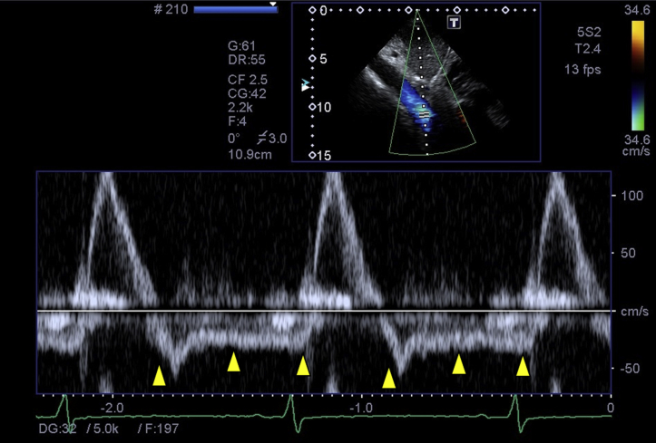
Pulsed-wave Doppler image of the abdominal aorta showing obvious holodiastolic flow reversal (arrowheads).
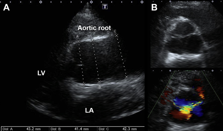
Two-dimensional long-axis view of zoomed aortic root (A) and color-compared short-axis view of AV in diastole (B). The AV did not show any organic abnormality, but insufficient coaptation due to tethering associated with dilation of aortic root was observed. LA, Left atrium; LV, left ventricle.

M-mode image of the AV showing premature opening of the valve in late diastole, just after the P wave of the electrocardiogram (arrowheads). LA, Left atrium.
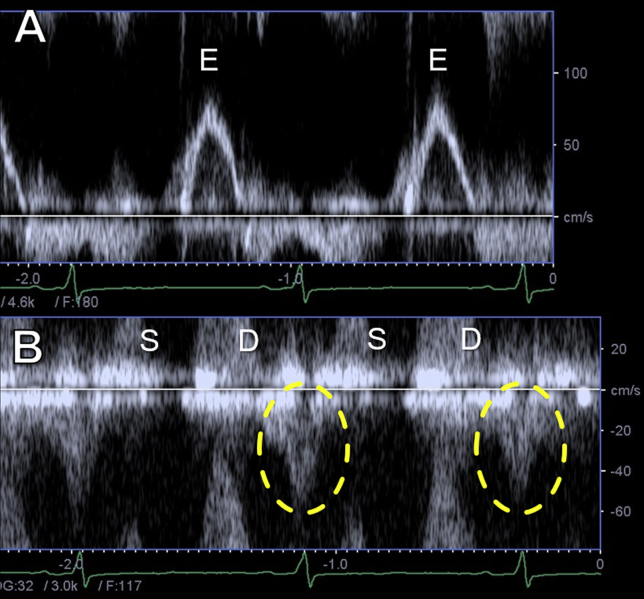
Pulsed-wave Doppler images of transmitral (A) and pulmonary venous (B) flow. Transmitral flow pattern showed monophasic transmitral flow with absence of late-diastolic wave (A). Pulmonary venous flow showed a prominent peak atrial-systolic backward flow (dashed circles) (B).

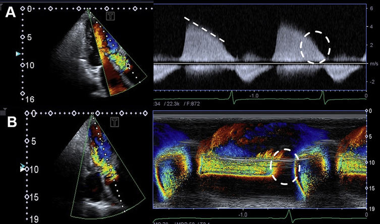
Continuous-wave Doppler image of AR showing extremely shortened pressure half-time (174 msec; dashed line) and abrupt decline of velocity just before atrial contraction to end-diastole (dashed circle) (A). Color M-mode Doppler image of AR showing discontinuation of regurgitation at late diastole (dashed circle) (B).

Estimated pressure waveforms of the left ventricle (LV) and aorta (A) and corresponding continuous-wave Doppler image of AR (B). Left ventricular EDP was estimated to be 44 mm Hg from the systemic diastolic blood pressure, and aortic–left ventricular pressure gradient just before atrial contraction was estimated to be 27 mm Hg from the continuous-wave Doppler image. Therefore, left ventricular pre-A pressure was estimated to be 17 mm Hg from their difference.
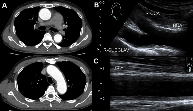
Chest enhanced computed tomographic imaging showing wall thickening of the aorta from the ascending aorta, through the aortic arch, to the descending aorta and the pulmonary artery (A). Carotid ultrasonography showing diffuse and homogeneous wall thickening with medium echogenicity of the aortic arch branches (B). Mean and maximal intima-media thickness of left common carotid artery (L-CCA) were 0.24 and 0.27 cm, respectively (C). BCA, Brachial artery; R-CCA, right common carotid artery; R-SUBCLAV, right subclavian artery.
References
-
- Weaver W.F., Wilson C.S., Rourke T., Caudill C.C. Mid-diastolic aortic valve opening in severe acute aortic regurgitation. Circulation. 1977;55:145–148. - PubMed
-
- Pietro D.A., Parisi A.F., Harrington J.J., Askenazi J. Premature opening of the aortic valve: an index of highly advanced aortic regurgitation. J Clin Ultrasound. 1978;6:170–172. - PubMed
-
- Cohen I.S., Wharton T.P., Jr., Neill W.A. Pathophysiologic observations on premature opening of the aortic valve utilizing a technique for multiplane echocardiographic analysis. Am Heart J. 1979;97:766–772. - PubMed
Publication types
LinkOut - more resources
Full Text Sources
Research Materials
