

A crystal structure of the human protein kinase Mps1 reveals an ordered conformation of the activation loop
- PMID: 30582207
- PMCID: PMC6590424
- DOI: 10.1002/prot.25651
A crystal structure of the human protein kinase Mps1 reveals an ordered conformation of the activation loop
Abstract
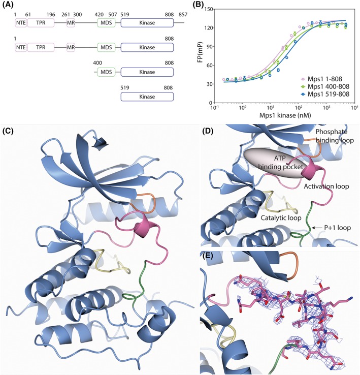
Monopolar spindle 1 (Mps1) is a dual-specificity protein kinase, orchestrating faithful chromosome segregation during mitosis. All reported structures of the Mps1 kinase adopt the hallmarks of an inactive conformation, which includes a mostly disordered activation loop. Here, we present a 2.4 Å resolution crystal structure of an "extended" version of the Mps1 kinase domain, which shows an ordered activation loop. However, the other structural characteristics of an active kinase are not present. Our structure shows that the Mps1 activation loop can fit to the ATP binding pocket and interferes with ATP, but less so with inhibitors binding, partly explain the potency of various Mps1 inhibitors.
Keywords: ATP; Mps1; TTK; X-ray crystallography; activation loop; mitotic kinase; spindle assembly checkpoint.
© 2018 The Authors. Proteins: Structure, Function, and Bioinformatics published by Wiley Periodicals, Inc.
Figures

Similar articles
-
Identification of the hot spot residues for pyridine derivative inhibitor CCT251455 and ATP substrate binding on monopolar spindle 1 (MPS1) kinase by molecular dynamic simulation.J Biomol Struct Dyn. 2019 Feb;37(3):611-622. doi: 10.1080/07391102.2018.1433552. Epub 2018 Mar 8. J Biomol Struct Dyn. 2019. PMID: 29380674
-
Leader of the SAC: molecular mechanisms of Mps1/TTK regulation in mitosis.Open Biol. 2018 Aug;8(8):180109. doi: 10.1098/rsob.180109. Open Biol. 2018. PMID: 30111590 Free PMC article. Review.
-
Understanding inhibitor resistance in Mps1 kinase through novel biophysical assays and structures.J Biol Chem. 2017 Sep 1;292(35):14496-14504. doi: 10.1074/jbc.M117.783555. Epub 2017 Jul 18. J Biol Chem. 2017. PMID: 28726638 Free PMC article.
-
Structural basis of reversine selectivity in inhibiting Mps1 more potently than aurora B kinase.Proteins. 2016 Dec;84(12):1761-1766. doi: 10.1002/prot.25174. Epub 2016 Oct 11. Proteins. 2016. PMID: 27699881
-
Molecular design and anticancer activities of small-molecule monopolar spindle 1 inhibitors: A Medicinal chemistry perspective.Eur J Med Chem. 2019 Aug 1;175:247-268. doi: 10.1016/j.ejmech.2019.04.047. Epub 2019 Apr 20. Eur J Med Chem. 2019. PMID: 31121430 Review.
Cited by
-
LAHMA: structure analysis through local annotation of homology-matched amino acids.Acta Crystallogr D Struct Biol. 2021 Jan 1;77(Pt 1):28-40. doi: 10.1107/S2059798320014473. Epub 2021 Jan 1. Acta Crystallogr D Struct Biol. 2021. PMID: 33404523 Free PMC article.
References
Publication types
MeSH terms
Substances
LinkOut - more resources
Full Text Sources
