Review
. 2019 Oct 20;31(12):874-897.
doi: 10.1089/ars.2018.7695. Epub 2019 Feb 25.

Redox Biology of Peroxisome Proliferator-Activated Receptor-γ in Pulmonary Hypertension

Affiliations
Review

Redox Biology of Peroxisome Proliferator-Activated Receptor-γ in Pulmonary Hypertension

Victor Tseng et al. Antioxid Redox Signal. .

Abstract

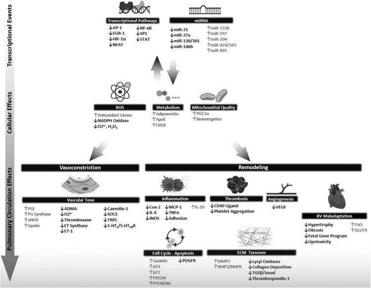
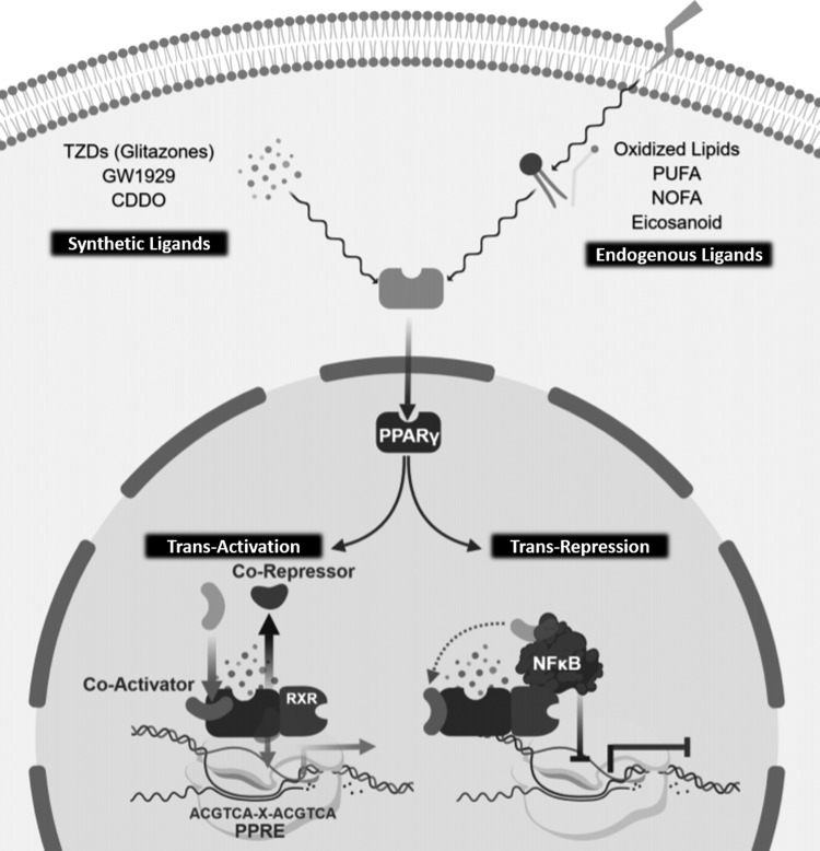
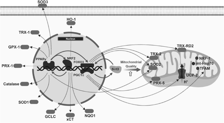
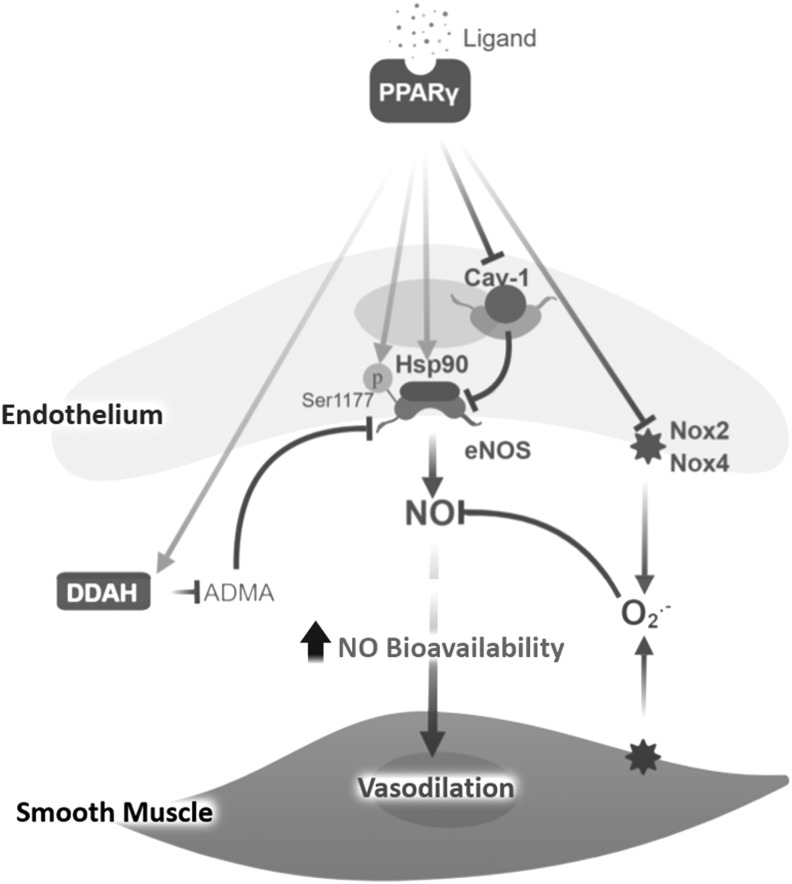
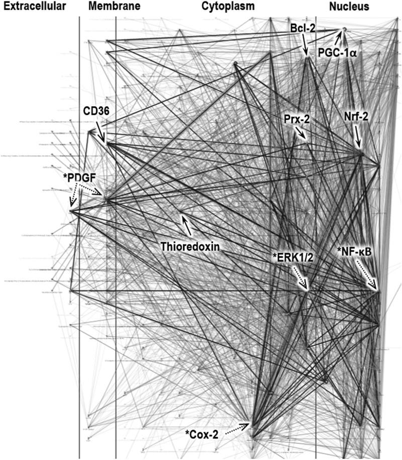
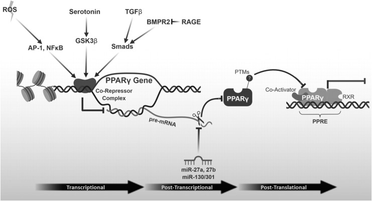
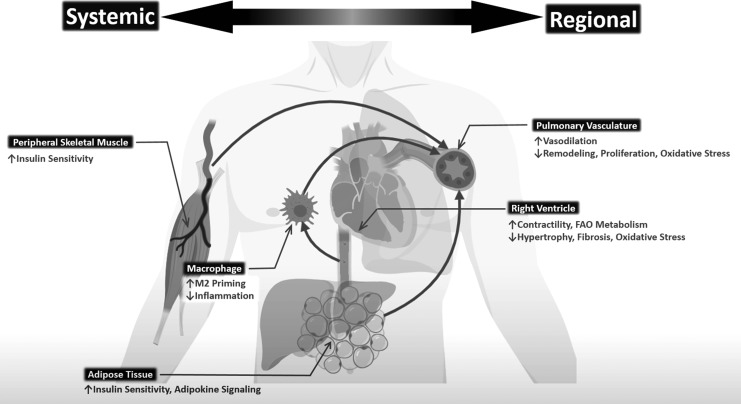
Significance: Peroxisome proliferator-activated receptor-gamma (PPARγ) maintains pulmonary vascular health through coordination of antioxidant defense systems, inflammation, and cellular metabolism. Insufficient PPARγ contributes to pulmonary hypertension (PH) pathogenesis, whereas therapeutic restoration of PPARγ activity attenuates PH in preclinical models. Recent Advances: Numerous studies in the past decade have elucidated the complex mechanisms by which PPARγ in the pulmonary vasculature and right ventricle (RV) protects against PH. The scope of PPARγ-interconnected pathways continues to expand and includes induction of antioxidant genes, transrepression of inflammatory signaling, regulation of mitochondrial biogenesis and bioenergetic integrity, control of cell cycle and proliferation, and regulation of vascular tone through interactions with nitric oxide and endogenous vasoactive molecules. Furthermore, PPARγ interacts with an extensive regulatory network of transcription factors and microRNAs leading to broad impact on cell signaling. Critical Issues: Abundant evidence suggests that targeting PPARγ exerts diverse salutary effects in PH and represents a novel and potentially translatable therapeutic strategy. However, progress has been slowed by an incomplete understanding of how specific PPARγ pathways are critically disrupted across PH disease subtypes and lack of optimal pharmacological ligands. Future Directions: Recent studies indicate that ligand-induced post-translational modifications of the PPARγ receptor differentially induce therapeutic benefits versus adverse side effects of PPARγ receptor activation. Strategies to selectively target PPARγ activity in diseased cells of pulmonary circulation and RV, coupled with development of ligands designed to specifically regulate post-translational PPARγ modifications, may unlock the full therapeutic potential of this versatile master transcriptional and metabolic regulator in PH.

Keywords: PPARγ; antioxidants; hypoxia; oxidative stress; pulmonary hypertension; thiazolidinedione.


Conflict of interest statement

Drs. Tseng, Sutliff, and Hart have no actual or potential conflicts of interest to disclose and no competing financial interests exist.

Figures

<b>FIG. 1.</b>
FIG. 1.
Impact of PPARγ on molecular pathways involved in PH pathogenesis. PPARγ modulates many pathways related to PH. Upstream, PPARγ regulates transcriptional events that control gene expression as well as noncoding RNAs that influence cellular antioxidant enzymes, ROS production, metabolism of fatty acids and glucose, and mitochondrial bioenergetic integrity. Simultaneously, at the level of the pulmonary vasculature, PPARγ contributes to the regulation of vascular tone and remodeling. For instance, many inputs leading to vasoconstriction are modulated by PPARγ, including ET signaling, NO production and scavenging, calcium handling in contractile SMCs, and the prostacyclin pathway. Vascular remodeling events mitigated by PPARγ include inflammatory cell recruitment and trafficking, thrombosis, angiogenesis, cell proliferation, and abnormal ECM turnover. Although not depicted in the figure, there is extensive bidirectional cross talk between these targets and pathways. Representative pathways involved in PH pathogenesis that are reduced by PPARγ agonism are depicted in black font, whereas processes that are stimulated by PPARγ agonism are depicted in gray. ADMA, asymmetric dimethylarginine; Akt, protein kinase B; AP-1, activator protein-1; BMP2, bone morphogenetic protein-2; BMPR2, BMP2 receptor; Cox-2, cyclooxygenase-2; CREB, cyclic AMP response element binding protein; ECM, extracellular matrix; EGR-1, early growth response-1; eNOS, endothelial nitric oxide synthase; ET, endothelin; FAO, fatty acid oxidation; Gadd45, growth arrest and DNA damage 45; GLUT4, glucose transporter-4; HIF-1, hypoxia inducible factor-1; 5-HT2BR, serotonin 2B receptor; IL-6, interleukin-6; iNOS, inducible nitric oxide synthase; MCP-1, monocyte chemotactic protein-1; miR, microRNA; MMP, matrix metalloproteinase; NFAT, nuclear factor of activated T cells; NF-κB, nuclear factor kappa-light-chain enhancer of activated B cells; NO, nitric oxide; PDCD4, programmed cell death protein 4; PDGF, platelet-derived growth factor; PG, prostaglandin; PGC1α, peroxisome proliferator-activated receptor-γ coactivator-1α; PH, pulmonary hypertension; PPARγ, peroxisome proliferator-activated receptor-gamma; PTEN, phosphatase and tensin homologue; ROS, reactive oxygen species; Smad, proteins similar to the gene products of the Drosophila gene “mothers against decapentaplegic” (Mad) and the C. elegans gene Sma; SMC, smooth muscle cell; SOCE, store-operated calcium entry; SP1, specificity protein-1; STAT, signal transducer and activator of transcription; TGFβ, transforming growth factor-beta; TNFα, tumor necrosis factor-alpha; TRPC, transient receptor potential cation channel; VCAM, vascular cell adhesion molecule; VEGF, vascular endothelial growth factor.
<b>FIG. 2.</b>
FIG. 2.
Transcriptional regulation by PPARγ. Ligation of the PPARγ receptor with either naturally occurring endogenous ligands or synthetic exogenous ligands induces conformational changes in PPARγ that promote heterodimerization with RXR, binding to PPRE in the promoter regions of target genes, and the recruitment of transcriptional coactivators, the derecruitment of transcriptional corepressors, and transcriptional activation. Alternatively, at other promoter sites, ligand-induced PPARγ activation sequesters transcription factors (e.g., NF-κB) away from their binding sites or recruits corepressors resulting in transrepression of gene expression. PPRE, PPAR response element; PUFA, polyunsaturated fatty acid; RXR, retinoid X receptor.
<b>FIG. 3.</b>
FIG. 3.
Interaction of PPARγ with master transcriptional regulators of cellular ROS detoxification. PPARγ induces transactivation of Nrf2 and PGC1α, which in turn stimulate a broad program of gene expression related to antioxidant response and mitochondrial biogenesis. GCLC, glutamate/cysteine ligase catalytic subunit-1; GPX-1, glutathione peroxidase-1; HO-1, heme oxygenase-1; mt-Hsp70, mitochondrial heat shock protein (mortalin, Grp75); mt-TFA, mitochondrial transcription factor A (TFAM); NQO-1, NADPH dehydrogenase quinone-1; NRF-1, nuclear (encoded) respiratory factor-1; Nrf2, nuclear factor erythroid 2-related factor 2; PRX, peroxiredoxin; SOD, superoxide dismutase; TRX-2, mitochondrial thioredoxin; TRX-RD2, thioredoxin reductase 2; xCT, cysteine/glutamate transporter (SLC7A11).
<b>FIG. 4.</b>
FIG. 4.
Regulation of NO bioavailability by PPARγ. Activation of PPARγ increases NO bioavailability and eNOS coupling through complex mechanisms unrelated to direct transcriptional regulation of eNOS. These mechanisms include transactivation of DDAH resulting in greater decomposition of ADMA, alleviating its inhibition of eNOS; stimulation of eNOS S1177 phosphorylation increasing eNOS activity; stimulation of Hsp90-eNOS interaction increasing eNOS activity; inhibition of Cav-1-eNOS interaction; transrepression of NADPH oxidase expression thereby reducing superoxide generation and increasing NO bioavailability. Cav-1, caveolin-1; DDAH, dimethylarginase dimethylaminohydrolase; Hsp90, heat shock protein 90; Nox, NADPH oxidase.
<b>FIG. 5.</b>
FIG. 5.
Pathway mapping of PPARγ-related redox processes. A network consisting of the top 500 nodes was constructed with MetaCore pathway analysis. The network was filtered for genes in heart and lung tissue and the following gene ontology processes: oxidation/reduction, cellular responses to oxidative stress, response to superoxide, response to hydrogen peroxide. Dark edges represent the tracing of the resulting subnetwork. The identity of central hubs was determined by visual inspection and annotation. Signaling nodes designated by * are downregulated by PPARγ, whereas those without * are upregulated by PPARγ. Each edge represents one regulatory connection. Bcl-2, B cell lymphoma-2; ERK, extracellular signal-regulated kinase.
<b>FIG. 6.</b>
FIG. 6.
Mechanisms of PPARγ reduction in pulmonary hypertension. Multiple studies indicate that PPARγ levels are reduced by transcriptional and post-transcriptional mechanisms in PH. Transcriptional suppression occurs via oxidative stress signaling coupled to AP-1 transcription factors, serotonergic activation of GSK3β, TGFβ activation, and loss of BMPR2 function. Post-transcriptional silencing of PPARγ is mediated by miR-27a, miR-27b, and miR-130/301, which are all upregulated in hypoxia and various forms of PH. GSK3β, glycogen synthase kinase 3-beta; PTMs, post-translational modifications; RAGE, receptor for advanced glycation end products.
<b>FIG. 7.</b>
FIG. 7.
Potential regional and systemic benefits of PPARγ activation in PH. PPARγ agonists or selective modulators may ameliorate many facets of PH, including pulmonary vasoconstriction and remodeling and RV failure. Broader systemic effects may include peripheral insulin sensitization, increased adipokine signaling, and anti-inflammatory programming of circulating macrophages. RV, right ventricle.

Similar articles

Cited by

References

    1. Adesina SE, Kang BY, Bijli KM, Ma J, Cheng J, Murphy TC, Hart CM, and Sutliff RL. Targeting mitochondrial reactive oxygen species to modulate hypoxia-induced pulmonary hypertension. Free Radic Biol Med 87: 36–47, 2015 - PMC - PubMed
    1. Adir Y. and Amir O. Pulmonary hypertension associated with left heart disease. Semin Respir Crit Care Med 34: 665–680, 2013 - PubMed
    1. Ahmadian M, Suh JM, Hah N, Liddle C, Atkins AR, Downes M, and Evans RM. PPARgamma signaling and metabolism: the good, the bad and the future. Nat Med 19: 557–566, 2013 - PMC - PubMed
    1. Akın A, Alehan D, Aykan HH, Özkutlu S, Özer S, and Karagöz T. Serum Bcl-2 values in children with pulmonary hypertension. Pediatr Cardiol 36: 579–583, 2015 - PubMed
    1. Al-Husseini A, Wijesinghe DS, Farkas L, Kraskauskas D, Drake JI, Van Tassel B, Abbate A, Chalfant CE, and Voelkel NF. Increased eicosanoid levels in the Sugen/chronic hypoxia model of severe pulmonary hypertension. PLoS One 10: e0120157, 2015 - PMC - PubMed

Publication types