Comparative Study
. 2019 Jan 18;124(2):263-278.
doi: 10.1161/CIRCRESAHA.118.314028.

Tissue Resident CCR2- and CCR2+ Cardiac Macrophages Differentially Orchestrate Monocyte Recruitment and Fate Specification Following Myocardial Injury

Affiliations
Comparative Study

Tissue Resident CCR2- and CCR2+ Cardiac Macrophages Differentially Orchestrate Monocyte Recruitment and Fate Specification Following Myocardial Injury

Geetika Bajpai et al. Circ Res. .

Abstract

Rationale: Recent advancements have brought to light the origins, complexity, and functions of tissue-resident macrophages. However, in the context of tissue injury or disease, large numbers of monocytes infiltrate the heart and are thought to contribute to adverse remodeling and heart failure pathogenesis. Little is understood about the diversity of monocytes and monocyte-derived macrophages recruited to the heart after myocardial injury, including the mechanisms that regulate monocyte recruitment and fate specification.

Objective: We sought to test the hypothesis that distinct subsets of tissue-resident CCR2- (C-C chemokine receptor 2) and CCR2+ macrophages orchestrate monocyte recruitment and fate specification after myocardial injury.

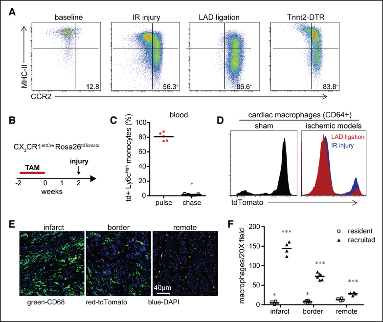
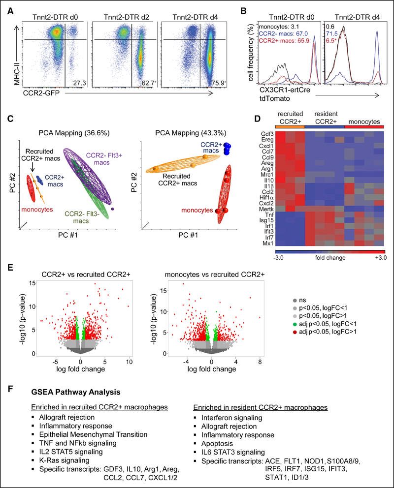
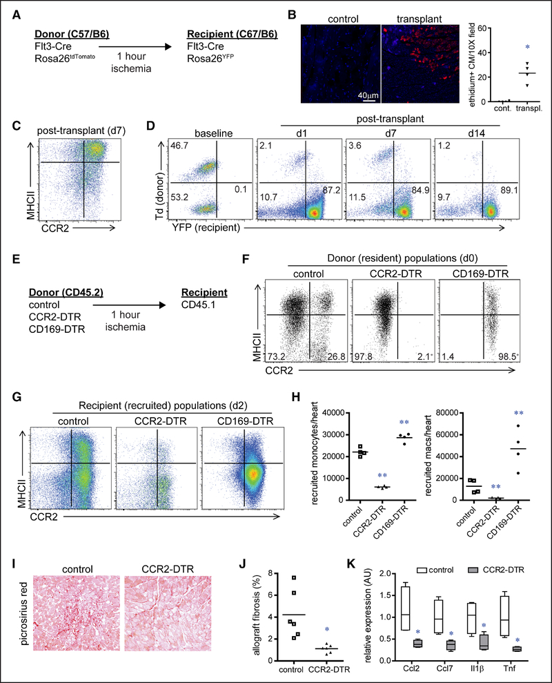
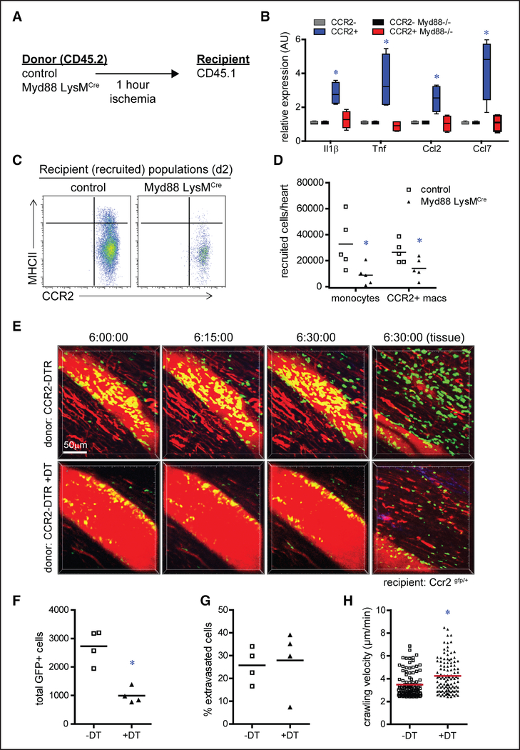
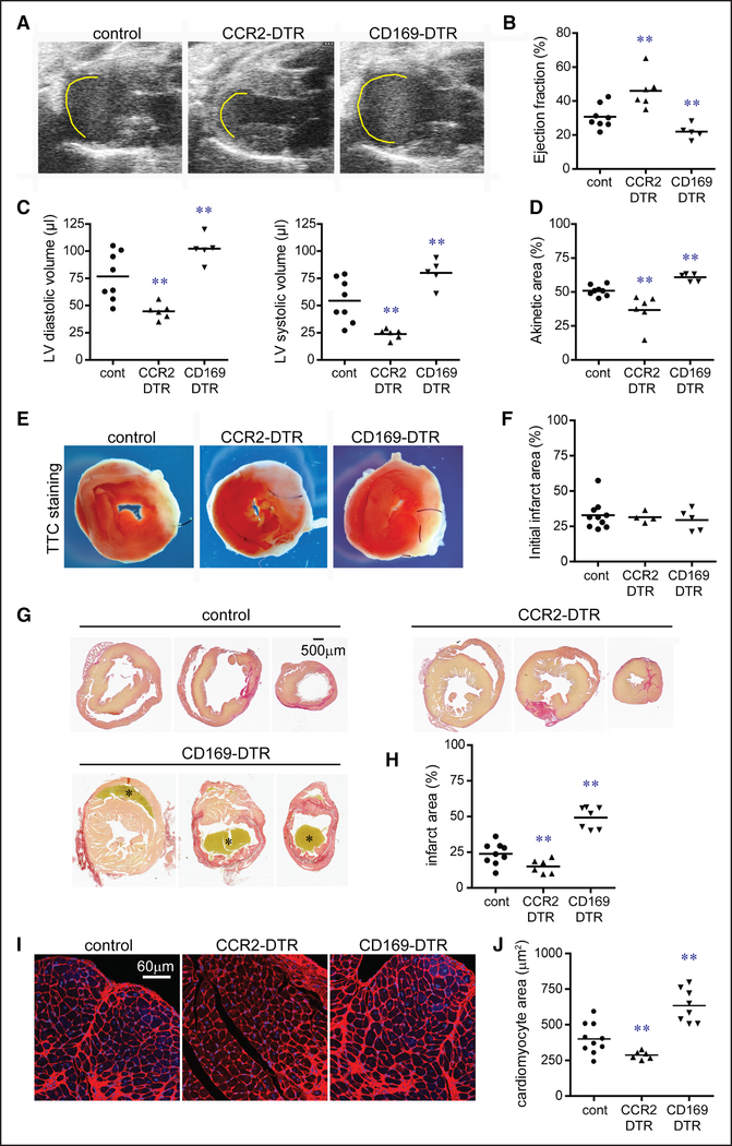
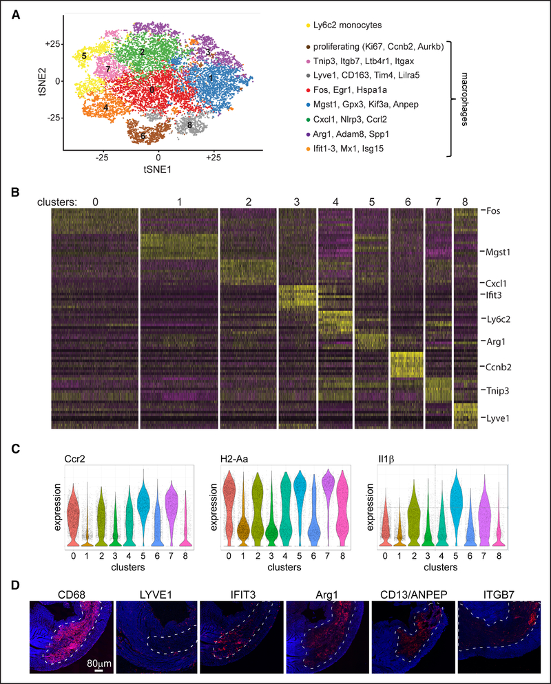
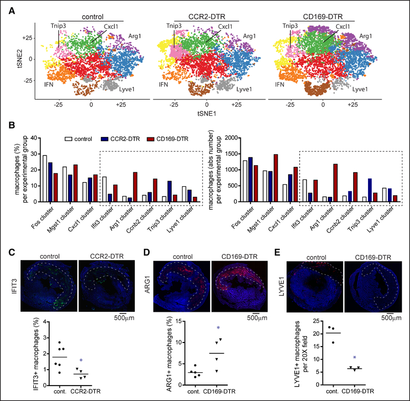
Methods and results: We reveal that in numerous mouse models of cardiomyocyte cell death (permanent myocardial infarction, reperfused myocardial infarction, and diphtheria toxin cardiomyocyte ablation), there is a shift in macrophage ontogeny whereby tissue-resident macrophages are predominately replaced by infiltrating monocytes and monocyte-derived macrophages. Using syngeneic cardiac transplantation to model ischemia-reperfusion injury and distinguish tissue-resident from recruited cell populations in combination with intravital 2-photon microscopy, we demonstrate that monocyte recruitment is differentially orchestrated by distinct subsets of tissue-resident cardiac macrophages. Tissue-resident CCR2+ macrophages promote monocyte recruitment through an MYD88 (myeloid differentiation primary response 88)-dependent mechanism that results in release of MCPs (monocyte chemoattractant proteins) and monocyte mobilization. In contrast, tissue-resident CCR2- macrophages inhibit monocyte recruitment. Using CD (cluster of differentiation) 169-DTR (diphtheria toxin receptor) and CCR2-DTR mice, we further show that selective depletion of either tissue-resident CCR2- or CCR2+ macrophages before myocardial infarction results in divergent effects on left ventricular function, myocardial remodeling, and monocyte recruitment. Finally, using single-cell RNA sequencing, we show that tissue-resident cardiac macrophages differentially instruct monocyte fate specification.

Conclusions: Collectively, these observations establish the mechanistic basis by which monocytes are initially recruited to the injured heart and provide new insights into the heterogeneity of monocyte-derived macrophages.

Keywords: inflammation; macrophages; monocytes; myocardial infarction; receptors, CCR2.


Figures

Figure 1.
Figure 1.. Myocardial injury triggers shifts in macrophage ontogeny.
A, Flow cytometry of cardiac monocyte and macrophage subsets under baseline conditions and 4 d after myocardial ischemia-reperfusion (IR) injury (90 min of ischemia), left anterior descending artery (LAD) ligation, and diphtheria toxin mediated cardiomyocyte ablation (Tnnt2 [troponin T2]-DTR [diphtheria toxin receptor]). Displayed frequencies indicate the percentage of CCR2+ (C-C chemokine receptor 2) monocytes and macrophages. n=4 per experimental group. B, Schematic describing the strategy to distinguish tissue resident from recruited macrophages. TAM: tamoxifen. C, Quantification of the percentage of tdTomato+ Ly6Chigh blood monocytes in CX3CR1ertCre (CX3C chemokine receptor 1) Rosa26tdTomato mice after 2 wk of TAM treatment (pulse) and 2 wk of TAM treatment followed by 2 wk of normal chow (chase). n=5 per experimental group. D, Flow cytometry showing the distribution of tdTomato+ cardiac macrophages (CD [cluster of differentiation] 64+) 4 d after sham surgery, IR injury, or LAD ligation in CX3CR1ertCre Rosa26tdTomato mice that underwent the TAM pulse-chase protocol. Similar results were obtained from 5 independent biological replicates. E, Immunostaining for CD68 (green), tdTomato (red), and DAPI (4’,6-diamidino-2-phenylindole; blue) showing the spatial distribution of resident and recruited macrophages 4 d after LAD ligation. ×200 magnification. F, Quantification of the absolute number of tissue-resident (tdTomato+) and recruited (tdTomato−) CD68+ macrophages in the infarct, remote, and border zones after LAD ligation. n=4 per experimental group. *P <0.05 compared with resident macrophages in the remote zone; ***P <0.05 compared with all other groups.
Figure 2.
Figure 2.. Recruited monocyte-derived macrophages are distinct from tissue-resident subsets.
A and B, Flow cytometry of cardiac monocyte and macrophage subsets in Tnnt2 (troponin T2)-DTR (diphtheria toxin receptor; A) and Tnnt2-DTR CX3CR1ertCre (CX3C chemokine receptor 1) Rosa26tdTomato (B) mice after diphtheria toxin administration and the TAM pulse-chase protocol. Displayed frequencies indicate the percentage of CCR2+ (C-C chemokine receptor 2) monocytes and macrophages (left) and the percentage of tdTomato+ monocytes and macrophages (right). n=4 per experimental group. C, Principal component analysis (PCA) of RNA sequencing data obtained from sorted monocyte and macrophage populations harvested from the hearts of Tnnt2-DTR Flt3-Cre Rosa26tdTomato mice after diphtheria toxin administration. Tissue-resident CCR2− and CCR2+ macrophages were isolated from hearts 36 h after diphtheria toxin (DT) treatment, recruited CCR2+ macrophages were isolated from hearts 4 d after DT treatment. Monocytes were harvested 36 h and 4 d after DT treatment. D, Heat map highlighting genes that were differentially expressed between tissue-resident CCR2+ macrophages, recruited CCR2+ macrophages, and CCR2+Ly6Chigh monocytes. E, Volcano plots showing the number of genes differentially expressed between tissue-resident CCR2+ vs recruited CCR2+ macrophages (left) and monocytes vs recruited CCR2+ macrophages (right). logFC: log based 2 fold change, adj p: adjusted P (false discovery rate analysis). F, Gene set enrichment analysis (GSEA) pathway analysis showing pathways and specific genes enriched in tissue-resident and recruited CCR2+ macrophages. *P <0.05 compared with baseline.
Figure 3.
Figure 3.. Tissue-resident CCR2− (C-C chemokine receptor 2) and CCR2+ cardiac macrophages differentially orchestrate monocyte recruitment.
A, Schematic describing the strategy to distinguish tissue-resident from recruited macrophages in our model of syngeneic heart transplantation. B, Ethidium homodimer-1 (red) staining showing cardiomyocyte cell death 2 h after heart transplantation. C, Flow cytometry showing the cell surface phenotype of CD (cluster of differentiation) 45+CD64+ monocytes and macrophages 7 d after heart transplantation. D, Flow cytometry characterizing the lineage of cardiac CD45+CD64+ monocytes and macrophages after heart transplantation: no reporter (donor, Flt3-Cre negative), dTomato+ (donor, Flt3-Cre positive), and YFP+ (yellow florescent protein; recipient Flt3-Cre positive). Displayed frequencies indicate the percentage of each population. n=4 per experimental group. E, Schematic describing the strategy to investigate whether tissue-resident (donor) macrophages influence recipient monocyte recruitment after syngeneic heart transplantation. F, Flow cytometry of CD45+CD64+ cardiac macrophages in diphtheria toxin-treated control, CCR2-DTR (diphtheria toxin receptor), and CD169-DTR donor hearts before transplantation. Displayed frequencies indicate the percentage of tissue-resident CCR2− and CCR2+ macrophages. *P <0.05 compared with baseline. n=4 per experimental group. G, Flow cytometry of recipient CD45.1+CD64+ monocytes and macrophages 2 d after heart transplantation. H, Quantification of the number of recipient monocytes (CD45+CD64+Ly6C+CCR2+MHC-II [major histocompatibility complex II]low) and macrophages (CD45+CD64+Ly6Clow) recruited to the heart 2 d after transplantation. I, Picrosirius red staining of cardiac allografts 28 d after transplantation (×100 magnification). J, Quantification of Picrosirius red staining in control and CCR2-DTR allografts. K, Quantitative RT-PCR (reverse transcription polymerase chain reaction) measuring chemokine and cytokine mRNA expression in control and CCR2-DTR allografts 4 d after transplantation. Data are displayed as a box and whiskers plot. Line indicates the mean value. n=4 per experimental group. *P <0.05 compared with control; **P <0.05 compared with all other groups.
Figure 4.
Figure 4.. Tissue-resident CCR2+ (C-C chemokine receptor 2) cardiac macrophages promote monocyte recruitment through an MYD88 (myeloid differentiation primary response 88)-dependent pathway regulating monocyte mobilization.
A, Schematic describing the strategy to investigate whether MYD88 is required for activation of tissue-resident (donor) cardiac macrophages after syngeneic heart transplantation. B, Quantitative RT-PCR (reverse transcription polymerase chain reaction) measuring chemokine and cytokine mRNA abundance in tissue-resident (CD [cluster of differentiation] 45.2+) CCR2− and CCR2+ macrophages isolated from control and Myd88 LysMCre (lysozyme M) donor hearts by FACS (fluorescence-activated cell sorting) 2 h after transplantation. Data are displayed as a box and whiskers plot. Line indicates the mean value. n=4 per experimental group. C, Flow cytometry of recipient CD45.1+CD64+ monocytes and macrophages isolated from control and CCR2-DTR (diphtheria toxin receptor) donor hearts day 2 after transplantation. D, Quantification of recipient monocytes (CD45+CD64+Ly6C+CCR2+MHC-II [major histocompatibility complex II]low) and macrophages (CD45+CD64+Ly6Clow) recruited to control and Myd88 LysMCre donor hearts 2 d after transplantation. E, Intravital 2-photon microscopy images obtained 6 h after transplantation of CCR2-DTR donor hearts into CCR2-GFP recipients. Control group: CCR2-DTR donors; experimental group: CCR2-DTR donors treated with diphtheria toxin (DT) before transplantation. Images are focused on coronary veins or adjacent myocardial tissue (far right). red: intravascular Qdot; green: recipient monocytes. F, Quantification of total number of CCR2+ cells. G, Quantification of the percent of CCR2+ cells that extravasated into the myocardium. H, Quantification of crawling velocity. *P <0.05 compared with control.
Figure 5.
Figure 5.. Tissue-resident cardiac macrophages govern outcomes after myocardial infarction.
A, Echocardiographic images of control, CCR2 (C-C chemokine receptor 2)-DTR (diphtheria toxin receptor), and CD (cluster of differentiation) 169-DTR hearts 28 d after closed-chest ischemia-reperfusion injury. Diphtheria toxin (DT) was administered before ischemia-reperfusion injury. Yellow line denotes akinetic myocardial segments. B–D, Quantification of ejection fraction (B), left ventricular (LV) diastolic and systolic volumes (C), and akinetic area (D) 28 d after ischemia-reperfusion injury. E, Triphenyltetrazolium chloride (TTC) staining of control, CCR2-DTR, and CD169-DTR hearts 48 h after ischemia-reperfusion injury. DT was administered before ischemia-reperfusion injury. White area denotes the infarcted region, and red area indicates viable myocardial tissue. F, Quantification of infarct area at 48 h based on TTC staining. G, Picrosirius red staining of control, CCR2-DTR, and CD169-DTR hearts 28 d after ischemia-reperfusion injury. DT was administered before ischemia-reperfusion injury. Red staining highlights the infarcted region, and yellow staining indicates viable myocardial tissue. Asterisks denote a thrombus. H, Quantification of infarct area 28 d after ischemia-reperfusion injury based on Picrosirius red staining. I, Wheat germ agglutinin (WGA; red) staining demonstrating differential effects on cardiomyocyte hypertrophy within the borderzone of control, CCR2-DTR, and CD169-DTR hearts 28 d after ischemia-reperfusion injury. DT was administered before ischemia-reperfusion injury. Blue: DAPI (4’,6-diamidino-2-phenylindole). ×200 magnification. J, Quantification of cardiomyocyte cross-sectional area 28 d after ischemia-reperfusion injury based on WGA staining. **P <0.05 compared with all other groups.
Figure 6.
Figure 6.. Single-cell RNA sequencing of monocytes and macrophages after myocardial infarction.
A, Unsupervised clustering of CD (cluster of differentiation) 45+Ly6G-CD11b+CD64+ cells isolated by flow cytometry from control, CCR2 (C-C chemokine receptor 2)-DTR (diphtheria toxin receptor), and CD169-DTR hearts 4 d after ischemia-reperfusion injury. Diphtheria toxin (DT) was administered before ischemia-reperfusion injury. Each experimental group consists of a pool of 4 biologically independent samples. Data are displayed as a tSNE (t-distributed stochastic neighbor embedding) plot. B, Seurat-generated heat map showing the top 10 genes by P expressed in each cluster. Selected genes are noted in the right column. C, Violin plots demonstrating that CCR2 and H2-Aa (MHC-II [major histocompatibility complex II]) expression does not resolve clusters identified by unsupervised clustering. Il1β expression is also displayed. D, Immunostaining of myocardial tissue 4 d after ischemia-reperfusion injury. 100× tile scans showing the relationship of macrophage subsets to the infarct area (dashed white line). Blue: DAPI (4’,6-diamidino-2-phenylindole).
Figure 7.
Figure 7.. Tissue-resident cardiac macrophages influence monocyte fate specification after myocardial infarction.
A, tSNE plots separated by experimental group highlighting differences in cell abundance within each cluster. Clusters showing the greatest differences between experimental groups are labeled. B, Quantification of the percentage and absolute number of macrophages assigned to each macrophage cluster in control, CCR2 (C-C chemokine receptor 2)-DTR (diphtheria toxin receptor), and CD (cluster of differentiation) 169-DTR hearts on day 4 after ischemia-reperfusion injury. Diphtheria toxin (DT) was administered before ischemia-reperfusion injury. Dotted box denotes clusters that showed differences in both the percentage and absolute number of macrophages between experimental groups. C–E, Immunostaining of control, CCR2-DTR, and CD169-DTR hearts 4 d after ischemia-reperfusion injury for IFIT3 (IFN-induced protein with tetratricopeptide repeats 3; C), ARG1 (arginase 1; D), and LYVE1 (E). DT was administered before ischemia-reperfusion injury. Dashed lines indicated the infarct area. Tile scan of ×100 magnification images. Blue: DAPI (4’,6-diamidino-2-phenylindole). Quantification of the number of IFIT3+, ARG1+, and LYVE1+ macrophages between experimental groups is shown below the immunostaining images. **P <0.05 compared with all other groups.

Comment in

References

    1. Dutta P, Courties G, Wei Y, et al. Myocardial infarction accelerates atherosclerosis. Nature. 2012;487:325–329. doi: 10.1038/nature11260 - DOI - PMC - PubMed
    1. Dutta P, Sager HB, Stengel KR, et al. Myocardial infarction activates CCR2(+) hematopoietic stem and progenitor cells. Cell Stem Cell. 2015;16:477–487. doi: 10.1016/j.stem.2015.04.008 - DOI - PMC - PubMed
    1. Frantz S, Nahrendorf M. Cardiac macrophages and their role in ischaemic heart disease. Cardiovasc Res. 2014;102:240–248. doi: 10.1093/cvr/cvu025 - DOI - PMC - PubMed
    1. Leuschner F, Rauch PJ, Ueno T, et al. Rapid monocyte kinetics in acute myocardial infarction are sustained by extramedullary monocytopoiesis. J Exp Med. 2012;209:123–137. doi: 10.1084/jem.20111009 - DOI - PMC - PubMed
    1. Shi C, Pamer EG. Monocyte recruitment during infection and inflammation. Nat Rev Immunol. 2011;11:762–774. doi: 10.1038/nri3070 - DOI - PMC - PubMed

Publication types

MeSH terms