

The Role of Three-Dimensional Magnetic Resonance Elastography in the Diagnosis of Nonalcoholic Steatohepatitis in Obese Patients Undergoing Bariatric Surgery
- PMID: 30582669
- PMCID: PMC6591099
- DOI: 10.1002/hep.30483
The Role of Three-Dimensional Magnetic Resonance Elastography in the Diagnosis of Nonalcoholic Steatohepatitis in Obese Patients Undergoing Bariatric Surgery
Abstract
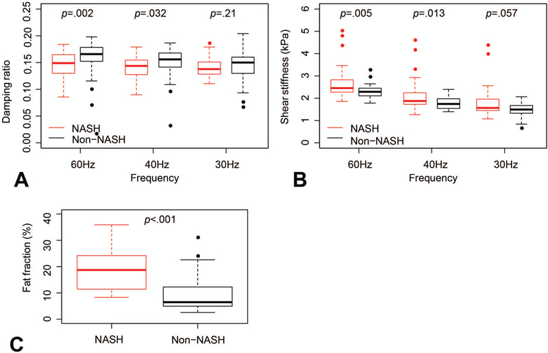
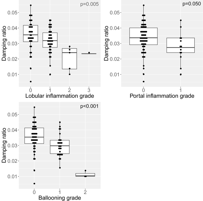
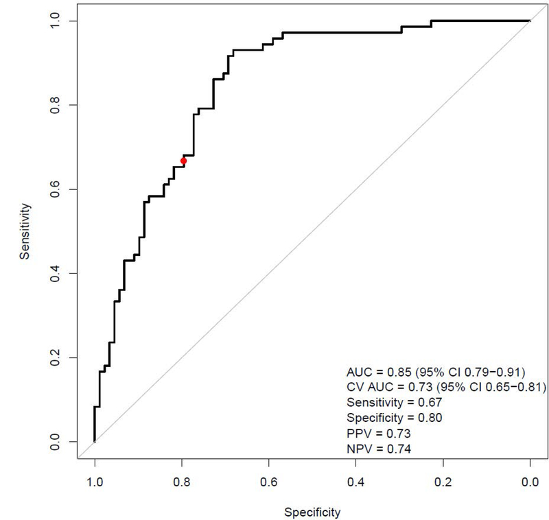
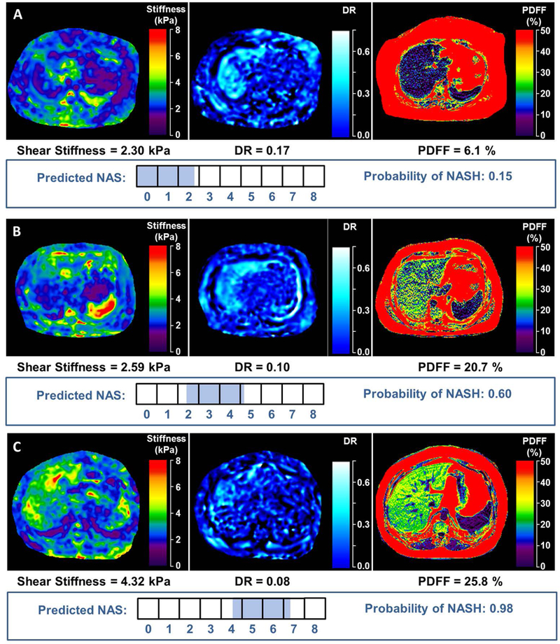
The lack of reliable, noninvasive methods to diagnose early nonalcoholic steatohepatitis (NASH) is a major unmet need. We aimed to determine the diagnostic accuracy of three-dimensional magnetic resonance elastography (3D-MRE), with shear stiffness measured at 60 Hz, damping ratio at 40 Hz, and magnetic resonance imaging proton density fat fraction (MRI-PDFF) in the detection of NASH in individuals undergoing bariatric surgery. Obese adults at risk for NASH were enrolled between 2015 and 2017 (prospective cohort, n = 88) and 2010 and 2013 (retrospective cohort, n = 87). The imaging protocol consisted of multifrequency 3D-MRE (mf3D-MRE) with shear waves delivered at different frequencies to explore parameters that best correlated with histologic NASH, and MRI-PDFF to estimate steatosis. The prospective cohort was used to establish the optimal mf3D-MRE technical parameters for NASH detection. The two cohorts were then combined to derive predictive models of NASH and disease activity by nonalcoholic fatty liver disease activity score (NAS) using the three imaging parameters that correlated with NASH. A total of 175 patients (median age 45, 81% women, and 81 [46%] with histologic NASH) were used for model derivation. From the complex shear modulus output generated by mf3D-MRE, the damping ratio at 40 Hz and shear stiffness at 60 Hz best correlated with NASH. The fat fraction obtained from MRI-PDFF correlated with steatosis (P < 0.05 for all). These three parameters were fit into a logistic regression model that predicted NASH with cross-validated area under the receiver operating characteristic curve (AUROC) = 0.73, sensitivity = 0.67, specificity = 0.80, positive predictive value = 0.73 and negative predictive value = 0.74, and disease activity by NAS with cross-validated AUROC = 0.82. Conclusion: The mf3D-MRE allows identification of imaging parameters that predict early NASH and disease activity. This imaging biomarker represents a promising alternative to liver biopsy for NASH diagnosis and monitoring. The results provide motivation for further studies in nonbariatric cohorts.
Trial registration: ClinicalTrials.gov NCT02565446.
© 2018 by the American Association for the Study of Liver Diseases.
Conflict of interest statement
Conflict of interest
The Mayo Clinic and Drs. Ehman and Yin have intellectual property and a financial interest related to this research. This research has been reviewed by the Mayo Clinic Conflict of Interest Review Board as is being conducted in compliance with Mayo Clinic Conflict of Interest policies.
Figures
References
-
- Younossi ZM, Koenig AB, Abdelatif D, Fazel Y, Henry L, Wymer M. Global epidemiology of nonalcoholic fatty liver disease-Meta-analytic assessment of prevalence, incidence, and outcomes. Hepatology 2016;64:73–84. - PubMed
Publication types
MeSH terms
Substances
Associated data
Grants and funding
LinkOut - more resources
Full Text Sources
Other Literature Sources
Medical
