Degeneration of dopaminergic circuitry influences depressive symptoms in Lewy body disorders
- PMID: 30582885
- PMCID: PMC6767514
- DOI: 10.1111/bpa.12697
Degeneration of dopaminergic circuitry influences depressive symptoms in Lewy body disorders
Abstract
Aims: Depression is commonly observed even in prodromal stages of Lewy body disorders (LBD), and is associated with cognitive impairment and a faster rate of cognitive decline. Given the role of dopamine in the development of movement disorders, but also in motivation and reward, we investigated neurodegenerative pathology in dopaminergic circuitry in Parkinson's disease (PD), PD with dementia (PDD) and dementia with Lewy bodies (DLB) patients in relation to depressive symptoms.
Methods: α-synuclein, hyperphosphorylated tau and amyloid-beta pathology was assessed in 17 DLB, 14 PDD and 8 PD cases within striatal and midbrain subregions, with neuronal cell density assessed in substantia nigra and ventral tegmental area. Additionally, we used a structural equation modeling (SEM) approach to investigate the extent to which brain connectivity might influence the deposition of pathological proteins within dopaminergic pathways.
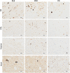
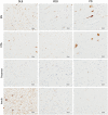
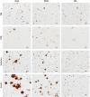
Results: A significantly higher α-synuclein burden was observed in the substantia nigra (P = 0.006), ventral tegmental area (P = 0.011) and nucleus accumbens (P = 0.031) in LBD patients with depression. Significant negative correlations were observed between cell density in substantia nigra with Lewy body (LB) Braak stage (P = 0.013), whereas cell density in ventral tegmental area showed negative correlations with LB Braak stage (P = 0.026) and neurofibrillary tangle Braak stage (P = 0.007).
Conclusions: Dopaminergic α-synuclein pathology appears to drive depression. Selective targeting of dopaminergic pathways may therefore provide symptomatic relief for depressive symptoms in LBD patients.
Keywords: dementia with Lewy bodies; depression; dopaminergic pathways; α-synuclein.
© 2018 The Authors Brain Pathology published by John Wiley & Sons Ltd on behalf of International Society of Neuropathology.
Figures


Similar articles
-
Distribution of cerebral amyloid deposition and its relevance to clinical phenotype in Lewy body dementia.Neurosci Lett. 2010 Dec 3;486(1):19-23. doi: 10.1016/j.neulet.2010.09.036. Epub 2010 Sep 17. Neurosci Lett. 2010. PMID: 20851165
-
Lewy- and Alzheimer-type pathologies in midbrain and cerebellum across the Lewy body disorders spectrum.Neuropathol Appl Neurobiol. 2016 Aug;42(5):451-62. doi: 10.1111/nan.12308. Epub 2016 Apr 1. Neuropathol Appl Neurobiol. 2016. PMID: 26810462
-
TIGAR inclusion pathology is specific for Lewy body diseases.Brain Res. 2019 Mar 1;1706:218-223. doi: 10.1016/j.brainres.2018.09.032. Epub 2018 Sep 26. Brain Res. 2019. PMID: 30267647
-
A hypothesis explaining Alzheimer's disease, Parkinson's disease, and dementia with Lewy bodies overlap.Alzheimers Dement. 2025 Jun;21(6):e70363. doi: 10.1002/alz.70363. Alzheimers Dement. 2025. PMID: 40528299 Free PMC article. Review.
-
Lewy body pathology in Alzheimer's disease.J Mol Neurosci. 2001 Oct;17(2):225-32. doi: 10.1385/jmn:17:2:225. J Mol Neurosci. 2001. PMID: 11816795 Review.
Cited by
-
Psychiatric symptoms of frontotemporal dementia and subcortical (co-)pathology burden: new insights.Brain. 2023 Jan 5;146(1):307-320. doi: 10.1093/brain/awac043. Brain. 2023. PMID: 35136978 Free PMC article.
-
Depression in Patients with Parkinson's Disease: Current Understanding of its Neurobiology and Implications for Treatment.Drugs Aging. 2022 Jun;39(6):417-439. doi: 10.1007/s40266-022-00942-1. Epub 2022 Jun 16. Drugs Aging. 2022. PMID: 35705848 Free PMC article. Review.
-
Relationship Between Depression and Neurodegeneration: Risk Factor, Prodrome, Consequence, or Something Else? A Scoping Review.Biomedicines. 2025 Apr 23;13(5):1023. doi: 10.3390/biomedicines13051023. Biomedicines. 2025. PMID: 40426852 Free PMC article. Review.
-
Neuroanatomical substrates of depression in dementia with Lewy bodies and Alzheimer's disease.Geroscience. 2024 Dec;46(6):5725-5744. doi: 10.1007/s11357-024-01190-4. Epub 2024 May 15. Geroscience. 2024. PMID: 38750385 Free PMC article.
-
Intricate mechanism of anxiety disorder, recognizing the potential role of gut microbiota and therapeutic interventions.Metab Brain Dis. 2024 Dec 13;40(1):64. doi: 10.1007/s11011-024-01453-1. Metab Brain Dis. 2024. PMID: 39671133 Review.
References
-
- Aarsland D, Ballard C, Larsen JP, McKeith I (2001) A comparative study of psychiatric symptoms in dementia with Lewy bodies and Parkinson's disease with and without dementia. Int J Geriatr Psych 16:528–536. - PubMed
-
- Alexander GE, Crutcher MD, DeLong MR (1990) Basal ganglia‐thalamocortical circuits: parallel substrates for motor, oculomotor, “prefrontal” and “limbic” functions. Prog Brain Res 85:119–146. - PubMed
-
- Alexopoulos GS, Abrams RC, Young RC, Shamoian CA (1988) Cornell scale for depression in Dementia. Biol Psychiatry 23:271–284. - PubMed

