Review
. 2019 Aug:148:38-59.
doi: 10.1016/j.addr.2018.12.011. Epub 2018 Dec 22.

Recent advances in nanotherapeutic strategies for spinal cord injury repair

Affiliations
Review

Recent advances in nanotherapeutic strategies for spinal cord injury repair

Young Hye Song et al. Adv Drug Deliv Rev. 2019 Aug.

Abstract

Spinal cord injury (SCI) is a devastating and complicated condition with no cure available. The initial mechanical trauma is followed by a secondary injury characterized by inflammatory cell infiltration and inhibitory glial scar formation. Due to the limitations posed by the blood-spinal cord barrier, systemic delivery of therapeutics is challenging. Recent development of various nanoscale strategies provides exciting and promising new means of treating SCI by crossing the blood-spinal cord barrier and delivering therapeutics. As such, we discuss different nanomaterial fabrication methods and provide an overview of recent studies where nanomaterials were developed to modulate inflammatory signals, target inhibitory factors in the lesion, and promote axonal regeneration after SCI. We also review emerging areas of research such as optogenetics, immunotherapy and CRISPR-mediated genome editing where nanomaterials can provide synergistic effects in developing novel SCI therapy regimens, as well as current efforts and barriers to clinical translation of nanomaterials.

Keywords: Nanomaterials; Nanotechnology; Regenerative medicine; Spinal cord injury.


Figures

Figure 1.
Figure 1.
Representative images of nanomaterials and fabrication overview schematics. A: SEM images of PLGA nanoparticles [191]. Scale bar: 5 μm. B: Field-emission scanning electron microscopy (SEM) image of electrospun silk nanofibers [74]. Scale bar: 5 μm. C: SEM images of self-assembled peptide nanofibers [77]. Scale bar: 300 nm. Schematic of IKVAV self-assembling peptides are from [87]. D: SEM image of multi-walled carbon nanotubes. Scale bar: 250 μm (inset: 25 μm). Image and schematic adapted from [90]. E: Transmission electron microscopy (TEM) images of CdTe quantum dot [113]. Quantum dots with multiple shell layers can be fabricated by ultraviolet light irradiation. Scale bar: 20 nm. Top right: High-resolution TEM images. Scale bar: 2 nm. Copyright permissions: A from Elsevier, B and C (schematic) from John Wiley and Sons, C (SEM image) from Royal Society of Chemistry. D is open access. E (TEM image) from American Chemical Society.
Figure 2.
Figure 2.
Nanomaterials improve cell, tissue, and behavior outcomes in rat models of SCI. A: Injection of dextran sulfate (DS)-minocycline hydrochloride (MH) complexes in agarose gel into the lesion i) reduce reactive glial cells 6 weeks after injury (red: CD68), ii) decrease the ratio of pro-inflammatory M1 macrophages to anti-inflammatory M2 macrophages, and iii) improve behavioral outcomes [162]. Scale bar: 1 mm. * denotes significant difference from untreated control; + from blank gel, and # from intraperitoneal (IP) injection of MH. B: Flavopiridol-loaded poly(lactic-co-glycolic acid) (PLGA) nanoparticles result in improved tissue sparing (H&E images), higher BBB scores, and lower errors in grid walking compared to blank PLGA nanoparticles [191]. Scale bar: 500 μm. C: Left: spinal cord neurons interacting with a multi-walled carbon nanotube (MWCNT) substrate. Red arrows indicate tight contacts between MWCNT and axonal membrane. Scale bar: 500 nm. Right: Total number of identified growth cones normalized to the number of detected fibers on multi-walled carbon nanotubes (solid circle) and control conditions (open circle) [99]. *p < 0.05. D: Top: luxol fast blue (LFB) and H&E images of spinal cord injected with saline (control) or QL6 self-assembled peptides. Scale bar: 300 μm. Middle: Reactive astrocyte staining with GFAP 8 weeks after injury. Scale: 250 μm. Bottom: Basso, Beattie and Bresnahan (BBB) locomotor score indicating improved functional recovery after QL6 injection. [253]. Copyright permissions: A from Elsevier, B from Elsevier, C from American Chemical Society, D is open access.
Figure 3.
Figure 3.
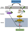
Pro-inflammatory responses via β-catenin signal transduction pathway. Binding of methylprednisolone induces lipoprotein receptor related protein-6 (LRP-6) phosphorylation, which in turn induces glycogen synthase kinase (GSK) phosphorylation and induces dissociation of β-catenin from a protein complex consisting of adenomatous polyposis coli (APC), Axin, and GSK. Subsequently, β-catenin is translocated into the nucleus where it binds to the transcription factors and regulate cell apoptosis. Adapted from [151].
Figure 4.
Figure 4.
Pathway for signaling from p75–Nogo receptor complex to RhoA. Binding of myelin derived inhibitors (Nogo, MAG, and OMgp) on the NgR causes strengthening of p75 and Rho GDI-Rho GTP complex resulting in actin stabilization and inhibition of neurite outgrowth. B) Binding of anti-Nogo Ab or competitive antagonist for the Nogo-66 receptor such as NEP 1-40 have shown to decrease myelin induced growth cone collapse and promote neurite outgrowth.
Figure 5.
Figure 5.
Glial scar mediated neurite inhibition. Glial scar acts as a barrier and axonal regeneration can be achieved after bacterial enzyme chondroitinase ABC (ChABC) treatment. Reactive glial cells, astrocytes, inflammatory cells such as microglia migrate to the lesion area following injury. They synthesize and secrete CSPGs around the lesion area and prevents regenerating axons to surpass. Treatment with ChABC removes the CSPGs in both the glial scar which facilitates the regenerating axons to pass through the lesion core to connect to the distal target. Figure adapted from Servier Medical Art.
Figure 6.
Figure 6.
BDNF-signaling pathway mechanism. Autophosphorylation tyrosine residues upon BDNF binding to TrkB acts through four different but overlapping intracellular signaling pathways. It sends pro-survival signals through phosphatidylinositol-3 (PI-3) kinase and serinethreonine-specific protein kinase B (Akt) pathway, stimulates axon growth through extracellular signal regulated kinase (ERK) and modulation of cyclic adenosine monophosphate (cAMP) pathway and influences synaptic plasticity and transmission through phospholipase C (PLC), inositol-3-phosphate (IP-3) and the calcium signaling pathway. Additionally, through the phosphorylation of N-methyl-D-aspartate (NMDA) receptor subunits, TrkB signaling may lead to an increase in calcium and sodium influx also contributing to the synaptic plasticity. Figure adapted from [312].
Figure 7.
Figure 7.
Future directions include minimally invasive delivery of injectable hydrogels from naturally-derived biomaterials into the injured spinal cords. These hydrogels can serve as not only a delivery vehicle for therapeutic nanomaterials, but also a source of pro-regenerative ECM proteins.

Similar articles

Cited by

References

    1. Bawa R, Audette GF, Rubinstein I, Handbook of Clinical Nanomedicine: Nanoparticles, Imaging, Therapy, and Clinical APplications, Pan Stanford Publishing, 2016.
    1. Echave P, Conlon IJ, Lloyd AC, Cell size regulation in mammalian cells, Cell Cycle. 6 (2007) 218–224. doi:10.4161/cc.6.2.3744. - DOI - PubMed
    1. Wan ACA, Ying JY, Nanomaterials for in situ cell delivery and tissue regeneration, Adv. Drug Deliv. Rev 62 (2010) 731–740. doi:10.1016/j.addr.2010.02.002. - DOI - PubMed
    1. Elliott Donaghue I, Tam R, Sefton MV, Shoichet MS, Cell and biomolecule delivery for tissue repair and regeneration in the central nervous system, J. Control. Release 190 (2014) 219–227. doi:10.1016/j.jconrel.2014.05.040. - DOI - PubMed
    1. Kabu S, Gao Y, Kwon BK, Labhasetwar V, Drug delivery, cell-based therapies, and tissue engineering approaches for spinal cord injury, J. Control. Release 219 (2015) 141–154. doi:10.1016/j.jconrel.2015.08.060. - DOI - PMC - PubMed

Publication types

MeSH terms

Substances