

EIF4A2 interacts with the membrane protein of transmissible gastroenteritis coronavirus and plays a role in virus replication
- PMID: 30583231
- PMCID: PMC7111847
- DOI: 10.1016/j.rvsc.2018.12.005
EIF4A2 interacts with the membrane protein of transmissible gastroenteritis coronavirus and plays a role in virus replication
Abstract
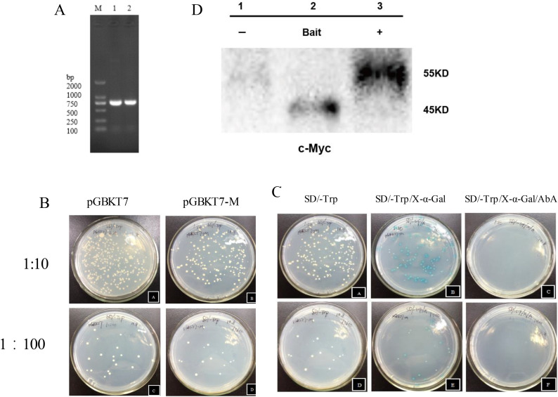
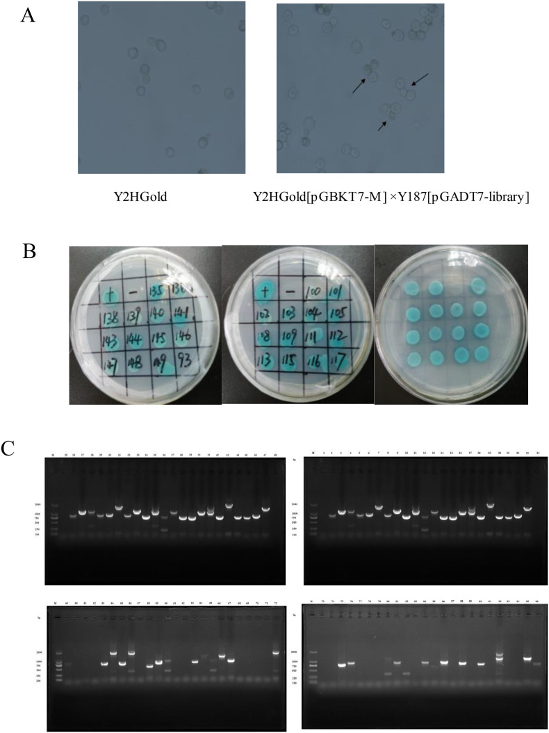
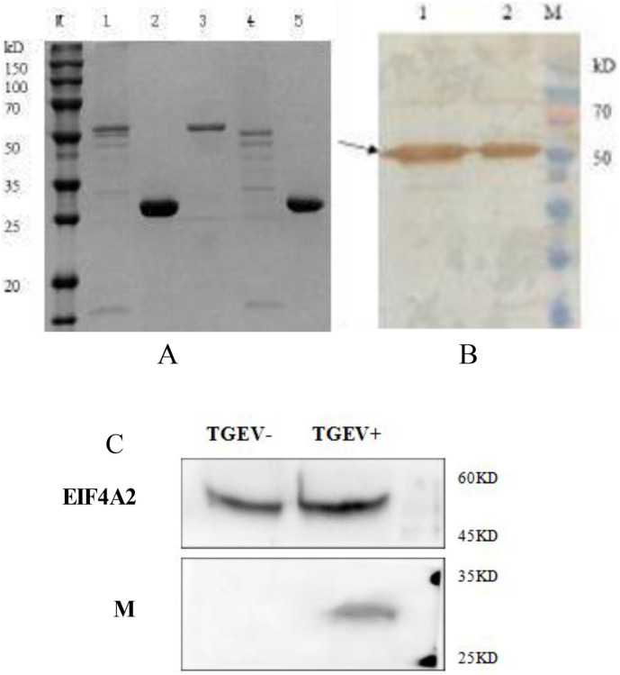
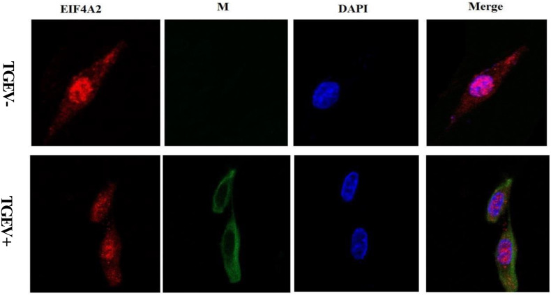
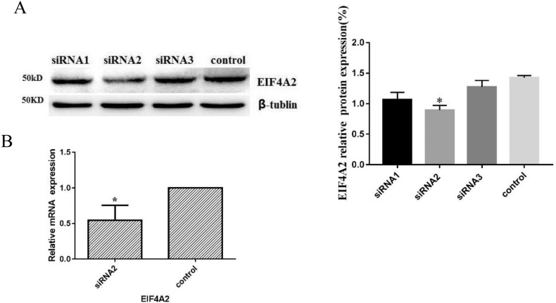
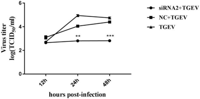
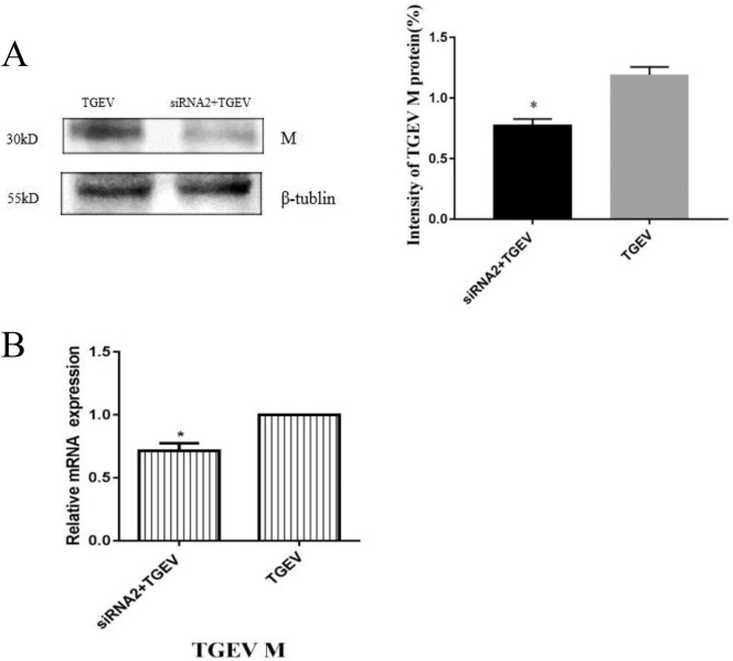
Transmissible gastroenteritis coronavirus (TGEV) is enteropathogenic coronavirus that causes diarrhea in pigs, and is associated with high morbidity and mortality in sucking piglets. The TGEV membrane (M) protein is a decisive protein for the proliferation of viral proteins, and is associated with virus assembly and budding. To identify the cellular proteins that interact with the TGEV M protein, yeast two-hybrid screening was employed, and seven cellular proteins were identified M-binding partners. Using the GST pull-down approach and a CO-IP assay, the M protein was found to interact with porcine intestinal cells via eukaryotic translation initiation factor 4-alpha (EIF4A2), an essential component of the cellular translational machinery. Additionally, confocal microscopy revealed that EIF4A2 and M were colocalized in the cytoplasm. Furthermore, the function of EIF4A2 in intestinal cells during TGEV infection was examined. A knockdown of EIF4A2 by siRNA markedly decreased M protein proliferation and TGEV replication in target cells. Thus demonstrating that EIF4A2 plays a significant role in TGEV replication. The present study provides mechanistic insight into the interaction between the TGEV M protein and intestinal cells which contributes to the understanding of coronavirus replication and may be useful for the development of novel therapeutic strategies for TGEV infection.
Keywords: Eukaryotic translation initiation factor 4-alpha (EIF4A2); Interaction; Membrane (M) protein; TGEV.
Copyright © 2018 Elsevier Ltd. All rights reserved.
Figures
Similar articles
-
UBXN1 interacts with the S1 protein of transmissible gastroenteritis coronavirus and plays a role in viral replication.Vet Res. 2019 Apr 27;50(1):28. doi: 10.1186/s13567-019-0648-9. Vet Res. 2019. PMID: 31029162 Free PMC article.
-
EF1A interacting with nucleocapsid protein of transmissible gastroenteritis coronavirus and plays a role in virus replication.Vet Microbiol. 2014 Aug 27;172(3-4):443-8. doi: 10.1016/j.vetmic.2014.05.034. Epub 2014 Jun 9. Vet Microbiol. 2014. PMID: 24974120 Free PMC article.
-
The PERK Arm of the Unfolded Protein Response Negatively Regulates Transmissible Gastroenteritis Virus Replication by Suppressing Protein Translation and Promoting Type I Interferon Production.J Virol. 2018 Jul 17;92(15):e00431-18. doi: 10.1128/JVI.00431-18. Print 2018 Aug 1. J Virol. 2018. PMID: 29769338 Free PMC article.
-
An overview of immunological and genetic methods for detecting swine coronaviruses, transmissible gastroenteritis virus, and porcine respiratory coronavirus in tissues.Adv Exp Med Biol. 1997;412:37-46. doi: 10.1007/978-1-4899-1828-4_4. Adv Exp Med Biol. 1997. PMID: 9191988 Review.
-
Transmissible gastroenteritis virus infection: a vanishing specter.Dtsch Tierarztl Wochenschr. 2006 Apr;113(4):157-9. Dtsch Tierarztl Wochenschr. 2006. PMID: 16716052 Review.
Cited by
-
Upper airway gene expression differentiates COVID-19 from other acute respiratory illnesses and reveals suppression of innate immune responses by SARS-CoV-2.medRxiv [Preprint]. 2020 May 19:2020.05.18.20105171. doi: 10.1101/2020.05.18.20105171. medRxiv. 2020. Update in: Nat Commun. 2020 Nov 17;11(1):5854. doi: 10.1038/s41467-020-19587-y. PMID: 32511476 Free PMC article. Updated. Preprint.
-
The Roles of Apoptosis in Swine Response to Viral Infection and Pathogenesis of Swine Enteropathogenic Coronaviruses.Front Vet Sci. 2020 Nov 26;7:572425. doi: 10.3389/fvets.2020.572425. eCollection 2020. Front Vet Sci. 2020. PMID: 33324698 Free PMC article. Review.
-
UBXN1 interacts with the S1 protein of transmissible gastroenteritis coronavirus and plays a role in viral replication.Vet Res. 2019 Apr 27;50(1):28. doi: 10.1186/s13567-019-0648-9. Vet Res. 2019. PMID: 31029162 Free PMC article.
-
Drug targets for COVID-19 therapeutics: Ongoing global efforts.J Biosci. 2020;45(1):87. doi: 10.1007/s12038-020-00067-w. J Biosci. 2020. PMID: 32661214 Free PMC article. Review.
-
Current understanding on molecular drug targets and emerging treatment strategy for novel coronavirus-19.Naunyn Schmiedebergs Arch Pharmacol. 2021 Jul;394(7):1383-1402. doi: 10.1007/s00210-021-02091-5. Epub 2021 May 7. Naunyn Schmiedebergs Arch Pharmacol. 2021. PMID: 33961065 Free PMC article. Review.
References
-
- Cologna R., Hogue B.G. Coronavirus nucleocapsid protein RNA interactions. Adv. Exp. Med. Biol. 1998;440:355–359. - PubMed
MeSH terms
Substances
LinkOut - more resources
Full Text Sources
Other Literature Sources
Molecular Biology Databases
Research Materials
