Review
. 2018 Dec 21;20(1):41.
doi: 10.3390/ijms20010041.

Extracellular Vesicles: New Players in Lymphomas

Affiliations
Review

Extracellular Vesicles: New Players in Lymphomas

Victor Navarro-Tableros et al. Int J Mol Sci. .

Abstract

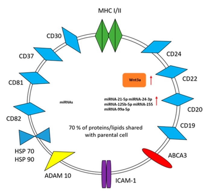
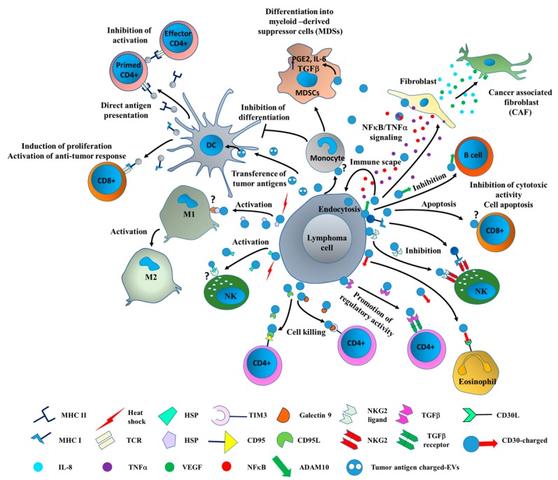
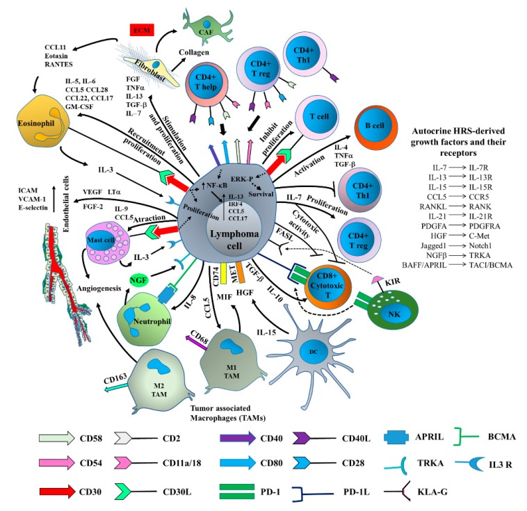
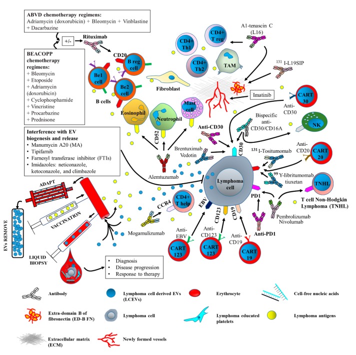
Lymphomas are heterogeneous diseases, and the term includes a number of histological subtypes that are characterized by different clinical behavior and molecular phenotypes. Valuable information on the presence of lymphoma cell-derived extracellular vesicles (LCEVs) in the bloodstream of patients suffering from this hematological cancer has recently been provided. In particular, it has been reported that the number and phenotype of LCEVs can both change as the disease progresses, as well as after treatment. Moreover, the role that LCEVs play in driving tumor immune escape has been reported. This makes LCEVs potential novel clinical tools for diagnosis, disease progression, and chemoresistance. LCEVs express surface markers and convey specific molecules in accordance with their cell of origin, which can be used as targets and thus lead to the development of specific therapeutics. This may be particularly relevant since circulating LCEVs are known to save lymphoma cells from anti-cluster of differentiation (CD)20-induced complement-dependent cytotoxicity. Therefore, effort should be directed toward investigating the feasibility of using LCEVs as predictive biomarkers of disease progression and/or response to treatment that can be translated to clinical use. The use of liquid biopsies in combination with serum EV quantification and cargo analysis have been also considered as potential approaches that can be pursued in the future. Upcoming research will also focus on the identification of specific molecular targets in order to generate vaccines and/or antibodies against LCEVs. Finally, the removal of circulating LCEVs has been proposed as a simple and non-invasive treatment approach. We herein provide an overview of the role of LCEVs in lymphoma diagnosis, immune tolerance, and drug resistance. In addition, alternative protocols that utilize LCEVs as therapeutic targets are discussed.

Keywords: extracellular vesicles; immunomodulation; innovative therapies; lymphoma.


Conflict of interest statement

The Authors declare no conflict of interest.

Figures

Figure 1
Figure 1
Lymphoma cell-derived extracellular vesicle (LCEV) cargo. LCEVs may contain nucleic acids, proteins, and lipids that resemble the content of their parental cell. The LCEV surface membrane is enriched with proteins that are involved in antigen presentation, signal transduction, adhesion, and drug resistance. Major histocompatibility complex (MHC) class I and II (MHC I/II); miRNA: micro-ribonucleic acid; ABCA3: ATP-binding cassette transporter A3; ICAM-1: Intercellular Adhesion Molecule 1; ADAM10: A Disintegrin and Metalloproteinase Domain-containing protein 10; HSP 70/90: Heat shock protein 70 and 90. Wnt3a: Wingless 3a. Red arrows indicate up-regulated molecules.
Figure 2
Figure 2
Cellular interaction in the lymphoma microenvironment (LME) driving immune escape. LCEVs contribute to tumor immune escape by coordinating the activation (black arrows) and/or inactivation (T bars) of different cells types in the tumor niche. LCEVs express different surface markers (cluster of differentiation: CD) and transport typical components resembling their cell of origin (lymphoma cell). Key molecules enriched in LCEVs interact with receptors expressed by target cells and activate intracellular signals that differentially modulate the activity of immune cells. The activation of DCs and fibroblasts further promote EV release and EV-mediated signaling events in neighboring cells. DC: Dendritic cells; NK: Natural killer cells.
Figure 3
Figure 3
Bidirectional interaction between lymphoma cells and LME components involved in tumor immune escape. Lymphoma cells avoid the immune attack by three main mechanisms: (1) direct interaction with immune cells through surface molecules, (2) production of immune suppressive cytokines, and (3) loss of target antigens. Lymphoma cells also releases molecules that promote tumor growth and stimulate new vessel formation. Dotted lines correspond to shared receptors. Arrows indicate a direct effect. T bars indicate the inhibitory activity.
Figure 4
Figure 4
Scheme of therapeutic approaches (currently used or under investigation) and liquid biopsy using LCEVs. Oncogenic drugs and specific antibodies targeting surface molecules expressed by LME components represent the main therapeutic approaches used in clinic. Current clinical trials are also evaluating new potential approaches using vaccinations with tumor antigens. ADAPT represents a potential approach to remove LCEVs from the bloodstream. Liquid biopsy has been proposed as screening method for diagnosis, disease progress, and response to therapy. New potential approaches are represented by the chimeric antigen receptor (CAR), which is introduced in immune cells to retarget their cytotoxicity toward specific tumor antigens (CART). CARs are synthetic proteins generated by the fusion of a single chain variable fragment (scFv) derived from a monoclonal antibody with the signaling and costimulatory machinery of the T-cell receptor (TCR) [107]. CART cells have been evaluated as promising new cell-based therapy approach using CD19 [108], CD30, CD123 [109], and the Epstein-Barr virus protein (EBV) as molecular targets [110]. ADAPT: Adaptive dynamic artificial poly-ligand targeting.

References

    1. Lynch R.C., Gratzinger D., Advani R.H. Clinical Impact of the 2016 Update to the WHO Lymphoma Classification. Curr. Treat. Options Oncol. 2017 doi: 10.1007/s11864-017-0483-z. - DOI - PubMed
    1. Swerdlow S.H., Campo E., Pileri S.A., Harris N.L., Stein H., Siebert R., Advani R., Ghielmini M., Salles G.A., Zelenetz A.D., et al. The 2016 revision to the World Health Organization (WHO) classification of lymphoid neoplasms. Blood. 2016 doi: 10.1182/blood-2016-01-643569. - DOI - PMC - PubMed
    1. Galardy P.J., Bedekovics T., Hermiston M.L. Targeting childhood, adolescent and young adult non-Hodgkin lymphoma: Therapeutic horizons. Br. J. Haematol. 2016 doi: 10.1111/bjh.14016. - DOI - PubMed
    1. Tarte K. Role of the microenvironment across histological subtypes of NHL. Hematology. 2017 doi: 10.1182/asheducation-2017.1.610. - DOI - PMC - PubMed
    1. Hanahan D., Weinberg R.A. Hallmarks of cancer: The next generation. Cell. 2011 doi: 10.1016/j.cell.2011.02.013. - DOI - PubMed

Substances