

Visualization of Annular Gap Junction Vesicle Processing: The Interplay Between Annular Gap Junctions and Mitochondria
- PMID: 30583492
- PMCID: PMC6337258
- DOI: 10.3390/ijms20010044
Visualization of Annular Gap Junction Vesicle Processing: The Interplay Between Annular Gap Junctions and Mitochondria
Abstract
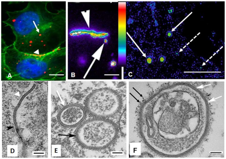
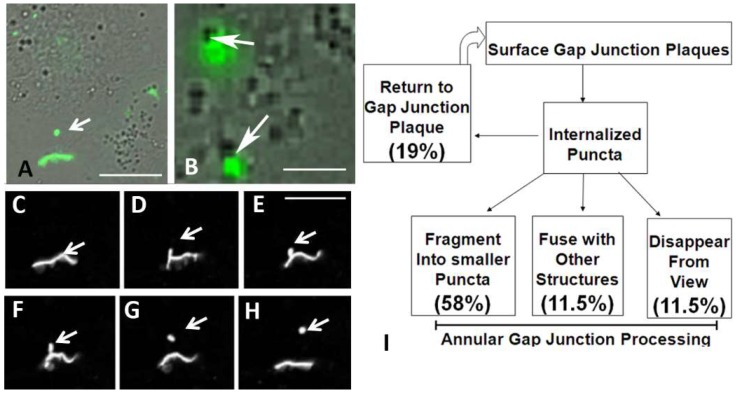
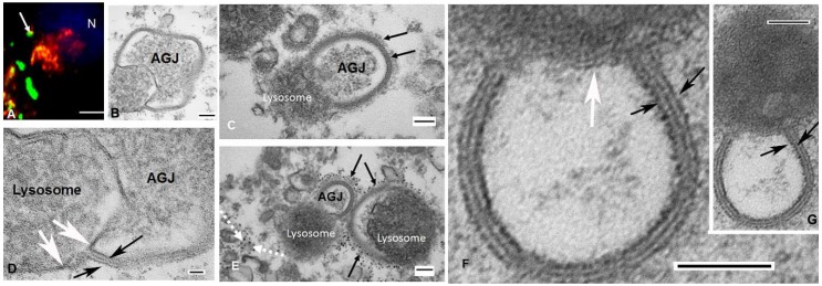
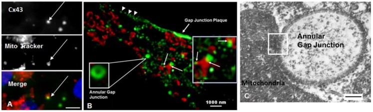
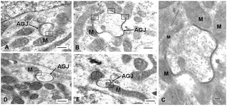
It is becoming clear that in addition to gap junctions playing a role in cell⁻cell communication, gap junction proteins (connexins) located in cytoplasmic compartments may have other important functions. Mitochondrial connexin 43 (Cx43) is increased after ischemic preconditioning and has been suggested to play a protective role in the heart. How Cx43 traffics to the mitochondria and the interactions of mitochondria with other Cx43-containing structures are unclear. In this study, immunocytochemical, super-resolution, and transmission electron microscopy were used to detect cytoplasmic Cx43-containing structures and to demonstrate their interactions with other cytoplasmic organelles. The most prominent cytoplasmic Cx43-containing structures-annular gap junctions-were demonstrated to form intimate associations with lysosomes as well as with mitochondria. Surprisingly, the frequency of associations between mitochondria and annular gap junctions was greater than that between lysosomes and annular gap junctions. The benefits of annular gap junction/mitochondrial associations are not known. However, it is tempting to suggest, among other possibilities, that the contact between annular gap junction vesicles and mitochondria facilitates Cx43 delivery to the mitochondria. Furthermore, it points to the need for investigating annular gap junctions as more than only vesicles destined for degradation.
Keywords: annular gap junction vesicle; connexin; gap junction; lysosome; mitochondria.
Conflict of interest statement
The authors declare no conflict of interest
Figures
Similar articles
-
Redistribution of connexin 43 during cell division.Cell Biol Int. 2016 Apr;40(4):387-96. doi: 10.1002/cbin.10576. Epub 2016 Jan 21. Cell Biol Int. 2016. PMID: 26724787 Free PMC article.
-
Molecular mechanisms regulating formation, trafficking and processing of annular gap junctions.BMC Cell Biol. 2016 May 24;17 Suppl 1(Suppl 1):22. doi: 10.1186/s12860-016-0087-7. BMC Cell Biol. 2016. PMID: 27230503 Free PMC article. Review.
-
Ubiquitylation of the gap junction protein connexin-43 signals its trafficking from early endosomes to lysosomes in a process mediated by Hrs and Tsg101.J Cell Sci. 2009 Nov 1;122(Pt 21):3883-93. doi: 10.1242/jcs.053801. Epub 2009 Oct 6. J Cell Sci. 2009. PMID: 19808888
-
Endocytosis of connexin protein in adrenal cells.Endocr Res. 2004 Nov;30(4):647-54. doi: 10.1081/erc-200043942. Endocr Res. 2004. PMID: 15666807
-
Analysis of the function and therapeutic strategy of connexin 43 from its subcellular localization.Biochimie. 2024 Mar;218:1-7. doi: 10.1016/j.biochi.2023.08.011. Epub 2023 Aug 22. Biochimie. 2024. PMID: 37611889 Review.
Cited by
-
Connexin/Innexin Channels in Cytoplasmic Organelles. Are There Intracellular Gap Junctions? A Hypothesis!Int J Mol Sci. 2020 Mar 21;21(6):2163. doi: 10.3390/ijms21062163. Int J Mol Sci. 2020. PMID: 32245189 Free PMC article.
-
Subcellular Localization of Connexin 26 in Cardiomyocytes and in Cardiomyocyte-Derived Extracellular Vesicles.Molecules. 2021 Nov 6;26(21):6726. doi: 10.3390/molecules26216726. Molecules. 2021. PMID: 34771134 Free PMC article.
-
Mitochondrial Connexins and Mitochondrial Contact Sites with Gap Junction Structure.Int J Mol Sci. 2023 May 20;24(10):9036. doi: 10.3390/ijms24109036. Int J Mol Sci. 2023. PMID: 37240383 Free PMC article. Review.
-
Mitochondrial transfer/transplantation: an emerging therapeutic approach for multiple diseases.Cell Biosci. 2022 May 19;12(1):66. doi: 10.1186/s13578-022-00805-7. Cell Biosci. 2022. PMID: 35590379 Free PMC article. Review.
-
Connexins in the Heart: Regulation, Function and Involvement in Cardiac Disease.Int J Mol Sci. 2021 Apr 23;22(9):4413. doi: 10.3390/ijms22094413. Int J Mol Sci. 2021. PMID: 33922534 Free PMC article. Review.
References
MeSH terms
Substances
Grants and funding
LinkOut - more resources
Full Text Sources
Miscellaneous
