

Transcriptional Programs and Regeneration Enhancers Underlying Heart Regeneration
- PMID: 30583498
- PMCID: PMC6463103
- DOI: 10.3390/jcdd6010002
Transcriptional Programs and Regeneration Enhancers Underlying Heart Regeneration
Abstract
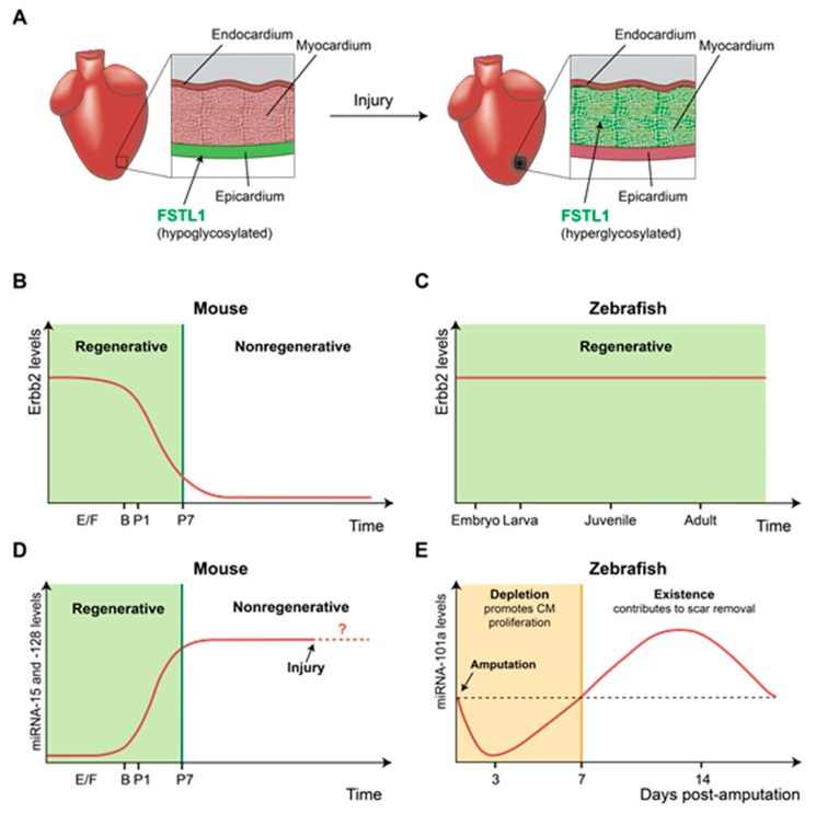
The heart plays the vital role of propelling blood to the entire body, which is essential to life. While maintaining heart function is critical, adult mammalian hearts poorly regenerate damaged cardiac tissue upon injury and form scar tissue instead. Unlike adult mammals, adult zebrafish can regenerate injured hearts with no sign of scarring, making zebrafish an ideal model system with which to study the molecular mechanisms underlying heart regeneration. Investigation of heart regeneration in zebrafish together with mice has revealed multiple cardiac regeneration genes that are induced by injury to facilitate heart regeneration. Altered expression of these regeneration genes in adult mammals is one of the main causes of heart regeneration failure. Previous studies have focused on the roles of these regeneration genes, yet the regulatory mechanisms by which the expression of cardiac regeneration genes is precisely controlled are largely unknown. In this review, we will discuss the importance of differential gene expression for heart regeneration, the recent discovery of cardiac injury or regeneration enhancers, and their impact on heart regeneration.
Keywords: development; enhancer; gene regulation; heart; regeneration; transcription; zebrafish.
Conflict of interest statement
The authors declare no conflict of interest.
Figures
Similar articles
-
A Roadmap to Heart Regeneration Through Conserved Mechanisms in Zebrafish and Mammals.Curr Cardiol Rep. 2021 Mar 2;23(4):29. doi: 10.1007/s11886-021-01459-6. Curr Cardiol Rep. 2021. PMID: 33655359 Free PMC article. Review.
-
Extensive scar formation and regression during heart regeneration after cryoinjury in zebrafish.Development. 2011 May;138(9):1663-74. doi: 10.1242/dev.060897. Epub 2011 Mar 23. Development. 2011. PMID: 21429987
-
Midkine-a Regulates the Formation of a Fibrotic Scar During Zebrafish Heart Regeneration.Front Cell Dev Biol. 2021 May 7;9:669439. doi: 10.3389/fcell.2021.669439. eCollection 2021. Front Cell Dev Biol. 2021. PMID: 34026760 Free PMC article.
-
Heart regeneration in zebrafish.Science. 2002 Dec 13;298(5601):2188-90. doi: 10.1126/science.1077857. Science. 2002. PMID: 12481136
-
Advances in Cardiac Development and Regeneration Using Zebrafish as a Model System for High-Throughput Research.J Dev Biol. 2021 Sep 25;9(4):40. doi: 10.3390/jdb9040040. J Dev Biol. 2021. PMID: 34698193 Free PMC article. Review.
Cited by
-
Molecular Pathways Modulating Sensory Hair Cell Regeneration in Adult Mammalian Cochleae: Progress and Perspectives.Int J Mol Sci. 2021 Dec 22;23(1):66. doi: 10.3390/ijms23010066. Int J Mol Sci. 2021. PMID: 35008497 Free PMC article. Review.
-
Zebra-Fishing for Regenerative Awakening in Mammals.Biomedicines. 2021 Jan 12;9(1):65. doi: 10.3390/biomedicines9010065. Biomedicines. 2021. PMID: 33445518 Free PMC article. Review.
-
Cardiac enhancers: Gateway to the regulatory mechanisms of heart regeneration.Semin Cell Dev Biol. 2025 Jun;170:103610. doi: 10.1016/j.semcdb.2025.103610. Epub 2025 Apr 10. Semin Cell Dev Biol. 2025. PMID: 40215762 Review.
-
The Role of ncRNAs in Cardiac Infarction and Regeneration.J Cardiovasc Dev Dis. 2023 Mar 15;10(3):123. doi: 10.3390/jcdd10030123. J Cardiovasc Dev Dis. 2023. PMID: 36975887 Free PMC article. Review.
-
Regeneration and developmental enhancers are differentially compatible with minimal promoters.Dev Biol. 2022 Dec;492:47-58. doi: 10.1016/j.ydbio.2022.09.007. Epub 2022 Sep 24. Dev Biol. 2022. PMID: 36167150 Free PMC article.
References
-
- Grivas J., Haag M., Johnson A., Manalo T., Roell J., Das T.L., Brown E., Burns A.R., Lafontant P.J. Cardiac repair and regenerative potential in the goldfish (Carassius auratus) heart. Comp. Biochem. Physiol. C Toxicol. Pharmacol. 2014;163:14–23. doi: 10.1016/j.cbpc.2014.02.002. - DOI - PMC - PubMed
Publication types
Grants and funding
- AHA16SDG30020001/American Heart Association
- AAC8355/School of Medicine and Public Health, University of Wisconsin-Madison
- AAC6429/Office of the Vice Chancellor for Research and Graduate Education, University of Wisconsin-Madison
- AAC8979/Stem Cell and Regenerative Medicine Center, University of Wisconsin-Madison
LinkOut - more resources
Full Text Sources
