. 2018 Dec 22;11(1):16.
doi: 10.3390/cancers11010016.

Mifepristone Overcomes Tumor Resistance to Temozolomide Associated with DNA Damage Repair and Apoptosis in an Orthotopic Model of Glioblastoma

Affiliations

Mifepristone Overcomes Tumor Resistance to Temozolomide Associated with DNA Damage Repair and Apoptosis in an Orthotopic Model of Glioblastoma

Monserrat Llaguno-Munive et al. Cancers (Basel). .

Abstract

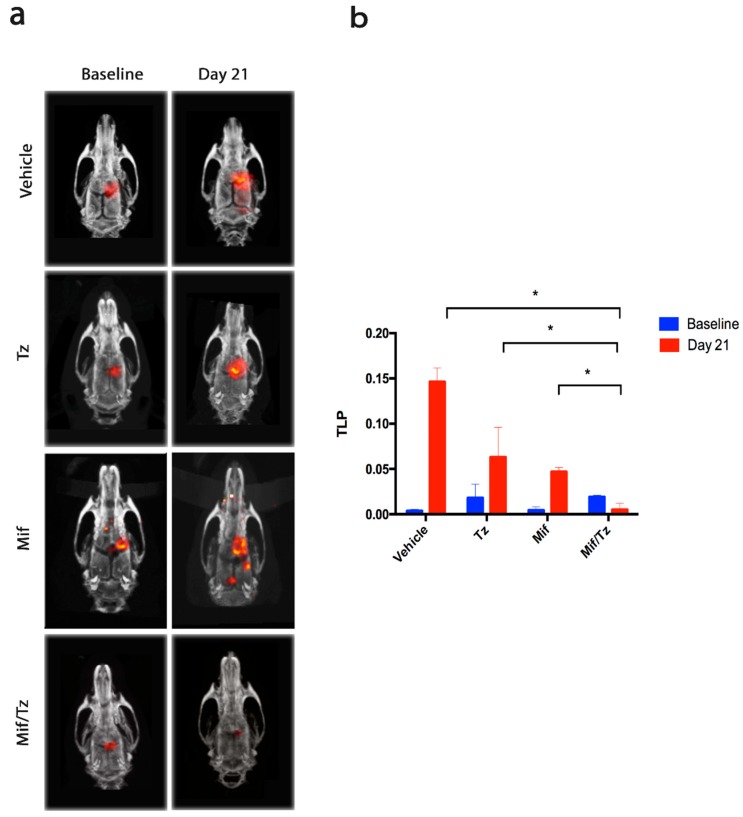
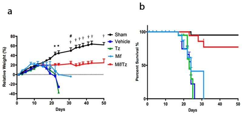
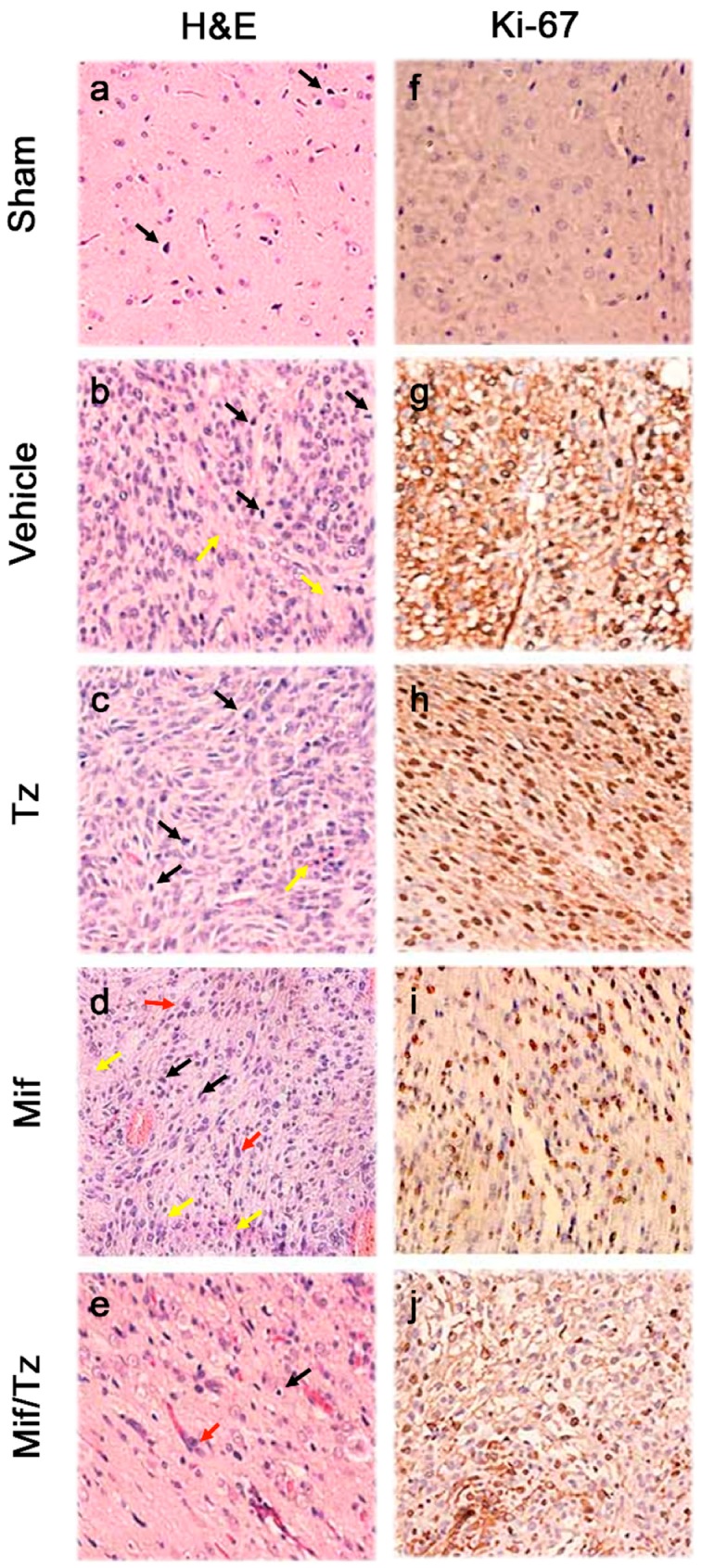
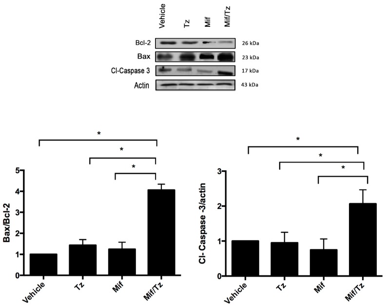
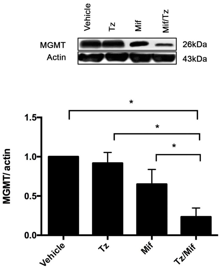
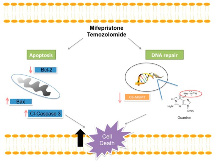
The standard treatment for glioblastoma multiforme (GBM) is surgery followed by chemo/radiotherapy. A major limitation on patient improvement is the high resistance of tumors to drug treatment, likely responsible for their subsequent recurrence and rapid progression. Therefore, alternatives to the standard therapy are necessary. The aim of the present study was to evaluate whether mifepristone, an antihormonal agent, has a synergistic effect with temozolomide (used in standard therapy for gliomas). Whereas the mechanism of temozolomide involves damage to tumor DNA leading to apoptosis, tumor resistance is associated with DNA damage repair through the O⁶-methylguanine-DNA-methyltransferase (MGMT) enzyme. Temozolomide/mifepristone treatment, herein examined in Wistar rats after orthotopically implanting C6 glioma cells, markedly reduced proliferation. This was evidenced by a decreased level of the following parameters: a proliferation marker (Ki-67), a tumor growth marker (18F-fluorothymidine uptake, determined by PET/CT images), and the MGMT enzyme. Increased apoptosis was detected by the relative expression of related proteins, (e.g. Bcl-2 (B-cell lymphoma 2), Bax (bcl-2-like protein 4) and caspase-3). Thus, greater apoptosis of tumor cells caused by their diminished capacity to repair DNA probably contributed significantly to the enhanced activity of temozolomide. The results suggest that mifepristone could possibly act as a chemo-sensitizing agent for temozolomide during chemotherapy for GBM.

Keywords: MGMT; apoptosis; drug resistance; glioblastoma; mifepristone; temozolomide.


Conflict of interest statement

The authors declare no conflict of interest.

Figures

Figure 1
Figure 1
Antitumor activity in the orthotopic rat model of a glioma. (a) PET/CT images showing 18F-FLT (18F-fluorothymidine) tumor uptake for the distinct groups. The images on the left represent the beginning of drug treatment (baseline, two weeks after tumor cell implantation, considered as day 0) and those on the right depict the end of the third week of treatment (day 21). Red reflects the uptake of (18F-FLT). (b) The proliferative activity of tumors, measured as total lesion proliferation (TLP). Data are expressed as the mean ± SEM of three animals. * A significant difference (p < 0.05) between the Mifeprostone/Temozolomide (Mif/Tz) group and the other groups with implantation of glioma cells (the vehicle only, Tz only and Mif only).
Figure 2
Figure 2
The effect of tumor growth in the orthotopic rat model of a glioma, evaluated by weight loss and overall survival. (a) Relative weight of rats in the five groups: sham surgery (without implanting glioma cells) (●), and the implantation of glioma cells followed by each of the treatments: vehicle only (), temozolomide only (), mifepristone only (), and the mifepristone/temozolomide combination (). Each point represents the mean ± SEM of six animals. * A significant difference (p < 0.05) between Mif/Tz and the other groups (Sham, Control, Tz only and Mif only) on day 24. # A significant difference (p < 0.05) between Mif/Tz and Mif only on day 31. † A significant difference (p < 0.05) between the Mif/Tz and the Sham group on day 31. (b) Survival analysis of the same groups for up to 50 days after implantation (day 0 = day of surgery).
Figure 3
Figure 3
Haematoxylin and eosin (H&E) staining and immunohistochemical analysis of glioma tissue. Sections of tumor tissue were stained with haematoxylin and eosin (ae): Mitosis (black arrows), nuclear pleomorphism (red arrows) and necrosis (yellow arrows). Immunostaining of Ki-67 (fj), showing few cell nuclei positive to this protein in the tumors of the Mif/Tz group compared to those of the individual treatments (Tz or Mif). The images are representative of three animals per treatment. Magnification 40×.
Figure 4
Figure 4
Expression of apoptotic proteins. Representative western blot results and densitometric analysis of Bcl-2, Bax and Cl-Caspase 3. Data are expressed as the mean ± SEM from three independent experiments. * Indicates a significant difference (p < 0.05) between Mif/Tz and the other groups with tumor cell implantation (given Tz, Mif or the vehicle).
Figure 5
Figure 5
Expression of MGMT (O6-methylguanine-DNA-methyltransferase). Representative western blot results and densitometric analysis of the MGMT protein. Data are expressed as the mean ± SEM of three independent experiments. * A significant difference (p < 0.05) between Mif/Tz and the other groups with tumor cell implantation (given Tz, Mif or the vehicle).
Figure 6
Figure 6
Schematic portrayal of the possible mechanisms of treatment with mifepristone/temozolomide. This combination decreased the level of anti-apoptotic protein Bcl-2 and increased the levels of pro-apoptotic proteins Bax and cl-caspase-3, leading to greater cellular apoptosis. As described elsewhere, temozolomide releases a compound, O6-guanine, that causes DNA damage and cell death. However, there are different mechanisms of repair or avoidance employed by tumor cells. For example, resistance to temozolomide treatment is reportedly related to the presence of the MGMT enzyme, which removes the methyl group from O6-guanine and thus restores the cellular replication of tumor cells. A lower expression of MGMT was herein detected when administering mifepristone/temozolomide versus temozolomide alone. The combination treatment perhaps allows methylation to occur in the purine bases of DNA at the O6 position of guanine without removing the methyl groups, and consequently contributes to an increase in the effect of temozolomide.

References

    1. Johnson D.R., O’Neill B.P. Glioblastoma survival in the United States before and during the temozolomide era. J. Neurooncol. 2012;107:359–364. doi: 10.1007/s11060-011-0749-4. - DOI - PubMed
    1. Cardoso F.L., Brites D., Brito M.A. Looking at the blood-brain barrier: Molecular anatomy and possible investigation approaches. Brain Res. Rev. 2010;64:328–363. doi: 10.1016/j.brainresrev.2010.05.003. - DOI - PubMed
    1. van Tellingen O., Yetkin-Arik B., de Gooijer M.C., Wesseling P., Wurdinger T., de Vries H.E. Overcoming the blood-brain tumor barrier for effective glioblastoma treatment. Drug Resist. Updat. 2015;19:1–12. doi: 10.1016/j.drup.2015.02.002. - DOI - PubMed
    1. Ramirez Y.P., Weatherbee J.L., Wheelhouse R.T., Ross A.H. Glioblastoma multiforme therapy and mechanisms of resistance. Pharmaceuticals. 2013;6:1475–1506. doi: 10.3390/ph6121475. - DOI - PMC - PubMed
    1. Denny B.J., Wheelhouse R.T., Stevens M.F., Tsang L.L., Slack J.A. NMR and molecular modeling investigation of the mechanism of activation of the antitumor drug temozolomide and its interaction with DNA. Biochemistry. 1994;33:9045–9051. doi: 10.1021/bi00197a003. - DOI - PubMed