

Secondary extramedullary plasmacytoma of sigmoid colon in a patient with multiple myeloma: a case report
- PMID: 30583721
- PMCID: PMC6305571
- DOI: 10.1186/s13256-018-1888-4
Secondary extramedullary plasmacytoma of sigmoid colon in a patient with multiple myeloma: a case report
Abstract
Background: Extramedullary plasmacytoma is an uncommon tumor that most often involves the nasopharynx or upper respiratory tract. Extramedullary plasmacytoma is a type of plasma cell neoplasm that can present as a primary tumor or secondary to another plasma cell neoplasm, such as multiple myeloma. Secondary extramedullary plasmacytoma is usually noted in the advanced stages of the disease. Involvement of the gastrointestinal tract occurs in approximately 10% of cases.
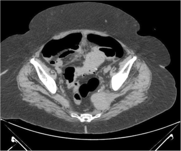
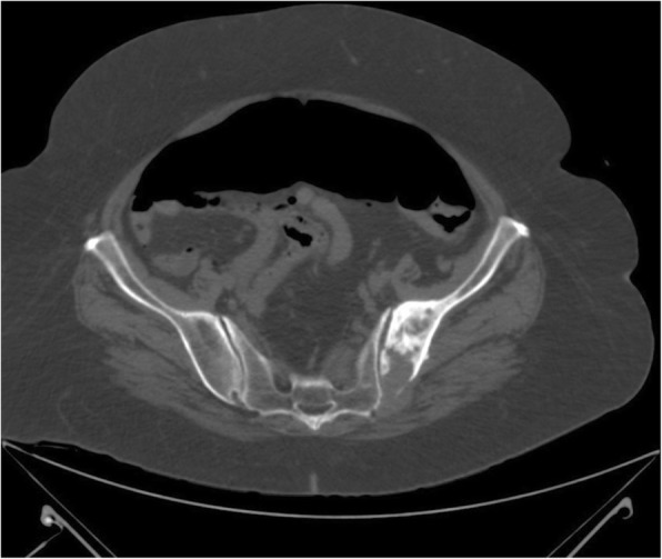
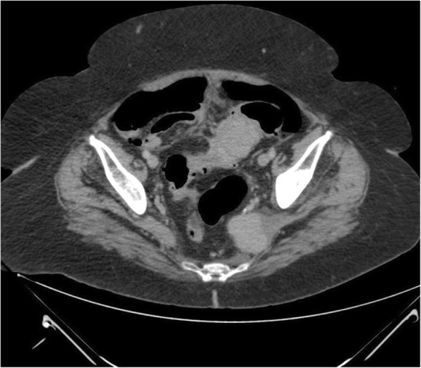
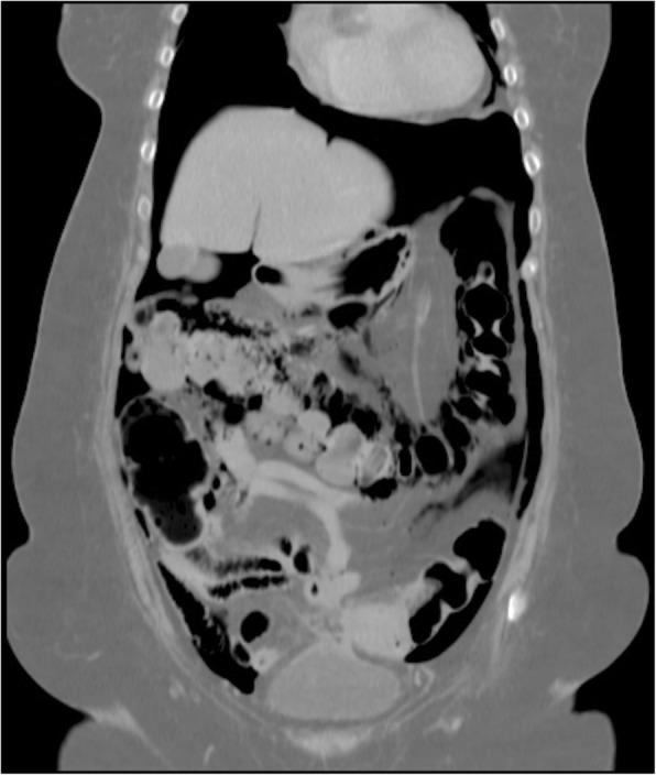
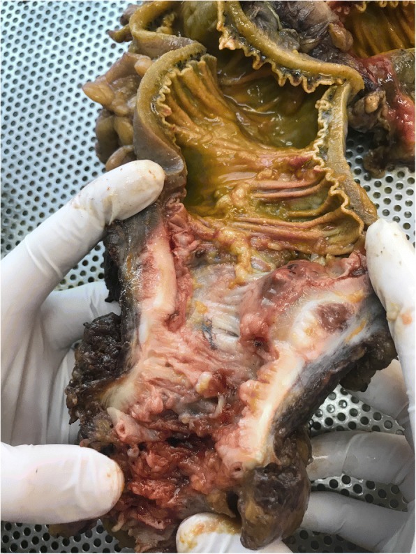
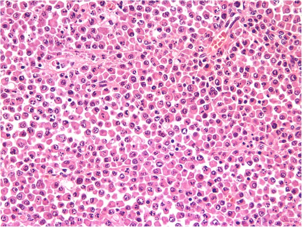
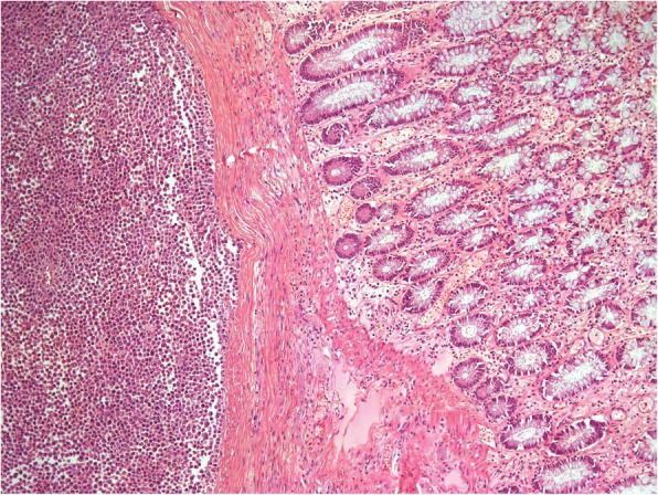
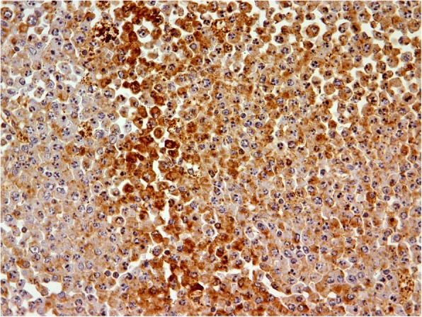
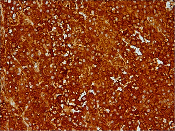
Case presentation: A 71-year-old Caucasian woman with known diverticular disease of the colon and multiple myeloma diagnosed 3 years previously, with monoclonal bands of immunoglobulin A, lambda light chains, and multiple osteolytic lesions, presented to our hospital with abdominal pain, abdominal discomfort, and pneumoperitoneum. She underwent left colectomy for diverticulitis with perforation, and an extramedullary secondary colonic plasmacytoma was found in histopathological examination of the sigmoid colon.
Conclusions: Plasmacytoma is known to occur in extraosseous sites. The stomach and small intestine are the most commonly involved sites in the gastrointestinal tract. Secondary extramedullary plasmacytoma of the colon is rare. Colonic plasmacytoma may have varying clinical presentations, such as inflammatory bowel disease and multiple colonic strictures. Although these cases are rare, treating physicians as well as radiologists, pathologists, and surgeons should be aware of this entity.
Keywords: Multiple myeloma; Oncology; Secondary extramedullary plasmacytoma; Sigmoid colon.
Conflict of interest statement
Ethics approval and consent to participate
Not applicable.
Consent for publication
Written informed consent was obtained from the patient for publication of this case report and any accompanying images. A copy of the written consent is available for review by the Editor-in-Chief of this journal.
Competing interests
The authors declare that they have no competing interests.
Publisher’s Note
Springer Nature remains neutral with regard to jurisdictional claims in published maps and institutional affiliations.
Figures
Similar articles
-
Plasmacytoma as a cause of small bowel obstruction in a virgin abdomen in a patient with multiple myeloma: a case report.J Med Case Rep. 2019 May 17;13(1):148. doi: 10.1186/s13256-019-2068-x. J Med Case Rep. 2019. PMID: 31097019 Free PMC article.
-
Primary isolated extramedullary plasmacytoma of colon.World J Surg Oncol. 2007 Apr 30;5:47. doi: 10.1186/1477-7819-5-47. World J Surg Oncol. 2007. PMID: 17470287 Free PMC article.
-
Primary extramedullary plasmacytoma of the sigmoid colon with perforation: a case report.Surg Case Rep. 2018 Apr 4;4(1):28. doi: 10.1186/s40792-018-0437-0. Surg Case Rep. 2018. PMID: 29619633 Free PMC article.
-
Plasmacytoma of the sigmoid colon associated with a diverticular stricture: case report and review of the literature.J R Coll Surg Edinb. 1997 Feb;42(1):47-9. J R Coll Surg Edinb. 1997. PMID: 9046147 Review.
-
Extramedullary presentation of multiple myeloma in the parotid gland as first evidence of the disease -- a review with case report.Niger Postgrad Med J. 2005 Mar;12(1):45-8. Niger Postgrad Med J. 2005. PMID: 15827597 Review.
Cited by
-
Extraosseous Plasmacytomas: A Radiologist's Perspective-A Narrative Review of the Literature.Diagnostics (Basel). 2024 Aug 16;14(16):1788. doi: 10.3390/diagnostics14161788. Diagnostics (Basel). 2024. PMID: 39202276 Free PMC article. Review.
-
Lower Gastrointestinal Involvement in Multiple Myeloma: A Case Report.Int Med Case Rep J. 2020 Aug 21;13:353-357. doi: 10.2147/IMCRJ.S266203. eCollection 2020. Int Med Case Rep J. 2020. PMID: 32922092 Free PMC article.
-
Intestinal perforation with abdominal abscess caused by extramedullary plasmacytoma of small intestine: A case report and literature review.World J Gastrointest Surg. 2022 Jun 27;14(6):611-620. doi: 10.4240/wjgs.v14.i6.611. World J Gastrointest Surg. 2022. PMID: 35979418 Free PMC article.
-
Extramedullary relapse in a patient with multiple myeloma: a rare cause of gastrointestinal perforation and massive bleeding.BMJ Case Rep. 2021 Nov 18;14(11):e243663. doi: 10.1136/bcr-2021-243663. BMJ Case Rep. 2021. PMID: 34794974 Free PMC article.
References
Publication types
MeSH terms
LinkOut - more resources
Full Text Sources
Medical
