

Automated classification of Alzheimer's disease and mild cognitive impairment using a single MRI and deep neural networks
- PMID: 30584016
- PMCID: PMC6413333
- DOI: 10.1016/j.nicl.2018.101645
Automated classification of Alzheimer's disease and mild cognitive impairment using a single MRI and deep neural networks
Abstract
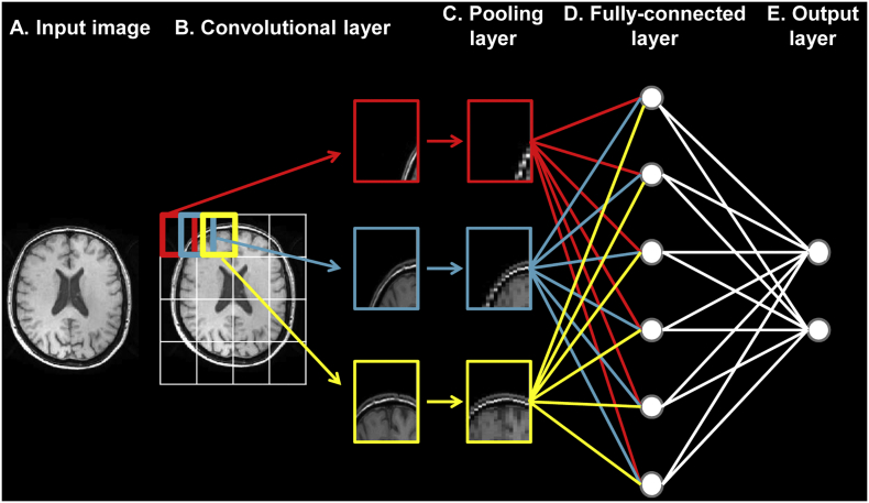
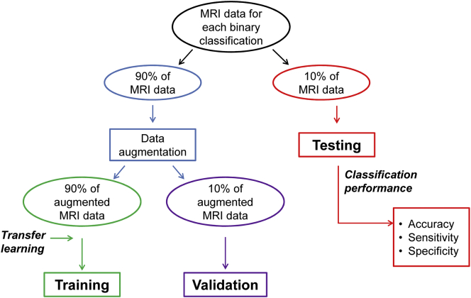
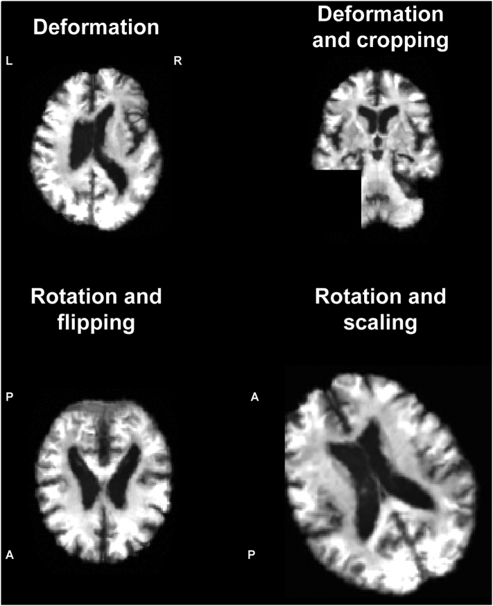
We built and validated a deep learning algorithm predicting the individual diagnosis of Alzheimer's disease (AD) and mild cognitive impairment who will convert to AD (c-MCI) based on a single cross-sectional brain structural MRI scan. Convolutional neural networks (CNNs) were applied on 3D T1-weighted images from ADNI and subjects recruited at our Institute (407 healthy controls [HC], 418 AD, 280 c-MCI, 533 stable MCI [s-MCI]). CNN performance was tested in distinguishing AD, c-MCI and s-MCI. High levels of accuracy were achieved in all the classifications, with the highest rates achieved in the AD vs HC classification tests using both the ADNI dataset only (99%) and the combined ADNI + non-ADNI dataset (98%). CNNs discriminated c-MCI from s-MCI patients with an accuracy up to 75% and no difference between ADNI and non-ADNI images. CNNs provide a powerful tool for the automatic individual patient diagnosis along the AD continuum. Our method performed well without any prior feature engineering and regardless the variability of imaging protocols and scanners, demonstrating that it is exploitable by not-trained operators and likely to be generalizable to unseen patient data. CNNs may accelerate the adoption of structural MRI in routine practice to help assessment and management of patients.
Keywords: Alzheimer's disease; Convolutional neural networks; Deep learning; Diagnosis; Mild cognitive impairment; Prediction.
Copyright © 2018 The Authors. Published by Elsevier Inc. All rights reserved.
Figures
References
-
- Albert M.S., DeKosky S.T., Dickson D., Dubois B., Feldman H.H., Fox N.C., Gamst A., Holtzman D.M., Jagust W.J., Petersen R.C., Snyder P.J., Carrillo M.C., Thies B., Phelps C.H. The diagnosis of mild cognitive impairment due to Alzheimer's disease: recommendations from the National Institute on Aging-Alzheimer's Association workgroups on diagnostic guidelines for Alzheimer's disease. Alzheimers Dement. 2011;7:270–279. - PMC - PubMed
-
- Ashburner J. A fast diffeomorphic image registration algorithm. NEUROIMAGE. 2007;38:95–113. - PubMed
-
- Bozzali M., Filippi M., Magnani G., Cercignani M., Franceschi M., Schiatti E., Castiglioni S., Mossini R., Falautano M., Scotti G., Comi G., Falini A. The contribution of voxel-based morphometry in staging patients with mild cognitive impairment. NEUROLOGY. 2006;67:453–460. - PubMed
-
- Dubois B., Feldman H.H., Jacova C., Hampel H., Molinuevo J.L., Blennow K., DeKosky S.T., Gauthier S., Selkoe D., Bateman R., Cappa S., Crutch S., Engelborghs S., Frisoni G.B., Fox N.C., Galasko D., Habert M.O., Jicha G.A., Nordberg A., Pasquier F., Rabinovici G., Robert P., Rowe C., Salloway S., Sarazin M., Epelbaum S., de Souza L.C., Vellas B., Visser P.J., Schneider L., Stern Y., Scheltens P., Cummings J.L. Advancing research diagnostic criteria for Alzheimer's disease: the IWG-2 criteria. Lancet Neurol. 2014;13:614–629. - PubMed
Publication types
MeSH terms
Grants and funding
LinkOut - more resources
Full Text Sources
Other Literature Sources
Medical
