. 2019 Jan 8;116(2):556-565.
doi: 10.1073/pnas.1808618116. Epub 2018 Dec 24.

LAMP-2B regulates human cardiomyocyte function by mediating autophagosome-lysosome fusion

Affiliations

LAMP-2B regulates human cardiomyocyte function by mediating autophagosome-lysosome fusion

Congwu Chi et al. Proc Natl Acad Sci U S A. .

Abstract

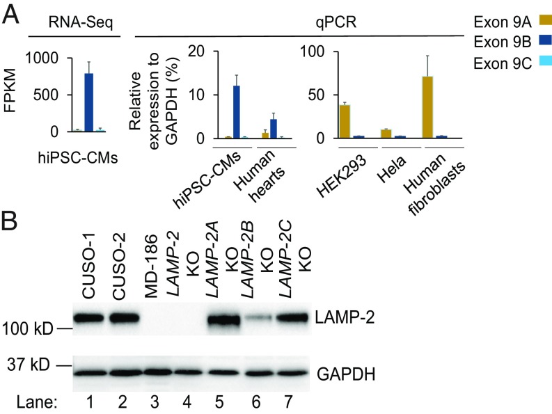
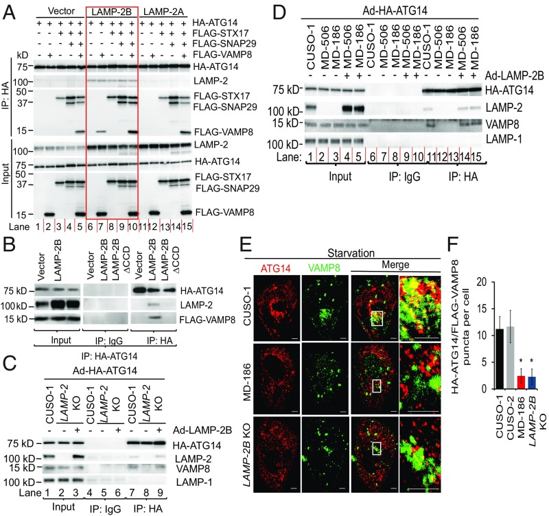
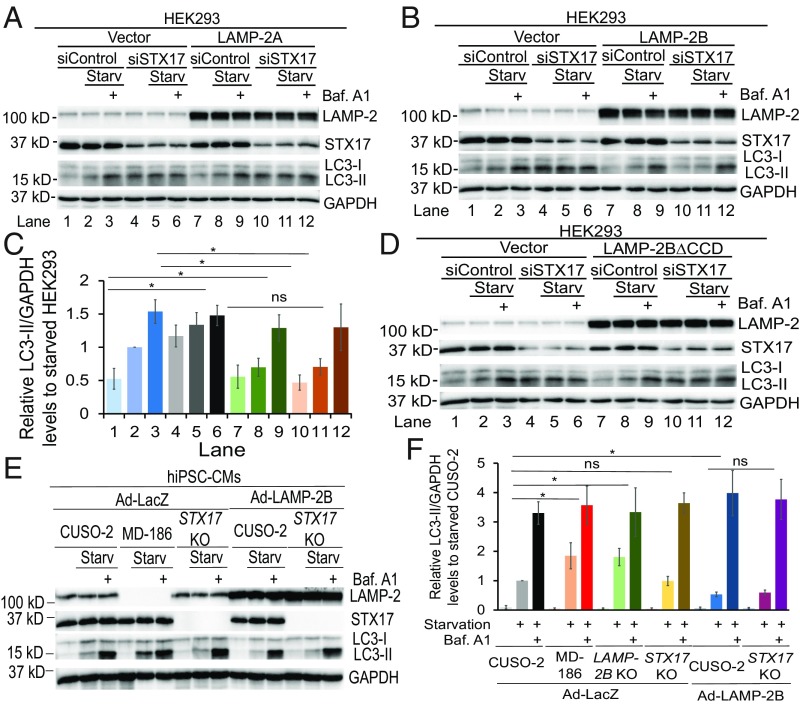
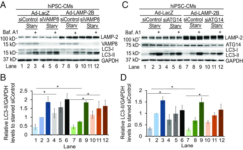
Mutations in lysosomal-associated membrane protein 2 (LAMP-2) gene are associated with Danon disease, which often leads to cardiomyopathy/heart failure through poorly defined mechanisms. Here, we identify the LAMP-2 isoform B (LAMP-2B) as required for autophagosome-lysosome fusion in human cardiomyocytes (CMs). Remarkably, LAMP-2B functions independently of syntaxin 17 (STX17), a protein that is essential for autophagosome-lysosome fusion in non-CMs. Instead, LAMP-2B interacts with autophagy related 14 (ATG14) and vesicle-associated membrane protein 8 (VAMP8) through its C-terminal coiled coil domain (CCD) to promote autophagic fusion. CMs derived from induced pluripotent stem cells (hiPSC-CMs) from Danon patients exhibit decreased colocalization between ATG14 and VAMP8, profound defects in autophagic fusion, as well as mitochondrial and contractile abnormalities. This phenotype was recapitulated by LAMP-2B knockout in non-Danon hiPSC-CMs. Finally, gene correction of LAMP-2 mutation rescues the Danon phenotype. These findings reveal a STX17-independent autophagic fusion mechanism in human CMs, providing an explanation for cardiomyopathy in Danon patients and a foundation for targeting defective LAMP-2B-mediated autophagy to treat this patient population.

Keywords: Danon disease; LAMP-2B; autophagosome–lysosome fusion; autophagy; cardiomyopathy.


Conflict of interest statement

Conflict of interest statement: E.M.S. is the recipient of a research grant from Novartis Pharmaceuticals that is not related to the present study.

Figures

Fig. 1.
Fig. 1.
LAMP-2B is the predominant LAMP-2 isoform expressed in human CMs. (A) Expression of the three LAMP-2 isoforms in different types of cells was examined by RNA-seq or qPCR. RNAs from three human hearts were used for qPCR. FPKM, fragments per kilobase of exon per million reads. (B) Immunoblotting analysis of LAMP-2 expression in indicated hiPSC-CMs. Quantification of LAMP-2 from three independent experiments is shown in SI Appendix, Fig. S3C.
Fig. 2.
Fig. 2.
LAMP-2B deficiency is sufficient to cause defects in autophagosome–lysosome fusion. (A) Autophagic flux in control and LAMP-2 isoform-specific knockout hiPSC-CMs under regular and starvation conditions was assayed. Cells were cultured in either regular or starvation medium with or without 400 nM bafilomycin A1 for 4 h followed by immunoblotting analysis of indicated proteins. Quantification of LC3-II/GAPDH from three independent experiments is shown in SI Appendix, Fig. S5G. (B) Autophagic flux in indicated hiPSC-CMs was assayed. Quantification of LC3-II/GAPDH from three independent experiments is shown in SI Appendix, Fig. S5H. (C) LAMP-2B KO hiPSC-CMs were infected with adenovirus carrying either LAMP-2A (Ad-LAMP-2A) or LAMP-2B (Ad-LAMP-2B). Three days later, autophagic flux was assayed. Quantification of LC3-II/GAPDH from three independent experiments is shown in SI Appendix, Fig. S6B. (D and E) Monitoring autophagic flux in indicated hiPSC-CMs by using mRFP-EGFP-LC3. Indicated hiPSC-CMs were infected with adenovirus carrying mRFP-EGFP-LC3. hiPSC-CMs were cultured in regular or starvation medium for 4 h followed by imaging. Representative confocal images and statistical analysis of GFP+, RFP+, and GFP, RFP+ puncta are shown in D and E, respectively. Fifteen to 20 cells for each cell line per condition were analyzed. Data are presented as mean + SD. *P < 0.05 as assessed by Student’s t test. (Scale bars, 10 μm.) (F and G) Representative confocal images of indicated hiPSC-CMs stained for LC3 (green), LysoTracker (red), and nuclei (blue). hiPSC-CMs were starved for 4 h, followed by immunostaining and confocal imaging sequentially. Statistical analysis of colocalization of LC3 puncta and LysoTracker in hiPSC-CMs (20 cells for each group) in G. Data are presented as mean + SD for LC3 puncta/lysosomes, or mean − SD for LC3 puncta alone. (Scale bars, 5 μm.)
Fig. 3.
Fig. 3.
The CCD of LAMP-2B is required for it to promote formation of the ATG14–VAMP8 complex. (A) HEK293 cells were transfected with HA-ATG14 plus empty vector, LAMP-2B shown in red box, or LAMP-2A with or without FLAG-STX17, FLAG-SNAP29, and FLAG-VAMP8. Two days later, cells were lysed and immunoprecipitated with anti-HA antibody. Immunoblotting was performed with anti-HA, anti-LAMP-2, and anti-FLAG antibodies. (B) HEK293 cells were transfected with HA-ATG14 and FLAG-VAMP8 with empty vector, LAMP-2B, or LAMP-2B∆CCD. Two days later, cells were lysed and immunoprecipitated with anti-HA antibody, followed by immunoblotting with anti-LAMP-2, anti-FLAG, or anti-HA antibodies. (C and D) Control, LAMP-2 KO (C), or Danon (D) hiPSC-CMs were infected with adenovirus carrying HA-ATG14 (Ad-HA-ATG14) with or without coinfection with adenovirus carrying LAMP-2B (Ad-LAMP-2B). Three days later, CMs were lysed and immunoprecipitated with anti-HA antibody, followed by immunoblotting analysis for indicated proteins. (E and F) Representative confocal images of indicated hiPSC-CMs stained for HA-ATG14 (red) and FLAG-VAMP8 (green) in E. hiPSC-CMs were infected with adenovirus carrying HA-ATG14 or FLAG-VAMP8. Three days later, hiPSC-CMs were starved for 2 h followed by immunostaining for HA-ATG14 and FLAG-VAMP8. White boxes are enlarged in Insets. Quantification of colocalization of ATG14 and VAMP8 is shown in F. n = 17–24 CMs, Student’s t test, *P < 0.0001. Data are presented as mean ± SD. (Scale bars, 5 μm.)
Fig. 4.
Fig. 4.
The CCD is required for LAMP-2B to promote autophagosome–lysosome fusion, independently of STX17. Starv, starvation. (AD) Immunoblotting analysis of indicated proteins in HEK293 cells. HEK293 cells transfected with empty vector, LAMP-2A, LAMP-2B, or LAMP-2B∆CCD were treated with siRNAs against luciferase (siControl) or STX17. Three days later, cells were cultured in regular or starvation medium for 4 h with or without 400 nM of bafilomycin A1 (Baf. A1). Western blotting was performed with indicated antibodies. Densitometry quantification of LC3-II/GAPDH from three independent experiments in A is shown in SI Appendix, Fig. S7A; B is shown in C; and D is shown in SI Appendix, Fig. S7B, with data being normalized to siControl-treated cells under starved conditions in each experiment. Data are presented as mean ± SD. *P < 0.05 as assessed by Student’s t test. ns, not significant. (E and F) Immunoblotting analysis of indicated proteins in indicated hiPSC-CMs. hiPSC-CMs infected with adenovirus carrying LacZ (Ad-LacZ) or LAMP-2B (Ad-LAMP-2B). Autophagic flux was assayed as described in AD. Densitometry quantification of LC3-II/GAPDH is shown in F, with data being normalized to control hiPSC-CMs under starved conditions in each experiment. Data are presented as mean ± SD. *P < 0.05 as assessed by Student’s t test. n = 4. ns, not significant.
Fig. 5.
Fig. 5.
Both VAMP8 and ATG14 are required for LAMP-2B to promote fusion between autophagosomes and endosomes/lysosomes for degradation. Immunoblotting analysis of indicated proteins in hiPSC-CMs. hiPSC-CMs infected with adenovirus carrying LacZ (Ad-LacZ) or LAMP-2B (Ad-LAMP-2B) were treated with siRNAs against luciferase, VAMP8 (in A and B) or ATG14 (in C and D). Three days later, cells were cultured in regular or starvation medium for 4 h with or without 400 nM bafilomycin A1 (Baf. A1). Western blotting was performed with anti-LAMP-2, anti-LC3, anti-VAMP8 or anti-ATG14, and anti-GAPDH antibodies. Quantification of LC3-II/GAPDH from three independent experiments is shown in B and D, with data being normalized to siControl-treated cells under starved conditions in each experiment. Data are presented as mean ± SD. *P < 0.05 as assessed by Student’s t test. Starv, starvation.
Fig. 6.
Fig. 6.
Correction of the LAMP-2 mutation rescues functional abnormalities in human CMs. (A and B) Autophagic flux measured by LC3-II in control, MD-186, and corrected MD-186C hiPSC-CMs was assayed. Quantification of LC3-II/GAPDH from three independent experiments is shown in B. Data are presented as mean ± SD. *P < 0.05 as assessed by Student’s t test. ns, not significant. (C and D) Monitoring autophagic flux in indicated hiPSC-CMs by using mRFP-EGFP-LC3. hiPSC-CMs were infected with adenovirus carrying mRFP-EGFP-LC3. hiPSC-CMs were cultured in regular or starvation medium for 4 h followed by imaging. Representative fluorescent images and statistical analysis of GFP+/RFP+ and GFP/RFP+ puncta are shown in C and D, respectively. Twenty cells for each condition were analyzed. Data are presented as mean ± SD. *P < 0.05 as assessed by Student’s t test. ns, not significant. (E) Cellular ATP content in control, MD-186, and MD-186C hiPSC-CMs was measured. Student’s t test, *P < 0.005, n = 3. Data are presented as mean ± SD. ns, not significant. (F) OCR of control, MD-186, and MD-186C hiPSC-CMs to indicate mitochondrial function was measured using the Seahorse XF Cell Mito Stress Test Kit. Representative time course data for indicated hiPSC-CMs are shown at Left. Data are presented as mean ± SD. Statistical analysis of OCR are shown at Right. Student’s t test, *P < 0.05, n = 3. Data are presented as mean ± SD. ns, not significant.
Fig. 7.
Fig. 7.
LAMP-2B deficiency causes mitochondrial and contractile abnormalities in human CMs. (A) ATP levels in indicated hiPSC-CMs is shown. n = 3, Student’s t test, *P < 0.01. Data are presented as mean + SD. (B and C) Mitochondrial function indicated by OCR of hiPSC-CMs was measured using the Seahorse XF Cell Mito Stress Test Kit. Representative time course data for indicated hiPSC-CMs are shown. Data are expressed as mean ± SD. Quantification of OCR is shown in C. n = 3. Data are presented as mean + SD, Student’s t test, *P < 0.05 versus control. ns, not significant. (D) Maximal tension generated by myofibrils isolated from hearts of control and patients with Danon disease. Nine to 16 myofibrils were isolated from cardiac tissue from each patient. Three donors and three Danon disease hearts were analyzed, respectively. Each point represents the maximal tension of a single myofibril of an individual patient. Data are presented as mean ± SD. P values were obtained from a one-way ANOVA with a Tukey’s multiple comparison test, *P < 0.05. (EG) Twitch force produced by single hiPSC-CMs on day 60. Force of control and LAMP-2 KO hiPSC-CMs measured from the six to seven independent experiments (with ∼25 cells each) are plotted together in E. Force of control and Danon hiPSC-CMs measured from the three independent experiments (with ∼25 cells each) are plotted together in F. Twitch force of Danon (MD-186) and genetics-corrected (MD-186C) hiPSC-CMs measured from the four to five independent experiments (with ∼25 cells each) are plotted together in G. Each data point represents the average force per post of an individual cell. Lines represent average force per post for each group. The reported P value was obtained from a one-way ANOVA with a Bonferroni post hoc test, *P < 0.05.

References

    1. Levine B, Kroemer G. Autophagy in the pathogenesis of disease. Cell. 2008;132:27–42. - PMC - PubMed
    1. Kitsis RN, Peng CF, Cuervo AM. Eat your heart out. Nat Med. 2007;13:539–541. - PubMed
    1. Lavandero S, Chiong M, Rothermel BA, Hill JA. Autophagy in cardiovascular biology. J Clin Invest. 2015;125:55–64. - PMC - PubMed
    1. Shirakabe A, Ikeda Y, Sciarretta S, Zablocki DK, Sadoshima J. Aging and autophagy in the heart. Circ Res. 2016;118:1563–1576. - PMC - PubMed
    1. Furuta N, Fujita N, Noda T, Yoshimori T, Amano A. Combinational soluble N-ethylmaleimide-sensitive factor attachment protein receptor proteins VAMP8 and Vti1b mediate fusion of antimicrobial and canonical autophagosomes with lysosomes. Mol Biol Cell. 2010;21:1001–1010. - PMC - PubMed

Publication types

MeSH terms

LinkOut - more resources