. 2018 Dec 18;10(1):7.
doi: 10.1038/s41419-018-1246-x.

Transgenic mice overexpressing human TNF-α experience early onset spontaneous intervertebral disc herniation in the absence of overt degeneration

Affiliations

Transgenic mice overexpressing human TNF-α experience early onset spontaneous intervertebral disc herniation in the absence of overt degeneration

Deborah J Gorth et al. Cell Death Dis. .

Abstract

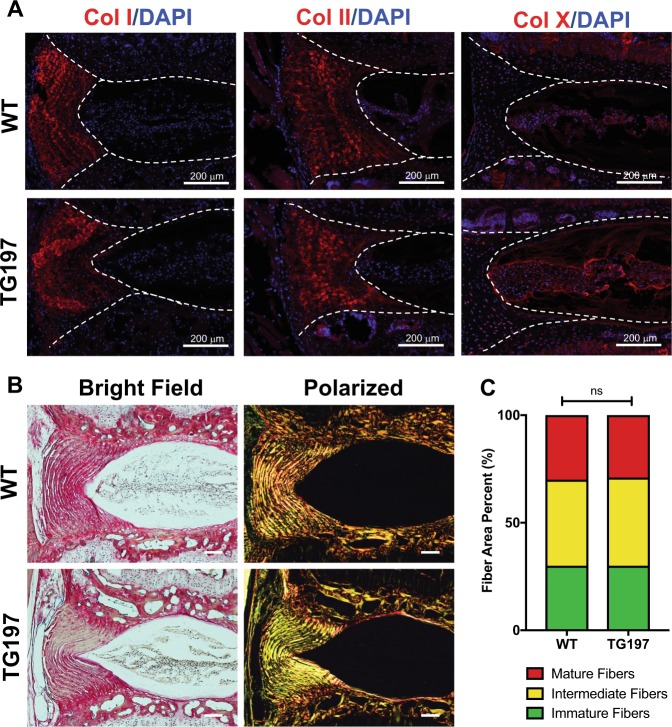
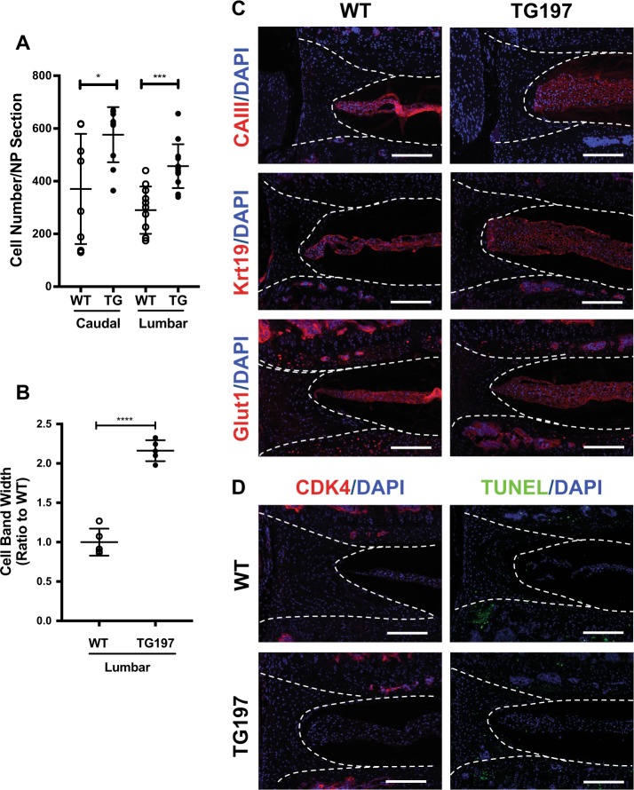
There is a well-established link between cytokine expression and the progression of intervertebral disc degeneration. Among these cytokines, interleukin-1β (IL-1β) and tumor necrosis factor-α (TNF-α) are the most commonly studied. To investigate whether systemic hTNF-α overexpression affects intervertebral disc health, we studied the spine phenotype of Tg197 mice, a widely used hTNF-α transgenic line. These mice were studied at 12-16 weeks of age using comprehensive histochemical and immunohistological analysis of the spinal motion segment. Micro-CT analysis was performed to quantify vertebral trabecular bone architecture. The Tg197 mice evidenced spontaneous annular tears and herniation with increased vascularity in subchondral bone and significant immune cell infiltration. The full-thickness annular tear without nucleus pulposus (NP) extrusion resulted in neutrophil, macrophage, and mast cell infiltration into the disc, whereas the disc with full-thickness tear and pronounced NP herniation showed additional presence of CD4+ and CD8+ T cells. While the observed defects involved failure of the annular, endplate, and vertebral junction, there were no obvious alterations in the collagen or aggrecan content in the NP and annulus fibrosus or the maturity of collagen fibers in Tg197 mice. Despite elevated systemic inflammation and pronounced loss of trabecular bone in the vertebrae, intact Tg197 discs were healthy and showed an increase in NP cell number. The NP cells in intact discs preserved expression of phenotypic markers: CAIII, Glut1, and Krt19. In conclusion, elevated systemic TNF-α increases the susceptibility of mice to spontaneous disc herniation and possibly radiculopathy, without adversely affecting intact intervertebral disc health.


Conflict of interest statement

The authors declare that they have no conflict of interest.

Figures

Fig. 1
Fig. 1. Immune cell response to spontaneous herniation in Tg197 caudal discs.
a, b’ Left column shows Safranin O/Fast Green/Hematoxylin staining of spontaneous caudal herniation. Inset box (a, b) shows the location of the higher magnification image beneath (a’, b’). Each row shows staining of successive sections of the same region, with white dashed outlines for positional context. Immunofluorescence staining of the annular tear shows CD68-positive cells (macrophages) (c, c’), Ly6-positive cells (neutrophils) (d, d’), and tryptase-positive cells (mast cells) (e, e’), Note: these sections were negative for CD4 and CD8, T cell markers (f, g’). The nucleus pulposus extrusion shows a large immune cell infiltration positive for all macrophage (h, h’), neutrophil (i, i’), mast cell (j, j’), and T cell markers, including both CD4 and CD8 (k, l’) (first and third row, scale bars = 200 μm; second and fourth row, scale bars = 50 μm). m Quantitation of the immune signal of each marker. n TUNEL staining of herniated disc sections. Insert box showing DAPI signal beneath cluster of cells displaying high intensity TUNEL signal on bottom image. o CD31 staining showing increased vascularity in the subchondral bony endplate of herniated discs
Fig. 2
Fig. 2. Tg197 discs are healthier or no different than wild-type controls.
ah Safranin O/Fast Green/Hematoxylin staining of coronal sections of wild-type and Tg197 mouse intervertebral discs. Low magnification images of lumbar Tg197 discs show thickening of the nucleus pulposus cell band. (top row, scale bars = 200 μm; middle rows, scale bars = 50 μm; bottom row, scale bars = 20 μm) i, j Distribution of histological grades using the modified Thomson scale for i caudal and j lumbar intervertebral discs. k, l Average modified Thompson score for k caudal and l lumbar intervertebral discs. m Aspect ratio of caudal and lumbar nucleus pulposus. n Bone volume/total volume of endplate in Tg197 and control mice (n = 5 mice/genotype). o Endplate scoring of caudal and lumbar discs. Data was collected from 3 discs per mouse (n = 10 mice/genotype). Significance between average grades was determined using unpaired t test. ns = not significant, **p ≤ 0.01, ****p ≤ 0.0001
Fig. 3
Fig. 3. Similarities in histology and immunohistochemistry of Tg197 and wild-type intervertebral discs.
a Immunofluorescence staining of collagen I, collagen II, and collagen X (red). Collagen I and II showed similar distributions in the annulus fibrosus, while collagen X staining was confined to the nucleus pulposus and slightly elevated in the Tg197 disc. Dotted lines were drawn to distinguish different intervertebral disc compartments. All staining was performed using at least three animals per group. b Picrosirius Red staining of 16-week caudal discs showing collagen deposition in the annulus fibrosus and the nucleus pulposus. Collagen fibers visualized under polarized light (right column) show organized lamellae in (scale bar 100 μm). c Quantification of the fiber content distribution (n = 5). Significance between fiber distribution determined using χ2 test. ns = not significant
Fig. 4
Fig. 4. Tg197 discs show altered inflammatory environment without changes in matrix composition.
a Chondroitin sulfate (CS) and aggrecan (Acan) staining show no differences between WT and Tg197 (n = 3 animals/genotype), but IL-6 and IL-1β levels are increased in Tg197 NP (n = 6 animals/genotype). b MMP-13, ARGxx, and DIPEN show no differences in between the two genotypes. c Quantification of staining for IL-6, IL-1β, ARGxx, and DIPEN performed using n = 6 mice/genotype. **p ≤ 0.01 Dotted lines in a, b were drawn to distinguish different intervertebral disc compartments. Scale bar = 200 μm
Fig. 5
Fig. 5. Tg197 lumbar and caudal discs are more cellular than wild-type controls, and the cells in Tg197 mice have comparable nucleus pulposus phenotypes.
a Both caudal and lumbar Tg197 discs are more cellular than wild-type controls (n = 5). b Width of lumbar disc cell band in Tg197 mice is more than two-fold larger than wild-type controls (n = 5). c Immunofluorescence staining of nucleus pulposus markers CAIII, Krt19, and Glut1. The staining between genotype was very similar. d Immunofluorescence staining of CDK4 shows no difference in cell proliferation. Tunnel assay of 16-week-old lumbar discs showed no difference in cell death. Dotted lines were drawn to distinguish different intervertebral disc compartments. All staining was performed using at least three animals per genotype. (scale bar = 200 μm) t test. *p ≤ 0.05, ***p ≤ 0.001, ****p ≤ 0.0001
Fig. 6
Fig. 6. Tg197 vertebrae show characteristic bone erosion and trabecular thinning.
a Representative μCT scans of caudal motion segments of 16-week-old spines showing gross trabecular thinning. bh Bone volume/trabecular volume (BV/TV), trabecular number (Tb.n), trabecular spacing (Tb.s), trabecular thickness (Tb.th), vertebral body length, disc height, and disc height index (DHI) of Tg197 and wild-type mice (mean ± SD) (n = 10 per genotype with 3 consecutive vertebrae/animal). Scatter plots show all data points and plotted as mean ± SD. t test. ns = not significant, **p ≤ 0.01, ****p ≤ 0.0001

References

    1. Katz JN. Lumbar disc disorders and low-back pain: socioeconomic factors and consequences. J. Bone Jt. Surg. Am. 2006;88(Suppl 2):21–24. - PubMed
    1. Murray CJL, et al. The state of US health, 1990-2010: burden of diseases, injuries, and risk factors. JAMA. 2013;310:591–608. doi: 10.1001/jama.2013.13805. - DOI - PMC - PubMed
    1. Vos T, et al. Global, regional, and national incidence, prevalence, and years lived with disability for 301 acute and chronic diseases and injuries in 188 countries, 1990-2013: a systematic analysis for the Global Burden of Disease Study 2013. Lancet. 2015;386:743–800. doi: 10.1016/S0140-6736(15)60692-4. - DOI - PMC - PubMed
    1. Arnbak B, et al. Prevalence of degenerative and spondyloarthritis-related magnetic resonance imaging findings in the spine and sacroiliac joints in patients with persistent low back pain. Eur. Radiol. 2015;26:1191–1203. doi: 10.1007/s00330-015-3903-0. - DOI - PubMed
    1. Livshits G, et al. Lumbar disc degeneration and genetic factors are the main risk factors for low back pain in women: the UK Twin Spine Study. Ann. Rheum. Dis. 2011;70:1740–1745. doi: 10.1136/ard.2010.137836. - DOI - PMC - PubMed

Publication types

MeSH terms