. 2019 Mar:92:287-415.
doi: 10.1016/j.simyco.2018.11.001. Epub 2018 Nov 17.

Sporocadaceae, a family of coelomycetous fungi with appendage-bearing conidia

Affiliations

Sporocadaceae, a family of coelomycetous fungi with appendage-bearing conidia

F Liu et al. Stud Mycol. 2019 Mar.

Abstract

Species of Sporocadaceae are endophytic, plant pathogenic or saprobic, and associated with a wide range of host plants. Recent molecular studies that have attempted to address familial and generic boundaries of fungi belonging to Sporocadaceae were based on a limited number of samples and DNA loci. The taxonomy of this group of fungi is therefore still not fully resolved. The aim of the present study is to provide a natural classification for the Sporocadaceae based on multi-locus phylogenetic analyses, using LSU, ITS, tef-1α, tub2 and rpb2 loci, in combination with morphological data. A total of 30 well-supported monophyletic clades in Sporocadaceae are recognised, representing 23 known and seven new genera. Typifications are proposed for the type species of five genera (Diploceras, Discosia, Monochaetia, Sporocadus and Truncatella) to stabilise the application of these names. Furthermore, Neotruncatella and Dyrithiopsis are synonymised under Hymenopleella, and the generic circumscriptions of Diploceras, Disaeta, Hymenopleella, Monochaetia, Morinia, Pseudopestalotiopsis, Sarcostroma, Seimatosporium, Synnemapestaloides and Truncatella are emended. A total of 51 new species, one nomina nova and 15 combinations are introduced.

Keywords: Bartalinia pini F. Liu, L. Cai & Crous; Discosia rubi F. Liu, L. Cai & Crous; Discosia tricellularis (Okane et al.) F. Liu, L. Cai & Crous; Discosia yakushimensis (Kaz. Tanaka et al.) F. Liu, L. Cai & Crous; Distononappendiculata F. Liu, L. Cai & Crous; Distononappendiculata banksiae (Crous & Summerell) F. Liu, L. Cai & Crous; Distononappendiculata casuarinae F. Liu, L. Cai & Crous; Distononappendiculata verrucata F. Liu, L. Cai & Crous; Diversimediispora F. Liu, L. Cai & Crous; Diversimediispora humicola F. Liu, L. Cai & Crous; Heterotruncatella F. Liu, L. Cai & Crous; Heterotruncatella acacigena F. Liu, L. Cai & Crous; Heterotruncatella aspera F. Liu, L. Cai & Crous; Heterotruncatella avellanea F. Liu, L. Cai & Crous; Heterotruncatella breviappendiculata F. Liu, L. Cai & Crous; Heterotruncatella constricta F. Liu, L. Cai & Crous; Heterotruncatella diversa F. Liu, L. Cai & Crous; Heterotruncatella grevilleae F. Liu, L. Cai & Crous; Heterotruncatella longissima F. Liu, L. Cai & Crous; Heterotruncatella lutea (H.J. Swart & D.A. Griffiths) F. Liu, L. Cai & Crous; Heterotruncatella proteicola F. Liu, L. Cai & Crous; Heterotruncatella quercicola F. Liu, L. Cai & Crous; Heterotruncatella restionacearum (S.J. Lee & Crous) F. Liu, L. Cai & Crous; Heterotruncatella singularis F. Liu, L. Cai & Crous; Heterotruncatella spadicea (S.J. Lee & Crous) F. Liu, L. Cai & Crous; Heterotruncatella spartii (Senan. et al.) F. Liu, L. Cai & Crous; Heterotruncatella synapheae F. Liu, L. Cai & Crous; Heterotruncatella vinaceobubalina F. Liu, L. Cai & Crous; Hymenopleella austroafricana F. Liu, L. Cai & Crous; Hymenopleella endophytica (Hyang B. Lee et al.) F. Liu, L. Cai & Crous; Hymenopleella lakefuxianensis (L. Cai et al.) F. Liu, L. Cai & Crous; Hymenopleella polyseptata F. Liu, L. Cai & Crous; Hymenopleella subcylindrica F. Liu, L. Cai & Crous; Monochaetia quercus F. Liu, L. Cai & Crous; Morinia acaciae (Crous) F. Liu, L. Cai & Crous; Morinia crini F. Liu, L. Cai & Crous; Multi-locus phylogeny; New taxa; Nonappendiculata F. Liu, L. Cai & Crous; Nonappendiculata quercina F. Liu, L. Cai & Crous; Parabartalinia F. Liu, L. Cai & Crous; Parabartalinia lateralis F. Liu, L. Cai & Crous; Pestalotia hypericina Ces.; Pestalotia monochaeta Desmazières; Pestalotiopsis hispanica F. Liu, L. Cai & Crous; Pestalotiopsis leucadendri F. Liu, L. Cai & Crous; Pestalotiopsis spathuliappendiculata F. Liu, L. Cai & Crous; Pestalotiopsis terricola F. Liu, L. Cai & Crous; Pseudopestalotiopsis elaeidis (C. Booth & J.S. Robertson) F. Liu, L. Cai & Crous; Pseudopestalotiopsis solicola F. Liu, L. Cai & Crous; Pseudosarcostroma F. Liu, L. Cai & Crous; Pseudosarcostroma osyridicola F. Liu, L. Cai & Crous; Robillarda australiana F. Liu, L. Cai & Crous; Sarcostroma africanum F. Liu, L. Cai & Crous; Sarcostroma australiense F. Liu, L. Cai & Crous; Sarcostroma diversiseptatum F. Liu, L. Cai & Crous; Sarcostroma leucospermi F. Liu, L. Cai & Crous; Sarcostroma longiappendiculatum F. Liu, L. Cai & Crous; Sarcostroma paragrevilleae F. Liu, L. Cai & Crous; Sarcostroma proteae F. Liu, L. Cai & Crous; Seimatosporium; Seimatosporium germanicum F. Liu, L. Cai & Crous; Seimatosporium soli F. Liu, L. Cai & Crous; Seimatosporium vitis-viniferae F. Liu, L. Cai & Crous; Sphaeria artocreas Tode; Sporocadus; Sporocadus biseptatus F. Liu, L. Cai & Crous; Sporocadus cornicola (Wijayaw. & Camporesi) F. Liu, L. Cai & Crous; Sporocadus cotini F. Liu, L. Cai & Crous; Sporocadus incanus F. Liu, L. Cai & Crous; Sporocadus lichenicola Corda; Sporocadus mali F. Liu, L. Cai & Crous; Sporocadus microcyclus F. Liu, L. Cai & Crous; Sporocadus multiseptatus F. Liu, L. Cai & Crous; Sporocadus rosarum (Henn.) F. Liu, L. Cai & Crous; Sporocadus rosigena F. Liu, L. Cai & Crous; Sporocadus rotundatus F. Liu, L. Cai & Crous; Sporocadus sorbi (Wijayaw. et al.) F. Liu, L. Cai & Crous; Sporocadus trimorphus F. Liu, L. Cai & Crous; Stilbospora angustata Pers; Synnemapestaloides juniperi F. Liu, L. Cai & Crous; Taxonomy; Truncatella spadicea S. Lee & Crous; Xenoseimatosporium F. Liu, L. Cai & Crous; Xenoseimatosporium quercinum (Goonas. et al.) F. Liu, L. Cai & Crous.


Figures
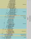
Fig. 1
Fig. 1
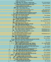
Overview phylogenetic tree of Sporocadaceae (50 % majority rule consensus) resulting from a Bayesian analysis of the combined LSU, ITS and rpb2 sequence alignment. Bayesian posterior probabilities (PP ≥ 0.95) are emphasised by thickened branches, maximum likelihood bootstrap support values (≥ 50 %) are shown at the nodes. The scale bar represents the expected number of changes per site. Genera are delimited in coloured boxes, with the genus name indicated to the right. Ex-type strains are represented in bold, and the generic type species are highlighted in red colour. Basionyms are indicated between apostrophes (‘’). Clades 1–3 are further analysed using combined LSU, ITS, rpb2, tef-1α and tub2 sequence alignment, corresponding to Fig. 2, Fig. 4, Fig. 7. The tree was rooted to Clypeosphaeria mamillana (CBS 140735).
Fig. 1
Fig. 1
Overview phylogenetic tree of Sporocadaceae (50 % majority rule consensus) resulting from a Bayesian analysis of the combined LSU, ITS and rpb2 sequence alignment. Bayesian posterior probabilities (PP ≥ 0.95) are emphasised by thickened branches, maximum likelihood bootstrap support values (≥ 50 %) are shown at the nodes. The scale bar represents the expected number of changes per site. Genera are delimited in coloured boxes, with the genus name indicated to the right. Ex-type strains are represented in bold, and the generic type species are highlighted in red colour. Basionyms are indicated between apostrophes (‘’). Clades 1–3 are further analysed using combined LSU, ITS, rpb2, tef-1α and tub2 sequence alignment, corresponding to Fig. 2, Fig. 4, Fig. 7. The tree was rooted to Clypeosphaeria mamillana (CBS 140735).
Fig. 2
Fig. 2
Phylogenetic tree resulting from a maximum likelihood analysis of the combined LSU, ITS, rpb2, tef-1α and tub2 sequence alignment (representing clade 1 of Fig. 1). Bayesian posterior probabilities (PP ≥ 0.95) are emphasised by thickened branches, maximum likelihood bootstrap support values (≥ 50 %) are shown at the nodes. The scale bar represents the expected number of changes per site. Genera are delimited in grey boxes. All taxa names in Allelochaeta consist of strain number, species name, host and location (underscore is used to separate species name and host). The taxon names of other genera consist of strain number, host/substrate and location. Ex-type strains are represented in bold, and the generic type species are highlighted in red colour. Basionyms, if present, are indicated between apostrophes (‘’). The tree was rooted to Lepteutypa fuckelii (CBS 140409).
Fig. 2
Fig. 2
Phylogenetic tree resulting from a maximum likelihood analysis of the combined LSU, ITS, rpb2, tef-1α and tub2 sequence alignment (representing clade 1 of Fig. 1). Bayesian posterior probabilities (PP ≥ 0.95) are emphasised by thickened branches, maximum likelihood bootstrap support values (≥ 50 %) are shown at the nodes. The scale bar represents the expected number of changes per site. Genera are delimited in grey boxes. All taxa names in Allelochaeta consist of strain number, species name, host and location (underscore is used to separate species name and host). The taxon names of other genera consist of strain number, host/substrate and location. Ex-type strains are represented in bold, and the generic type species are highlighted in red colour. Basionyms, if present, are indicated between apostrophes (‘’). The tree was rooted to Lepteutypa fuckelii (CBS 140409).
Fig. 3
Fig. 3
Phylogenetic tree of Discosia resulting from a maximum likelihood analysis of the ITS sequence alignment. Bayesian posterior probabilities (PP ≥ 0.95) are emphasised by thickened branches, maximum likelihood bootstrap support values (≥ 50 %) are shown at the nodes. The scale bar represents the expected number of changes per site. All taxon names consist of strain number, host/substrate and location. Ex-type strains are represented in bold, and the generic type species are highlighted in red colour. The tree was rooted to Seimatosporium rosae (CBS 139823).
Fig. 4
Fig. 4
Phylogenetic tree resulting from a maximum likelihood analysis of the combined LSU, ITS, rpb2, tef-1α and tub2 sequence alignment (representing clade 2 of Fig. 1). Bayesian posterior probabilities (PP ≥ 0.95) are emphasised by thickened branches, maximum likelihood bootstrap support values (≥ 50 %) are shown at the nodes. The scale bar represents the expected number of changes per site. Genera are delimited in grey boxes. All taxa names in genera Pestalotiopsis, Pseudopestalotiopsis, Neopestalotiopsis and Seiridium consist of strain number, species name, host and location (underscore is used to separate species name and host). Species names in other genera are aligned to the right. Ex-type strains are represented in bold, and the generic type species are highlighted in red colour. Basionyms, if present, are indicated between apostrophes (‘’). The tree was rooted to Lepteutypa fuckelii (CBS 140409).
Fig. 5
Fig. 5
Phylogenetic tree of Pestalotiopsis resulting from a maximum likelihood analysis of the combined ITS, tef-1α and tub2 sequence alignment. Bayesian posterior probabilities (PP ≥ 0.95) are emphasised by thickened branches, maximum likelihood bootstrap support values (≥ 50 %) are shown at the nodes. The scale bar represents the expected number of changes per site. All taxon names consist of strain number, host and location. Species names are aligned to the right. Ex-type strains are represented in bold. The tree was rooted to Pseudopestalotioposis theae (MFLUCC 12-0055).
Fig. 5
Fig. 5
Phylogenetic tree of Pestalotiopsis resulting from a maximum likelihood analysis of the combined ITS, tef-1α and tub2 sequence alignment. Bayesian posterior probabilities (PP ≥ 0.95) are emphasised by thickened branches, maximum likelihood bootstrap support values (≥ 50 %) are shown at the nodes. The scale bar represents the expected number of changes per site. All taxon names consist of strain number, host and location. Species names are aligned to the right. Ex-type strains are represented in bold. The tree was rooted to Pseudopestalotioposis theae (MFLUCC 12-0055).
Fig. 6
Fig. 6
Phylogenetic tree of Pseudopestalotiopsis resulting from a maximum likelihood analysis of the combined ITS, tef-1α and tub2 sequence alignment. Bayesian posterior probabilities (PP ≥ 0.95) are emphasised by thickened branches, maximum likelihood bootstrap support values (≥ 50 %) are shown at the nodes. The scale bar represents the expected number of changes per site. All taxon names consist of strain number, host and location. Species names are aligned to the right. Ex-type strains are represented in bold. Basionyms, if present, are indicated between apostrophes (‘’). The tree was rooted to Neopestalotiopsis protearum (CBS 114178) and Neo. rosae (CBS 101057).
Fig. 7
Fig. 7
Phylogenetic tree resulting from a maximum likelihood analysis of the combined LSU, ITS, rpb2, tef-1α and tub2 sequence alignment (representing clade 3 of Fig. 1). Bayesian posterior probabilities (PP ≥ 0.95) are emphasised by thickened branches, maximum likelihood bootstrap support values (≥ 50 %) are shown at the nodes. The scale bar represents the expected number of changes per site. Genera are delimited in grey boxes. All taxon names consist of strain number, host and location. Species names are aligned to the right. Ex-type strains are represented in bold, and the generic type species are highlighted in red colour. Basionyms, if present, are indicated between apostrophes (‘’). The tree was rooted to Beltrania pseudorhombica (CPC 23656).
Fig. 8
Fig. 8
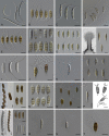
An illustration of the diversity of conidia and ascospores in different genera of Sporocadaceae (the generic order is corresponding to the topology of Fig. 1). A.Allelochaeta (CBS 144171, CBS 144191, CBS 131119, CBS 144181, from Crous et al. 2018). B.Sarcostroma (CBS 143879). C.Xenoseimatosporium (CBS 129171). D.Diploceras (CBS 492.97). E.Disaeta (CBS 143903). F.Sporocadus (NBRC 32625). G.Seimatosporium (CBS 139823). H.Synnemapestaloides (MAFF 239201, from K. Tanaka). I.Discosia (CBS 124848). J.Pestalotiopsis (CBS 143892). K.Ciliochorella (HHUF 2800, from K. Tanaka). L.Monochaetia (CBS 199.82). M. Ascus, ascospores and conidia of Seiridium (CBS 140403, Jaklitsch et al. 2016; CBS 343.97, from Bonthond et al. 2018). N.Nonappendiculata (CBS 116061). O.Heterotruncatella (CBS 143883, CBS 143901, CBS 143908, CBS 143897). P.Morinia (CBS 143888; F090354, Collado et al. 2006). Q. Asci, ascospores and conidia of Hymenopleella (from left to right: HKUCC 7303, Jeewon et al. 2003a; CBS 140410, from Jaklitsch et al. 2016; CBS 143886; CBS 647.74). R.Bartalinia (CBS 143891). S.Parabartalinia (CBS 399.71). T.Truncatella (CBS 144025). U–W.Broomella (MFLUCC 13-0798, U. Ascus; V. Ascospores; W. Conidia; reproduced with permission of A.D.A.C., originally published in Cryptogamie, Mycologie in Li et al. 2015). X.Hyalotiella (MFLUCC 13-0397, reproduced with permission of A.D.A.C. [Association des amis des cryptogames, Paris - France], originally published in Cryptogamie, Mycologie in Li et al. 2015). Y.Diversimediispora (CBS 302.86). Z.Pseudosarcostroma (CBS 103.76). AA.Robillarda (CBS 143882). AB. Asci, ascospores and conidia of Strickeria (CBS 140411, Jaklitsch et al. 2016). AC.Distononappendiculata (CBS 144032). AD.Immersidiscosia (MAFF 104197, from K. Tanaka). Scale bars = 10 μm.
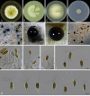
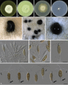
Fig. 8
Fig. 8
An illustration of the diversity of conidia and ascospores in different genera of Sporocadaceae (the generic order is corresponding to the topology of Fig. 1). A.Allelochaeta (CBS 144171, CBS 144191, CBS 131119, CBS 144181, from Crous et al. 2018). B.Sarcostroma (CBS 143879). C.Xenoseimatosporium (CBS 129171). D.Diploceras (CBS 492.97). E.Disaeta (CBS 143903). F.Sporocadus (NBRC 32625). G.Seimatosporium (CBS 139823). H.Synnemapestaloides (MAFF 239201, from K. Tanaka). I.Discosia (CBS 124848). J.Pestalotiopsis (CBS 143892). K.Ciliochorella (HHUF 2800, from K. Tanaka). L.Monochaetia (CBS 199.82). M. Ascus, ascospores and conidia of Seiridium (CBS 140403, Jaklitsch et al. 2016; CBS 343.97, from Bonthond et al. 2018). N.Nonappendiculata (CBS 116061). O.Heterotruncatella (CBS 143883, CBS 143901, CBS 143908, CBS 143897). P.Morinia (CBS 143888; F090354, Collado et al. 2006). Q. Asci, ascospores and conidia of Hymenopleella (from left to right: HKUCC 7303, Jeewon et al. 2003a; CBS 140410, from Jaklitsch et al. 2016; CBS 143886; CBS 647.74). R.Bartalinia (CBS 143891). S.Parabartalinia (CBS 399.71). T.Truncatella (CBS 144025). U–W.Broomella (MFLUCC 13-0798, U. Ascus; V. Ascospores; W. Conidia; reproduced with permission of A.D.A.C., originally published in Cryptogamie, Mycologie in Li et al. 2015). X.Hyalotiella (MFLUCC 13-0397, reproduced with permission of A.D.A.C. [Association des amis des cryptogames, Paris - France], originally published in Cryptogamie, Mycologie in Li et al. 2015). Y.Diversimediispora (CBS 302.86). Z.Pseudosarcostroma (CBS 103.76). AA.Robillarda (CBS 143882). AB. Asci, ascospores and conidia of Strickeria (CBS 140411, Jaklitsch et al. 2016). AC.Distononappendiculata (CBS 144032). AD.Immersidiscosia (MAFF 104197, from K. Tanaka). Scale bars = 10 μm.
Fig. 9
Fig. 9
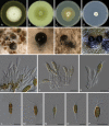
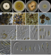
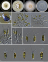
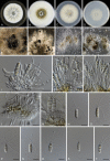
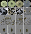
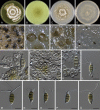
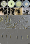
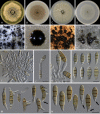
Bartalinia bella (ex-type CBS 464.61). A–D. Colonies on MEA, CMA, PDA and SNA, respectively. E–H. Conidiomata on MEA, CMA, PDA and SNA, respectively. I–J. Conidiophores. K–M. Conidia. Scale bars = 10 μm.
Fig. 10
Fig. 10
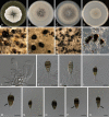
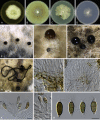
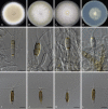
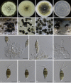
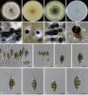
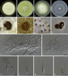
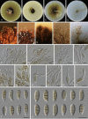
Bartalinia pini (CBS 143891/CPC 24328). A–D. Colonies on MEA, CMA, PDA and SNA, respectively. E–F. Conidiomata on MEA. G–H. Conidiomata on CMA and PDA. I–M. Conidiophores, conidiogenous cells and conidia. N–P. Conidia. Scale bars = 10 μm.
Fig. 11
Fig. 11
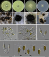
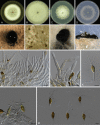
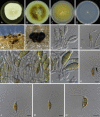
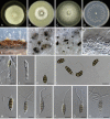
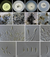
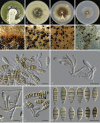
Diploceras hypericinum (A–I. CBS 143885/CPC 21115, J–N. CBS 492.97). A–D. Colonies on MEA, CMA, PDA and SNA, respectively. E–F. Conidiomata on MEA. G–H. Conidiomata on CMA. I–K. Conidiophores, conidiogenous cells. L–N. Conidia. Scale bars = 10 μm.
Fig. 12
Fig. 12
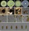
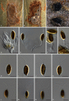
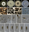
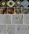
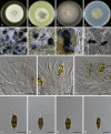
Disaeta arbuti (CBS 143903/CPC 28304). A–D. Colonies on MEA, CMA, PDA and SNA, respectively. E–H. Conidiomata on MEA, CMA, PDA and SNA, respectively. I–L. Conidiophores, conidiogenous cells and conidia. M. Conidia. Scale bars = 10 μm.
Fig. 13
Fig. 13
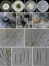
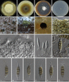
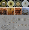
Discosia artocreas (CBS 124848). A–D. Colonies on MEA, CMA, PDA and SNA, respectively. E. Conidiomata on MEA. F–J. Conidiophores, conidiogenous cells and conidia. K. Conidia. Scale bars = 10 μm.
Fig. 14
Fig. 14
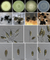
Discosia rubi (CBS 143893/CPC 25062). A–D. Colonies on MEA, CMA, PDA and SNA, respectively. E–H. Conidiomata on MEA, CMA, PDA and SNA, respectively. I–K. Conidiophores, conidiogenous cells and conidia. L–Q. Conidia. Scale bars = 10 μm.
Fig. 15
Fig. 15
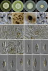
Distononappendiculata banksiae (CBS 143906/CPC 28968). A–D. Colonies on MEA, CMA, PDA and SNA, respectively. E–F. Conidiomata on MEA. G. Conidiomata on CMA. H. Conidiomata on PDA. I–K. Conidiophores, conidiogenous cells and conidia. L. Conidia with verruculose wall. M. Conidia with septal pores (arrow). N. Conidia. Scale bars = 10 μm.
Fig. 16
Fig. 16
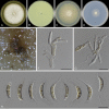
Distononappendiculata casuarinae (CBS 143884/CPC 17253). A–D. Colonies on MEA, CMA, PDA and SNA, respectively. E– F. Conidiomata on MEA and CMA. G–J. Conidiophores, conidiogenous cells and conidia. K. Conidia. Scale bars = 10 μm.
Fig. 17
Fig. 17
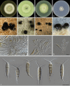
Distononappendiculata verrucata (CBS 144032/CPC 29074). A–D. Colonies on MEA, CMA, PDA and SNA. E. Conidiomata on PDA. F–J. Conidiophores and conidiogenous cells bearing conidia. K–L. Conidia. Scale bars = 10 μm.
Fig. 18
Fig. 18
Diversimediispora humicola (CBS 302.86). A–D. Colonies on MEA, CMA, PDA and SNA, respectively. E–H. Conidiomata on MEA, CMA, PDA and SNA, respectively. I. Conidiophores. J–L. Conidiogenous cells and conidia. M–Q. Conidia (arrows point to the verruculose wall of the first median cell from the basal cell). Scale bars = 10 μm.
Fig. 19
Fig. 19
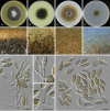
Heterotruncatella acacigena (CBS 143880/CPC 15130). A–D. Colonies on MEA, CMA, PDA and SNA, respectively. E–H. Conidiomata on MEA, CMA, PDA and SNA, respectively. I–J. Conidiophores. K–L. Conidiogenous cells and conidia. M. Conidia (arrows point to the untypical basal appendages). Scale bars = 10 μm.
Fig. 20
Fig. 20
Heterotruncatella aspera (CBS 143907/CPC 28992). A–D. Colonies on MEA, CMA, PDA and SNA, respectively. E–F. Conidiomata on MEA and CMA. G–H. Conidiomata on PDA. I–K. Conidiophores and conidiogenous cells. L–P. Conidia. Scale bars = 10 μm.
Fig. 21
Fig. 21
Heterotruncatella avellanea (CBS 143896/CPC 25377). A–D. Colonies on MEA, CMA, PDA and SNA, respectively. E–G. Conidiomata on MEA, CMA and PDA, respectively. H–J. Conidiophores and conidiogenous cells. K–L. Conidia. Scale bars = 10 μm.
Fig. 22
Fig. 22
Heterotruncatella breviappendiculata (CBS 143883/CPC 17239). A–D. Colonies on MEA, CMA, PDA and SNA, respectively. E–G. Conidiomata on MEA, CMA and PDA, respectively. H. Conidial lines on pine needle. I. Vertical section of conidioma. J–L. Conidiophores, conidiogenous cells and conidia. M. Conidia. Scale bars = 10 μm.
Fig. 23
Fig. 23
Heterotruncatella constricta (CBS 143901/CPC 27578). A–D. Colonies on MEA, CMA, PDA and SNA, respectively. E–H. Conidiomata on MEA, CMA, PDA and SNA, respectively. I–J. Conidiophores. K–N. Conidiogenous cells and conidia (arrow points to the annellations). O. Conidia. Scale bars = 10 μm.
Fig. 24
Fig. 24
Heterotruncatella diversa (CBS 143908/CPC 29040). A–D. Colonies on MEA, CMA, PDA and SNA, respectively. E–H. Conidiomata on MEA, CMA, PDA and SNA, respectively. I–L. Conidiophores, conidiogenous cells and conidia. M. Conidia. Scale bars = 10 μm.
Fig. 25
Fig. 25
Heterotruncatella grevilleae (CBS 143881/CPC 16997). A–D. Colonies on MEA, CMA, PDA and SNA, respectively. E–H. Conidiomata on MEA, CMA, PDA and SNA, respectively. I–L. Conidiophores, conidiogenous cells and conidia. M. Conidia. Scale bars = 10 μm.
Fig. 26
Fig. 26
Heterotruncatella longissima (CBS 143910/CPC 29114). A–D. Colonies on MEA, CMA, PDA and SNA, respectively. E–F. Conidiomata on MEA and SNA. G–H. Conidiogenous cells and conidia. I–L. Conidia. Scale bars = 10 μm.
Fig. 27
Fig. 27
Heterotruncatella lutea (CBS 349.73). A–D. Colonies on MEA, CMA, PDA and SNA, respectively. E–H. Conidiomata on MEA, CMA, PDA and SNA, respectively. I–K. Conidiophores and conidiogenous cells on SNA. L. Conidia on SNA. M. Conidiogeneses. N. Conidia on MEA. Scale bars = 10 μm.
Fig. 28
Fig. 28
Heterotruncatella proteicola (CBS 144020/CPC 13700). A–D. Colonies on MEA, CMA, PDA and SNA, respectively. E–H. Conidiogenous cells bearing conidia. I–L. Conidia. Scale bars = 10 μm.
Fig. 29
Fig. 29
Heterotruncatella quercicola (CBS 143895/CPC 25365). A–D. Colonies on MEA, CMA, PDA and SNA, respectively. E. Conidiomata on barley leaves. F. Colonies on filter paper. G–J. Conidiophores, conidiogenous cells and conidia. K–N. Conidia (arrow points to branched apical appendage). Scale bars = 10 μm.
Fig. 30
Fig. 30
Heterotruncatella singularis (HPC 721). A–B. Disease symptoms on the front and back of the leaves. C. Conidiomata. D–G. Conidiophores and conidiogenous cells bearing conidia. H–O. Conidia. Scale bars = 10 μm.
Fig. 31
Fig. 31
Heterotruncatella spadicea (CBS 118145). A–D. Colonies on MEA, CMA, PDA and SNA, respectively. E. Conidioma on CMA. F–I. Conidiophores and conidiogenous cells bearing conidia. J–K. Conidia. Scale bars = 10 μm.
Fig. 32
Fig. 32
Heterotruncatella spartii (CBS 143894/CPC 25363). A–D. Colonies on MEA, CMA, PDA and SNA, respectively. E–H. Conidiomata on MEA, CMA, PDA and SNA respectively. I–K. Conidiophores, conidiogenous cells and conidia. L–M. Conidia with 3–5 appendages. Scale bars = 10 μm.
Fig. 33
Fig. 33
Heterotruncatella synapheae (CBS 143909/CPC 29096). A–D. Colonies on MEA, CMA, PDA and SNA, respectively. E–F. Conidiogenous cells and conidia. G–H. Conidia. Scale bars = 10 μm.
Fig. 34
Fig. 34
Heterotruncatella vinaceobubalina (CBS 143897/CPC 26201). A–D. Colonies on MEA, CMA, PDA and SNA, respectively. E–G. Conidiomata on MEA, CMA and PDA, respectively. H–J. Conidiophores and conidiogenous cells. K. Conidia (thick arrows point to the atypical basal appendages; thin arrows point to the spathulate tips). Scale bars = 10 μm.
Fig. 35
Fig. 35
Hyalotiella transvalensis (CBS 303.65). A–D. Colonies on MEA, CMA, PDA and SNA, respectively. E. Vertical section of conidioma. F–H. Conidiophores and conidiogenous cells. I–L. Conidia. Scale bars: E = 20 um, F–L = 10 μm.
Fig. 36
Fig. 36
Hymenopleella austroafricana (CBS 143886/CPC 21940). A–D. Colonies on MEA, CMA, PDA and SNA, respectively. E–H. Conidiomata on MEA, CMA, PDA and SNA, respectively. I–K. Conidiophores, conidiogenous cells and conidia. L. Conidia. Scale bars = 10 μm.
Fig. 37
Fig. 37
Hymenopleella polyseptata (CBS 143887/CPC 21944). A–D. Colonies on MEA, CMA, PDA and SNA, respectively. E–F. Conidiomata on CMA. G. Conidiophores. H–I. Conidiogenous cells bearing conidia. J. 4-septate conidia. K. 5-septate conidia. L. 6-septate conidia. M. 7-septate conidia. Scale bars = 10 μm.
Fig. 38
Fig. 38
Hymenopleella subcylindrica (CBS 647.74). A–D. Colonies on MEA, CMA, PDA and SNA, respectively. E–G. Conidiomata on MEA, CMA and SNA, respectively. H–J. Conidiophores and conidiogenous cells bearing conidia. K. Conidia on SNA. L. Conidia on MEA. Scale bars = 10 μm.
Fig. 39
Fig. 39
Monochaetia monochaeta (CBS 199.82). A–D. Colonies on MEA, CMA, PDA and SNA, respectively. E–H. Conidiomata on MEA, CMA, PDA and SNA, respectively. I–J. Conidiophores. K–M. Conidiogenous cells bearing conidia. N–V. Conidia. Scale bars = 10 μm.
Fig. 40
Fig. 40
Phylogenetic tree of Monochaetia resulting from a Bayesian analysis of the ITS sequence alignment. Bayesian posterior probabilities (PP ≥ 0.95) are emphasised by thickened branches, maximum likelihood bootstrap support values (≥ 50 %) are shown at the nodes. The scale bar represents the expected number of changes per site. All taxon names consist of strain number, host and location, and with GenBank accession number in the bracket. Species names are aligned to the right. Ex-type strains are represented in bold.
Fig. 41
Fig. 41
Monochaetia quercus (CBS 144034/CPC 29514). A–D. Colonies on MEA, CMA, PDA and SNA, respectively. E–F. Conidiomata. G–J. Conidiophores and conidiogenous cells. K–P. Conidia. Scale bars = 10 μm.
Fig. 42
Fig. 42
Morinia crini (CBS 143888/CPC 21978). A–D. Colonies on MEA, CMA, PDA and SNA, respectively. E–F. Conidiomata on CMA. G–H. Conidiomata on PDA and SNA. I–K. Conidiophores and conidiogenous cells. L. Conidia. Scale bars = 10 μm.
Fig. 43
Fig. 43
Nonappendiculata quercina (CBS 116061). A–D. Colonies on MEA, CMA, PDA and SNA, respectively. E–F. Conidiomata on OA and SNA. G–K. Conidiophores, conidiogenous cells and conidia. L. Conidia. Scale bars = 10 μm.
Fig. 44
Fig. 44
Parabartalinia lateralis (CBS 399.71). (I–K, Q–T. on OA. L–P. on SNA). A–D. Colonies on MEA, CMA, PDA and SNA, respectively. E–F. Conidiomata on MEA and CMA. G–H. Conidiomata on SNA. I–M. Conidiophores and conidiogenous cells bearing conidia. N–T. Conidia. Scale bars = 10 μm.
Fig. 45
Fig. 45
Pestalotiopsis disseminata (CBS 143904/CPC 28705). A–D. Colonies on MEA, CMA, PDA and SNA, respectively. E. Conidiomata on MEA. F–I. Conidiophores, conidiogenous cells and conidia. J–M. Conidia. Scale bars = 10 μm.
Fig. 46
Fig. 46
Pestalotiopsis hispanica (CBS 115391). A–D. Colonies on MEA, CMA, PDA and SNA, respectively. E. Conidioma on PDA. F. Conidiomata on SNA. G–H. Conidiophores, conidiogenous cells and conidia. I–L. Conidia. Scale bars = 10 μm.
Fig. 47
Fig. 47
Pestalotiopsis leucadendri (CBS 121417). A–D. Colonies on MEA, CMA, PDA and SNA, respectively. E. Conidiomata on PDA. F–I. Conidiophore, conidiogenous cells and conidia. J–M. Conidia. Scale bars = 10 μm.
Fig. 48
Fig. 48
Pestalotiopsis sp. 1 (CBS 111576). A–D. Colonies on MEA, CMA, PDA and SNA, respectively. E–H. Conidiomata on MEA, CMA, PDA and SNA, respectively. I–K. Conidiophores, conidiogenous cells and conidia. L–O. Conidia. Scale bars = 10 μm.
Fig. 49
Fig. 49
Pestalotiopsis sp. 2 (CBS 114489). A–D. Colonies on MEA, CMA, PDA and SNA, respectively. E–G. Conidiomata on CMA, pine needle and barley leaves, respectively. H. Conidiomatal wall. I–K. Conidiophores and conidiogenous cells. L–Q. Conidia. Scale bars = 10 μm.
Fig. 50
Fig. 50
Pestalotiopsis sp. 3 (CBS 143892/CPC 24759). A–D. Colonies on MEA, CMA, PDA and SNA, respectively. E–H. Conidiomata on MEA, PDA, SNA and CMA, respectively. I–K. Conidiophores, conidiogenous cells and conidia. L–O. Conidia. Scale bars = 10 μm.
Fig. 51
Fig. 51
Pestalotiopsis sp. 4 (CBS 143905/CPC 28896). A–D. Colonies on MEA, CMA, PDA and SNA, respectively. E–H. Conidiomata on MEA, CMA, PDA and SNA, respectively. I–L. Conidiophores, conidiogenous cells and conidia. M–P. Conidia. Scale bars = 10 μm.
Fig. 52
Fig. 52
Pestalotiopsis sp. 5 (CBS 143900/CPC 27562). A–D. Colonies on MEA, CMA, PDA and SNA, respectively. E. Conidiomata on CMA. F. Conidiomata on PDA. G–I. Conidiophores, conidiogenous cells and conidia. J–L. Conidia. Scale bars = 10 μm.
Fig. 53
Fig. 53
Pestalotiopsis sp. 6 (CBS 143902/CPC 27649). A–D. Colonies on MEA, CMA, PDA and SNA, respectively. E–H. Conidiomata on MEA, CMA, PDA and SNA, respectively. I–J. Conidiophores and conidiogenous cells. K–P. Conidia. Scale bars = 10 μm.
Fig. 54
Fig. 54
Pestalotiopsis spathuliappendiculata (CBS 144035/CPC 29602). A–D. Colonies on MEA, CMA, PDA and SNA, respectively. E. Conidiomata on pine needle. F–G. Conidial masses on CMA. H–J. Conidiophores and conidiogenous cells bearing conidia. K–M. Conidia on pine needle. N–R. Conidia on CMA. Scale bars = 10 μm.
Fig. 55
Fig. 55
Pestalotiopsis terricola (CBS 141.69). A–D. Colonies on MEA, CMA, PDA and SNA, respectively. E–H. Conidiomata on MEA, CMA, PDA and SNA, respectively. I–L. Conidiophores, conidiogenous cells and conidia. M–R. Conidia. Scale bars = 10 μm.
Fig. 56
Fig. 56
Pseudopestalotiopsis solicola (CBS 386.97). A–D. Colonies on MEA, CMA, PDA and SNA, respectively. E–H. Conidiomata on MEA, CMA, PDA and SNA, respectively. I–L. Conidiophores and conidiogenous cells. M–Q. Conidia. Scale bars = 10 μm.
Fig. 57
Fig. 57
Pseudosarcostroma osyridicola (CBS 103.76). A–D. Colonies on MEA, CMA, PDA and SNA, respectively. E. Conidial masses on SNA. F–G. Immature conidiomatal wall and conidiogenous cells. H–I. Conidiogenous cells and conidia. J. Conidia. Scale bars = 10 μm.
Fig. 58
Fig. 58
Robillarda australiana (CBS 143882/CPC 17187). A–D. Colonies on MEA, CMA, PDA and SNA, respectively. E–H. Conidiomata on MEA, CMA, PDA and SNA, respectively. I–K. Conidiophores and conidiogenous cells (arrows point to protuberances). L–N. Conidia. Scale bars = 10 μm.
Fig. 59
Fig. 59
Sarcostroma africanum (CBS 143879/CPC 13920). A–D. Colonies on MEA, CMA, PDA and SNA, respectively. E. Conidomata on SNA. F–G. Conidiophores and conidiogenous cells. H. Conidia. Scale bars = 10 μm.
Fig. 60
Fig. 60
Sarcostroma australiense (CBS 144160/CPC 25389). A–D. Colonies on MEA, CMA, PDA and SNA, respectively. E–H. Conidiomata on MEA, CMA, PDA and SNA, respectively. I–K. Conidiophores and conidiogenous cells bearing conidia. L–M. 2–3-septate conidia. N. Rarely 7-septate conidium. Scale bars = 10 μm.
Fig. 61
Fig. 61
Sarcostroma diversiseptatum (CBS 189.81). A–D. Colonies on MEA, CMA, PDA, and SNA, respectively. E–G. Conidiophores, conidiogenous cells and conidia (arrows point to the annellations). H–I. Conidia. Scale bars = 10 μm.
Fig. 62
Fig. 62
Sarcostroma grevilleae (CBS 101.71). A–D. Colonies on MEA, CMA, PDA and SNA, respectively. E–H. Conidiomata on MEA, CMA, PDA and SNA, respectively. I–M. Conidiophores, conidiogenous cells and conidia. N. Mature and immature conidia. Scale bars = 10 μm.
Fig. 63
Fig. 63
Sarcostroma leucospermi (CBS 111290). A–D. Colonies on MEA, CMA, PDA and SNA, respectively. E–I. Conidiophores, conidiogenous cells and conidia (arrows point to annellations). J–K. Conidia. Scale bars = 10 μm.
Fig. 64
Fig. 64
Sarcostroma longiappendiculatum (CBS 143890/CPC 23411). A–D. Colonies on MEA, CMA, PDA and SNA, respectively. E–H. Conidiomata on MEA, CMA, PDA and SNA, respectively. I–J. Conidiophores. K. Conidia. Scale bars = 10 μm.
Fig. 65
Fig. 65
Sarcostroma paragrevilleae (CBS 114142). A–D. Colonies on MEA, CMA, PDA and SNA, respectively. E. Conidiomata on OA. F–I. Conidiophores, conidiogenous cells and conidia. J. Conidia. Scale bars = 10 μm.
Fig. 66
Fig. 66
Sarcostroma proteae (CBS 113610). A-D. Colonies on MEA, CMA, PDA, OA respectively. E–F. Conidiomata on SNA. G–J. Conidiophores, conidiogenous cells and conidia (arrow points to the annellation). K. Conidia on OA. L. Conidia on SNA. Scale bars = 10 μm.
Fig. 67
Fig. 67
Seimatosporium germanicum (CBS 437.87). A–D. Colonies on MEA, CMA, PDA and SNA, respectively. E–H. Conidiomata on MEA, CMA, PDA and SNA, respectively. I–L. Conidiophores, conidiogenous cells and conidia. M. Conidia with one or two appendages. Scale bars = 10 μm.
Fig. 68
Fig. 68
Seimatosporium rosae (A–F, M–O. ex-epitype CBS 139823) and Sei. pistaciae (G–L, P–R. ex-holotype CBS 138865). A, G. Colonies on MEA. B, H. Colonies on CMA. C, I. Colonies on PDA. D–F, J–L. Conidiomata on MEA, CMA and PDA, respectively. M, N, P, Q. Conidiophores. O, R. Conidia. Scale bars = 10 μm.
Fig. 69
Fig. 69
Seimatosporium soli (CBS 941.69). A–D. Colonies on MEA, CMA, PDA and SNA, respectively. E. Conidiomata on OA. F–G. Conidiogenous cells. H. Conidia. Scale bars = 10 μm.
Fig. 70
Fig. 70
Seimatosporium vitis-viniferae (CBS 123004). A–D. Colonies on MEA, CMA, PDA and SNA, respectively. E, F, I. Conidiomata on MEA, CMA and SNA, respectively. G–H. Conidiomata on PDA (G. near the inoculation, H. near the edge). J–K. Conidiogenous cells and conidia. L. Conidia. Scale bars = 10 μm.
Fig. 71
Fig. 71
Sporocadus biseptatus (CBS 110324). A–D. Colonies on MEA, CMA, PDA and SNA, respectively. E–H. Conidiomata on MEA, CMA, PDA and SNA, respectively. I–K. Conidiophores, conidiogenous cells and conidia. L. Conidia. Scale bars = 10 μm.
Fig. 72
Fig. 72
Sporocadus cornicola (CBS 143889/CPC 23235). A–D. Colonies on MEA, CMA, PDA and SNA, respectively. E–H. Conidiomata on MEA, CMA, PDA and SNA, respectively. I. Conidiophores. J–K. Conidiogenous cells and conidia. L. Conidia on SNA. M. Microcyclic conidiogenesis on MEA. N. Conidia on MEA (arrows point to conidial protrusions). Scale bars = 10 μm.
Fig. 73
Fig. 73
Sporocadus cotini (CBS 139966). A–D. Colonies on MEA, CMA, PDA and SNA, respectively. E–H. Conidiomata on MEA, CMA, PDA and SNA, respectively. I–K. Conidiophores, conidiogenous cells and conidia. L. Conidia. Scale bars = 10 μm.
Fig. 74
Fig. 74
Sporocadus incanus (CBS 123003). A–D. Colonies on MEA, CMA, PDA and SNA, respectively. E–H. Conidiomata on MEA, CMA, PDA and SNA, respectively. I–J. Conidiogenous cells and conidia. K. Conidia. Scale bars = 10 μm.
Fig. 75
Fig. 75
Sporocadus lichenicola (A–E, G, K–L. CBS 354.90. F, H–J. NBRC 32625). A–D. Colonies on MEA, CMA, PDA and SNA, respectively. E–F. Conidiomata on SNA. G–I. Conidiophores, conidiogenous cells, and conidia. J–L. Conidia. Scale bars = 10 μm.
Fig. 76
Fig. 76
Sporocadus mali (CBS 446.70). A–D. Colonies on MEA, CMA, PDA and SNA, respectively. E–F. Conidiomata on SNA. G–J. Conidiophores, conidiogenous cells and conidia (arrows points to annellations). K. Conidia. Scale bars = 10 μm.
Fig. 77
Fig. 77
Sporocadus microcyclus (CBS 424.95). A–D. Colonies on MEA, CMA, PDA and SNA, respectively. E–F. Conidiomata on MEA. G–I. Conidiomata on CMA, PDA and SNA, respectively. J–M. Conidiophores, conidiogenous cells and conidia. N–S. Microcyclic conidiogeneses. T. Conidia with constrictions. U. Conidia. Scale bars = 10 μm.
Fig. 78
Fig. 78
Sporocadus multiseptatus (CBS 143899/CPC 26606). A–D. Colonies on MEA, CMA, PDA and SNA, respectively. E–F. Conidiomata on MEA. G. Conidiomata on SNA. H–K. Conidiophores, conidiogenous cells and conidia. M. Conidia. Scale bars = 10 μm.
Fig. 79
Fig. 79
Sporocadus rosigena (CBS 466.96). A–D. Colonies on MEA, CMA, PDA and SNA, respectively. E–H. Conidiomata on MEA, CMA, PDA and SNA, respectively. I–L. Conidiophores, conidiogenous cells and conidia. M. Conidia. Scale bars = 10 μm.
Fig. 80
Fig. 80
Sporocadus rotundatus (CBS 616.83). A–D. Colonies on MEA, CMA, PDA and SNA, respectively. E–H. Conidiomata on MEA, CMA, PDA and SNA, respectively. I–K. Conidiophores, conidiogenous cells and conidia. L. Conidia (arrows points to the germination pores). Scale bars = 10 μm.
Fig. 81
Fig. 81
Sporocadus sp. 1 (CBS 506.71). A–D. Colonies on MEA, CMA, PDA and SNA, respectively. E–G. Conidiomata on MEA, CMA and SNA, respectively. H–K. Conidiophores, conidiogenous cells and conidia. L. Mature conidia on CMA. M. Immature conidia on SNA. Scale bars = 10 μm.
Fig. 82
Fig. 82
Sporocadus trimorphus (CBS 114203). A–D. Colonies on MEA, CMA, PDA and SNA, respectively. E–F. Conidiomata on OA. G–J. Conidiophores, conidiogenous cells and conidia. K. Conidia. Scale bars = 10 μm.
Fig. 83
Fig. 83
Synnemapestaloides juniperi (CBS 477.77). A–D. Colonies on MEA, CMA, PDA and SNA, respectively. E–G. Conidiomata on MEA, CMA and SNA, respectively. H–J. Conidiophores, conidiogenous cells and conidia. K. Conidia without appendages. L. Conidia with very short apical appendages (arrows). M. Conidia with very short apical and basal appendages (arrows). Scale bars = 10 μm.
Fig. 84
Fig. 84
Truncatella angustata (CBS 338.32). A. Conidiomata on OA. B. Conidial masses on SNA. C–E. Conidiophores and conidiogenous cells. F–I. Conidia. Scale bars = 10 μm.
Fig. 85
Fig. 85
Truncatella angustata (CBS 144025/CPC 21359). A–D. Colonies on MEA, CMA, PDA and SNA, respectively. E. Conidiomata on MEA. F–G. Conidiomata on CMA. H. Conidiomata on SNA. I–K. Conidiophores and conidiogenous cells. L–O. Conidia. Scale bars = 10 μm.
Fig. 86
Fig. 86
Xenoseimatosporium quercinum (CBS 129171). A–D. Colonies on MEA, CMA, PDA and SNA, respectively. E. Conidiomata on OA. F–H. Conidiophores and conidiogenous cells. I–M. Conidia (arrow points to the rough appendage). Scale bars = 10 μm.

Similar articles

  • Sporocadaceae revisited.
    Razaghi P, Raza M, Han SL, Ma ZY, Cai L, Zhao P, Chen Q, Phurbu D, Liu F. Razaghi P, et al. Stud Mycol. 2024 Dec;109:155-272. doi: 10.3114/sim.2024.109.03. Epub 2024 Jun 27. Stud Mycol. 2024. PMID: 39717655 Free PMC article.
  • Redisposition of acremonium-like fungi in Hypocreales.
    Hou LW, Giraldo A, Groenewald JZ, Rämä T, Summerbell RC, Huang GZ, Cai L, Crous PW. Hou LW, et al. Stud Mycol. 2023 Jun;105:23-203. doi: 10.3114/sim.2023.105.02. Epub 2023 Jun 2. Stud Mycol. 2023. PMID: 38895703 Free PMC article.
  • Resolving the Phoma enigma.
    Chen Q, Jiang JR, Zhang GZ, Cai L, Crous PW. Chen Q, et al. Stud Mycol. 2015 Sep;82:137-217. doi: 10.1016/j.simyco.2015.10.003. Epub 2015 Nov 26. Stud Mycol. 2015. PMID: 26955202 Free PMC article.
  • Fusarium: more than a node or a foot-shaped basal cell.
    Crous PW, Lombard L, Sandoval-Denis M, Seifert KA, Schroers HJ, Chaverri P, Gené J, Guarro J, Hirooka Y, Bensch K, Kema GHJ, Lamprecht SC, Cai L, Rossman AY, Stadler M, Summerbell RC, Taylor JW, Ploch S, Visagie CM, Yilmaz N, Frisvad JC, Abdel-Azeem AM, Abdollahzadeh J, Abdolrasouli A, Akulov A, Alberts JF, Araújo JPM, Ariyawansa HA, Bakhshi M, Bendiksby M, Ben Hadj Amor A, Bezerra JDP, Boekhout T, Câmara MPS, Carbia M, Cardinali G, Castañeda-Ruiz RF, Celis A, Chaturvedi V, Collemare J, Croll D, Damm U, Decock CA, de Vries RP, Ezekiel CN, Fan XL, Fernández NB, Gaya E, González CD, Gramaje D, Groenewald JZ, Grube M, Guevara-Suarez M, Gupta VK, Guarnaccia V, Haddaji A, Hagen F, Haelewaters D, Hansen K, Hashimoto A, Hernández-Restrepo M, Houbraken J, Hubka V, Hyde KD, Iturriaga T, Jeewon R, Johnston PR, Jurjević Ž, Karalti I, Korsten L, Kuramae EE, Kušan I, Labuda R, Lawrence DP, Lee HB, Lechat C, Li HY, Litovka YA, Maharachchikumbura SSN, Marin-Felix Y, Matio Kemkuignou B, Matočec N, McTaggart AR, Mlčoch P, Mugnai L, Nakashima C, Nilsson RH, Noumeur SR, Pavlov IN, Peralta MP, Phillips AJL, Pitt JI, Polizzi G, Quaedvlieg W, Rajeshkumar KC, Restrepo S, Rhaiem A, Robert J, Robert V, Ro… See abstract for full author list ➔ Crous PW, et al. Stud Mycol. 2021 Aug 17;98:100116. doi: 10.1016/j.simyco.2021.100116. eCollection 2021 Apr. Stud Mycol. 2021. PMID: 34466168 Free PMC article.
  • The phoma-like dilemma.
    Hou LW, Groenewald JZ, Pfenning LH, Yarden O, Crous PW, Cai L. Hou LW, et al. Stud Mycol. 2020 May 21;96:309-396. doi: 10.1016/j.simyco.2020.05.001. eCollection 2020 Jun. Stud Mycol. 2020. PMID: 32904212 Free PMC article.

Cited by

References

    1. Ariyawansa H.A., Hyde K.D., Jayasiri S.C. Fungal diversity notes 111–252—taxonomic and phylogenetic contributions to fungal taxa. Fungal Diversity. 2015;75:27–274.
    1. Barber P.A., Crous P.W., Groenewald J.Z. Reassessing Vermisporium (Amphisphaeriaceae), a genus of foliar pathogens of eucalypts. Persoonia. 2011;27:90–118. - PMC - PubMed
    1. Batista A.C., Lima JA de, Vasconcelos CT de. Fungos exóticos achados na atmosfera do Recife. Publicações do Instituto de Micologia da Universidade do Recife. 1960;276:1–18.
    1. Bonar L. Studies on some California fungi. Mycologia. 1928;20:292–300.
    1. Bonthond G., Sandoval-Denis M., Groenewald J.Z. Seiridium (Sporocadaceae): an important genus of plant pathogenic fungi. Persoonia. 2018;40:96–118. - PMC - PubMed

LinkOut - more resources