Case Reports
. 2018 Nov 21:2018:9821738.
doi: 10.1155/2018/9821738. eCollection 2018.

Pseudoaneurysm of the Perforating Peroneal Artery following Ankle Arthroscopy

Affiliations
Case Reports

Pseudoaneurysm of the Perforating Peroneal Artery following Ankle Arthroscopy

Ichiro Tonogai et al. Case Rep Orthop. .

Abstract

The use of standard anterolateral and anteromedial portals in ankle arthroscopy results in reduced risk of vascular complications. Anatomical variations of the arterial network of the foot and ankle might render the vessels more susceptible to injury during procedures involving the anterior ankle joint. The literature, to our knowledge, reports only one case of a pseudoaneurysm involving the peroneal artery after ankle arthroscopy. Here, we report the unusual case of a 48-year-old man in general good health with the absence of the anterior tibial artery and posterior tibial artery. The patient presented with a pseudoaneurysm of the perforating peroneal artery following ankle arthroscopy for traumatic osteoarthritis associated with nonunion of the medial malleolus. The perforating peroneal artery injury was repaired by performing end-to-end anastomosis. The perforating peroneal artery is at higher risk for iatrogenic injury during ankle arthroscopy in the presence of abnormal arterial variations of the foot and ankle, particularly the absence of the anterior tibial artery and posterior tibial artery. Before ankle arthroscopy, surgeons should therefore carefully observe the course of the perforating peroneal artery on enhanced 3-dimensional computed tomography, especially in patients with a history of trauma to the ankle joint.


Figures

Figure 1
Figure 1
(a) Plain radiography and (b) 3-dimensional computed tomography image indicating nonunion of the medial malleolus. Arrow shows nonunion site of the medial malleolus.
Figure 2
Figure 2
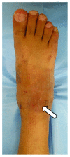
Photograph of the ankle and foot showing swelling around the anterolateral portal (arrow) 4 weeks after ankle arthroscopy.
Figure 3
Figure 3
Enhanced three-dimensional computed tomography (CT) images show the perforating peroneal artery (arrow) with the absence of the anterior tibial artery and posterior tibial artery (a) and the pseudoaneurysm of the perforating peroneal artery (arrow) at the lateral aspect of the ankle joint (b). Enhanced CT images also show the size of the pseudoaneurysm (arrow) 15 × 20 × 30 mm on the sagittal (c) and axial planes (d).
Figure 4
Figure 4
Color and duplex Doppler ultrasonography showing flow through the perforating peroneal artery (arrow) at the anterolateral ankle joint, suggesting a pseudoaneurysm (arrowhead) leaking into the ankle joint. Flow toward and away from the transducer is indicated by red and blue, respectively.
Figure 5
Figure 5
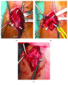
Photographic images taken during repair of the perforating peroneal artery injury. (a) Pseudoaneurysm (arrow) caused by disruption of the perforating peroneal artery wall on the anterolateral side of the ankle joint. Arrow indicates the main trunk of the perforating artery proximally and distally. (b) Pseudoaneurysm is cut (arrowhead) followed by removal of the hematoma-like content within the pseudoaneurysm. Arrow indicates the main trunk of the perforating artery proximally and distally. (c) Repair of the injured perforating peroneal artery wall using end-to-end anastomosis (arrow), without compromising blood supply to the foot.

Similar articles

Cited by

References

    1. Sarrafian S. K. Anatomy of the Foot and Ankle: Descriptive, Topographic, Functional. 2nd. Philadelphia, PA: Lippincott Williams & Wilkins; 1993.
    1. Yu J. L., Ho E., Wines A. P. Pseudoaneurysms around the foot and ankle: case report and literature review. Foot and Ankle Surgery. 2013;19(3):194–198. doi: 10.1016/j.fas.2012.08.003. - DOI - PubMed
    1. Tonogai I., Matsuura T., Iwame T., et al. Pseudoaneurysm of the anterior tibial artery following ankle arthroscopy in a soccer player. Case Reports in Orthopedics. 2017;2017:8. doi: 10.1155/2017/2865971.2865971 - DOI - PMC - PubMed
    1. Sadat U., See T., Cousins C., Hayes P., Gaunt M. Peroneal artery pseudoaneurysm—a case report and literature review. BMC Surgery. 2007;7(1):p. 4. doi: 10.1186/1471-2482-7-4. - DOI - PMC - PubMed
    1. Maguire D. W., Huffer J. M., Ahlstrand R. A., Crummy Jr A. B. Traumatic aneurysm of perforating peroneal artery. The Journal of Bone and Joint Surgery. American Volume. 1972;54(2):409–412. doi: 10.2106/00004623-197254020-00021. - DOI - PubMed

Publication types

LinkOut - more resources