Case Reports
. 2018 Jun 20;8(2):e2018028.
doi: 10.4322/acr.2018.028. eCollection 2018 Apr-Jun.

The diagnosis of multiple opportunistic infections in advanced stage AIDS: when Ockham's Razor doesn't cut it

Affiliations
Case Reports

The diagnosis of multiple opportunistic infections in advanced stage AIDS: when Ockham's Razor doesn't cut it

Marcos Vinicius Cardoso Pinheiro et al. Autops Case Rep. .

Abstract

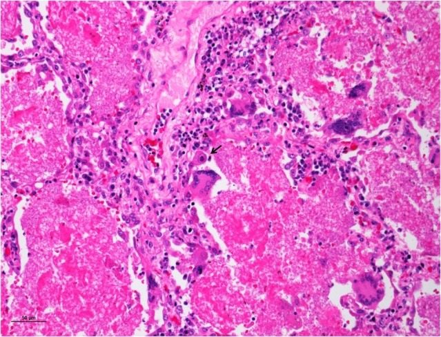
In the advanced stage of AIDS, the diagnosis of the opportunistic infections may be challenging due to the high risk of performing invasive diagnostic methods in a patient with a critical clinical condition, as well as the correct interpretation of the results of microbiological exams. One of the challenges for the diagnosis and treatment of the opportunistic infections is that they may occur concomitantly in the same patient and they may mimic each other, leading to a high discrepancy between clinical and autopsy diagnoses. We describe the case of a 52-year-old man who was hospitalized because of weight loss, anemia, cough, and hepatosplenomegaly. During the investigation, the diagnosis of AIDS was made, and the patient developed respiratory failure and died on the fourth day of hospitalization. At autopsy, disseminated non-tuberculosis mycobacteriosis was found, affecting mainly the organs of the reticuloendothelial system. Also, severe and diffuse pneumonia caused by multiple agents (Pneumocystis jirovecii , Histoplasma capsulatum, suppurative bacterial infection, non-tuberculosis mycobacteria, and cytomegalovirus) was seen in a morphological pattern that could be called "collision pneumonia." The lesson from this case, revealed by the autopsy, is that in advanced AIDS, patients often have multiple opportunistic infections, so the principle of Ockham's razor-that a single diagnosis is most likely the best diagnosis-fails in this clinical context.

Keywords: Acquired Immunodeficiency Syndrome; Cytomegalovirus; Histoplasma; Mycobacterium; Pneumocystis.


Conflict of interest statement

Conflict of interest: None

Figures

Figure 1
Figure 1. A – Chest radiography taken on the first day of hospitalization showing mediastinal enlargement, consolidation in the right superior and middle lobes, and sparse interstitial and alveolar infiltrates in the remaining fields; B – Gross examination of the right lung: diffuse consolidation (hepatization of the lung). Note the adhesions of the visceral pleura to the diaphragm and to the soft tissues of the chest are partially fixed.
Figure 2
Figure 2. Micrography of the lung. Diffuse pneumonia with “collision” of different infectious diseases: pneumocystosis (blue arrowhead), suppurative pneumonia (black arrow) and histoplasmosis (blue arrow). Note the septal edema and focal alveolar hemorrhage (H&E, 300X).
Figure 3
Figure 3. Micrography of the lung. The typical pneumocystosis foamy alveolar exudate associated with some multinuclear giant cells, type II pneumocytes hyperplasia, thickened alveolar septum, and an alveolar epithelial cell with cytomegalic cytopathic changes (arrow) (H&E, 300X).
Figure 4
Figure 4. Micrography of the lung. Foci of plate-like dystrophic calcification within the typical pneumocystosis foamy alveolar exudate (H&E, 300X). The inset shows typical P. jirovecii cup-shaped yeasts, measuring 4-6 µm, some with the central intracystic bodies at Grocott’s stain (400X).
Figure 5
Figure 5. Micrography of the lung. Pneumocystosis with two histologic patterns in the same field: on the left the typical foamy alveolar exudate, lined by an alveolar septum with type II pneumocyte hyperplasia, and an alveolar cell with cytomegalovirus cytopathic changes (arrow); on the right, a granulomatous reaction, surrounding foci of dystrophic calcifications (H&E, 300X).
Figure 6
Figure 6. Micrography of the lung. Severe pneumocystosis characterized by confluent foamy exudate, forming a “cystic” parenchymal cavitation in the center. At the periphery, alveoli filled by foamy exudate can be seen (arrowheads) (H&E, 300X).
Figure 7
Figure 7. Micrography of the lung. Alveolar macrophages with tiny Histoplasma capsulatum yeasts in their cytoplasm (arrow), admixed in the typical pneumocystosis foamy alveolar exudate. Note an alveolar epithelial cell with cytomegalic cytopathic changes (arrowhead), septal congestion, alveolar hemorrhage, and a discrete mixed inflammatory exudate in the alveolar space (H&E, 300X).
Figure 8
Figure 8. Micrography of the lungs. A – Area of acute histoplasmosis pneumonia, showing numerous alveolar macrophages with small yeasts forms in their cytoplasms (H&E, 300X); B – The Grocott’s stain shows small nucleated yeasts within the alveolar macrophages (1,000X); C and D – Scarce peribronchiolar areas and alveolar septa infiltrated by engorged macrophages, with pale bluish cytoplasm (arrows in C), with plenty acid-fast bacilli in the cytoplasm (arrows in D) (C: H&E, 300X; D: Ziehl-Neelsen, 400X).
Figure 9
Figure 9. A – Gross examination of the liver: congestion and various yellowish small nodules scattered in the hepatic parenchyma (arrows); B – Micrography of the liver: sheets of histiocytes infiltrating the liver, and sinusoidal dilatation in the centrilobular area (H&E, 40X); C – Micrography of the liver: engorged and bluish histiocytes forming aggregates in the portal tract, lobules and sinusoids with some multinucleated giant cells (inset) (H&E, 300X); D – Micrography of the liver: numerous acid-fast bacilli within the histiocytes and multinucleated giant cells cytoplasm (arrows). The infected cells are isolated or grouped in ill-defined granulomas in the lobules and sinusoids (H&E, 300X).
Figure 10
Figure 10. A and B – Gross examination of the spleen: the volume was increased. The splenic surface had irregular yellowish areas and adhesions to the omentum (arrows in A). The cut surface showed wedge-shaped areas of necrosis, corresponding to the external yellowish aspect of the organ (arrow in B). The red pulp was extremely friable. C and D – Micrography of the spleen: the entire splenic parenchyma was infiltrated by sheets of engorged and bluish histiocytes, causing vasculitis, ischemic necrosis (arrow in C) and thrombosis (arrowhead in C and arrow in D) (C, H&E 40X and D, H&E 200X). The Ziehl–Neelsen stain revealed acid-fast bacilli in the histiocyte cytoplasm (not shown).
Figure 11
Figure 11. A and B – Micrography of the small intestine: the mucosa and submucosa infiltrated by engorged macrophages with plenty acid-fast bacilli in the cytoplasm (A, H&E 40X; B, Ziehl–Neelsen 100X); C and D – Micrography of the kidney: interstitial chronic nephritis composed of sheets of engorged macrophages (arrow in C) with plenty acid-fast bacilli in the cytoplasm (arrows in D) (A, H&E 40X; B, Ziehl–Neelsen 100X).
Figure 12
Figure 12. A to D – Micrography showing aspects of non-tuberculous disseminated mycobacteriosis: sheets of engorged and bluish histiocytes infiltrating the bone marrow (A). The periadrenal fat tissue (B, arrows and inset) and an anthracotic thoracic lymph node (C, arrow). The Ziehl–Neelsen stain in all these sites revealed plenty of acid-fast bacilli within histiocytes’ cytoplasm (D). (A, H&E, 100X; B, H&E, 100X; C, H&E, 100X; D, Ziehl–Neelsen, 100X).

Similar articles

Cited by

References

    1. Brasil . Ministério da Saúde . HIV/AIDS 2017 . Brasília: : Ministério da Saúde; ; 2017. . ( Boletim epidemiológico - Ano V nº 1- 27ª a 53ª semanas epidemiológicas - julho a dezembro de 2016, da 01ª a 26ª semanas epidemiológicas - janeiro a junho de 2017 ) [ cited 2017 Oct 12 ]. Available from: http://www.aids.gov.br/pt-br/pub/2017/boletim-epidemiologico-hivaids-201...
    1. Grangeiro A , Escuder MM , Pereira JC . Late entry into HIV care: lessons from Brazil, 2003 to 2006 . BMC Infect Dis . 2012. ; 12 ( 1 ): 99 . 10.1186/1471-2334-12-99 . - DOI - PMC - PubMed
    1. Brasil . Ministério da Saúde . HIV/AIDS 2015 . Brasília: : Ministério da Saúde; ; 2015. . ( Boletim epidemiológico - HIV/AIDS. Ano IV nº 1 - 27ª à 53ª semana epidemiológica - julho a dezembro de 2014 da 01ª à 26ª semana epidemiológica - janeiro a junho de 2015 ). [ cited 2017 Jan 26 ]. Available from: http://www.aids.gov.br/sites/default/files/anexos/publicacao/2015/58534/...
    1. Coelho L , Veloso VG , Grinsztejn B , Luz PM . Trends in overall opportunistic illnesses, Pneumocystis carinii pneumonia, cerebral toxoplasmosis and Mycobacterium avium complex incidence rates over the 30 years of the HIV epidemic: a systematic review . Braz J Infect Dis . 2014. ; 18 ( 2 ): 196 - 210 . 10.1016/j.bjid.2013.10.003 . - DOI - PMC - PubMed
    1. Brasil . Ministério da Saúde . Perspectivas brasileiras para o fim da tuberculose como problema de saúde pública . Boletim Epidemiológico , 2016. ; 47 ( 13 ), 1 - 15 . [ cited 2017 Jan 26 ]. Available from http://portalsaude.saude.gov.br/images/pdf/2016/marco/24/2016-009-Tuberc...

Publication types