. 2018 Dec 3;4(6):e289.
doi: 10.1212/NXG.0000000000000289. eCollection 2018 Dec.

Mutation in POLR3K causes hypomyelinating leukodystrophy and abnormal ribosomal RNA regulation

Affiliations

Mutation in POLR3K causes hypomyelinating leukodystrophy and abnormal ribosomal RNA regulation

Imen Dorboz et al. Neurol Genet. .

Abstract

Objective: To identify the genetic cause of hypomyelinating leukodystrophy in 2 consanguineous families.

Methods: Homozygosity mapping combined with whole-exome sequencing of consanguineous families was performed. Mutation consequences were determined by studying the structural change of the protein and by the RNA analysis of patients' fibroblasts.

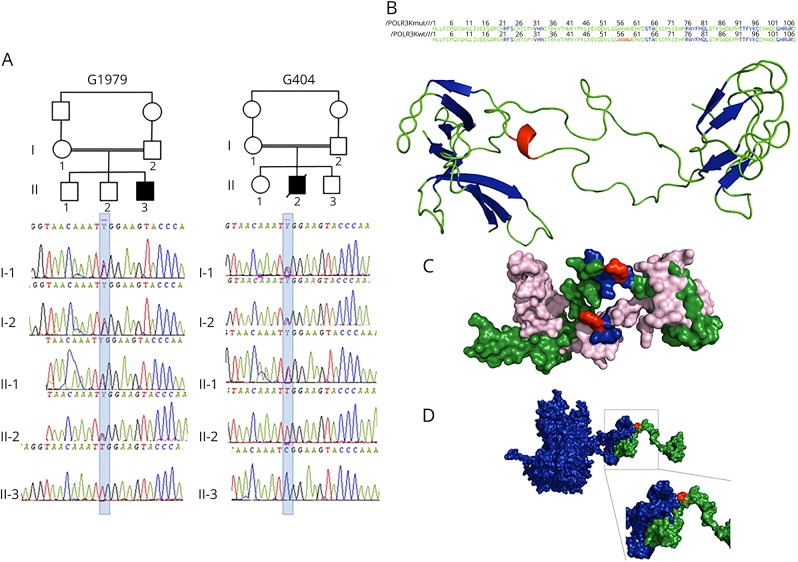
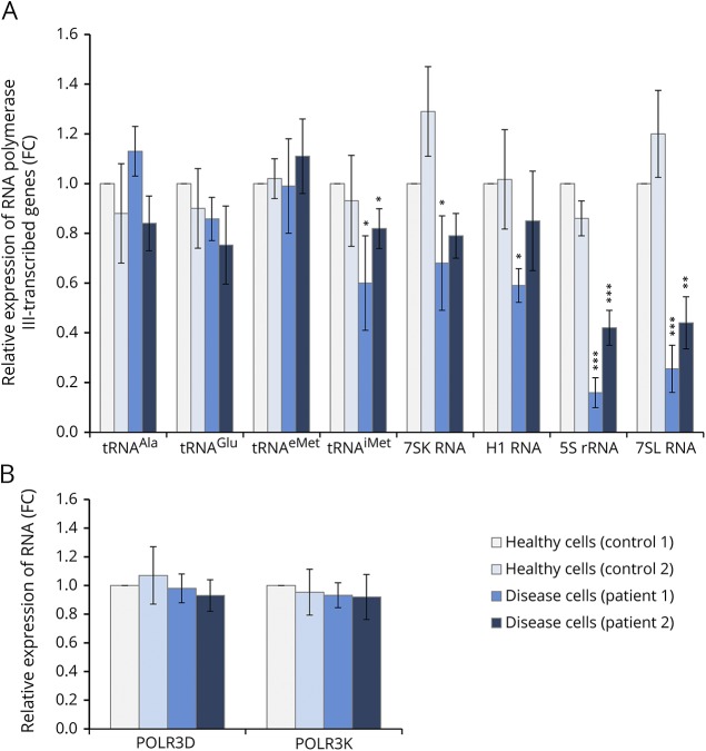
Results: We identified a biallelic mutation in a gene coding for a Pol III-specific subunit, POLR3K (c.121C>T/p.Arg41Trp), that cosegregates with the disease in 2 unrelated patients. Patients expressed neurologic and extraneurologic signs found in POLR3A- and POLR3B-related leukodystrophies with a peculiar severe digestive dysfunction. The mutation impaired the POLR3K-POLR3B interactions resulting in zebrafish in abnormal gut development. Functional studies in the 2 patients' fibroblasts revealed a severe decrease (60%-80%) in the expression of 5S and 7S ribosomal RNAs in comparison with control.

Conclusions: These analyses underlined the key role of ribosomal RNA regulation in the development and maintenance of the white matter and the cerebellum as already reported for diseases related to genes involved in transfer RNA or translation initiation factors.


Figures

Figure 1
Figure 1. POLR3K mutated families, structural model of POLR3K and of POLR3K-POLR3B interactions
(A) Pedigree and electropherograms of family G1979 (patient 1) and family G404 (patient 2). (B) Amino acid (AA) sequence and 2-dimensional structure of the mutated and wild-type POLR3K. The sheets, loops, and α-helix motifs are colored in blue, green, and red, respectively. The loop (AA 34–55), α-helix (AA 56–59), and loop (AA60–63) motifs of the wild-type protein are replaced by a unique loop (34–63) in the mutated protein. (C) Three-dimensional structure of the wild-type (in green) and mutated (in pink) POLR3K. The AAs at position 41 (arginine in the wild type and tryptophan in the mutant) are in red. The residues located within 4 Å around the Arg41, responsible for the stability of the protein (Asn40, Lys42, and Tyr43), are colored in blue. The Trp41 change induces a modification in the interactions of Tyr43 with Asn40 and Lys42 decreasing protein stability. (D) Three-dimensional structure of POLR3K (in green) and POLR3B (in blue) interactions. The mutated residue 41 colored in red is important in POLR3K-POLR3B interactions: the N-terminal part (1–41) of POLR3K interacting with the C-terminal part (1079–1133) of POLR3B.
Figure 2
Figure 2. MRI progression in patient 1
The diffuse hypomyelinating aspect of the white matter characterized by an isosignal T1, hypersignal T2, and flair of the white matter in comparison with the gray matter did not change during the 8 years of evolution (MRI performed at age 2, 4, and 10 years) despite the clinical progression of the disease observed after age 4 years. In contrast, a progressive atrophy is observed in the supratentorial and subtentorial structures between ages 4 and 10 years: deeper cortical sulci, increased subarachnoid spaces, frontal ventricular dilatation and white matter atrophy, corpus callosum atrophy (18% loss in the anterior part and 35% in the posterior part), and cerebellar atrophy (20% loss of the vermis and cerebellar hemispheres).
Figure 3
Figure 3. RNA levels determined by RT-qPCR analysis
Total RNAs were isolated from healthy (control cells) or POLR3K mutated (disease cells) patients' fibroblasts. Relative gene expression levels were normalized with β-actin and PPIA genes as an internal control and compared with control cells. Each bar represents the mean ± SD of at least 3 independent experiments. *Indicated a significant difference of disease cells compared with healthy cells. *p < 0.05, **p < 0.01, ***p < 0.001. (A) POLR3K and POLR3D mRNA expression levels. (B) Expression of RNA polymerase III–transcribed genes.

References

    1. Bernard G, Chouery E, Putorti ML, et al. . Mutations of POLR3A encoding a catalytic subunit of RNA polymerase Pol III cause a recessive hypomyelinating leukodystrophy. Am J Hum Genet 2011;89:415–423. - PMC - PubMed
    1. Tétreault M, Choquet K, Orcesi S, et al. . Recessive mutations in POLR3B, encoding the second largest subunit of Pol III, cause a rare hypomyelinating leukodystrophy. Am J Hum Genet 2011;89:652–705. - PMC - PubMed
    1. Dieci G, Fiorino G, Castelnuovo M, Teichmann M, Pagano A. The expanding RNA polymerase III transcriptome. Trends Genet 2007;23:614–622. - PubMed
    1. Dumay-Odelot H, Durrieu-Gaillard S, Da Silva D, Roeder RG, Teichmann M. Cell growth- and differentiation-dependent regulation of RNA polymerase III transcription. Cell Cycle 2010;9:3687–3699. - PMC - PubMed
    1. Dumay-Odelot H, Durrieu-Gaillard S, El Ayoubi L, Parrot C, Teichmann M. Contributions of in vitro transcription to the understanding of human RNA polymerase III transcription. Transcription 2014;5:e27526. - PMC - PubMed