. 2019 Apr;13(4):811-828.
doi: 10.1002/1878-0261.12434. Epub 2019 Feb 2.

High affinity binding of H3K14ac through collaboration of bromodomains 2, 4 and 5 is critical for the molecular and tumor suppressor functions of PBRM1

Affiliations

High affinity binding of H3K14ac through collaboration of bromodomains 2, 4 and 5 is critical for the molecular and tumor suppressor functions of PBRM1

Lili Liao et al. Mol Oncol. 2019 Apr.

Abstract

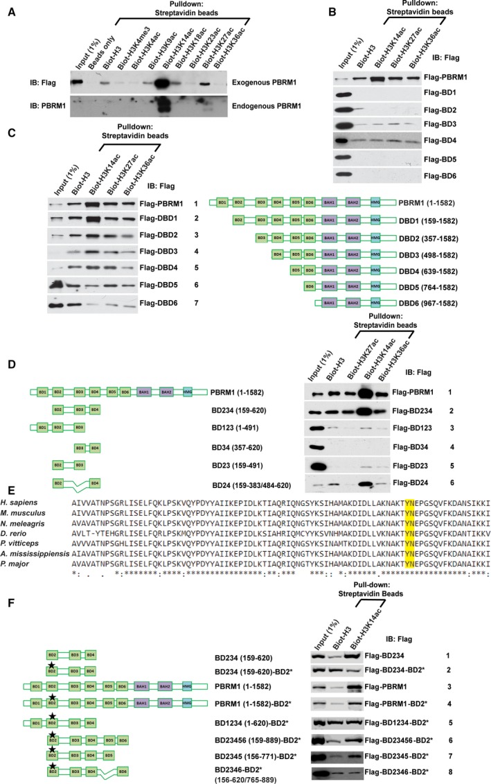
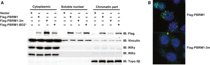
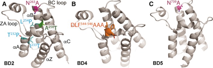
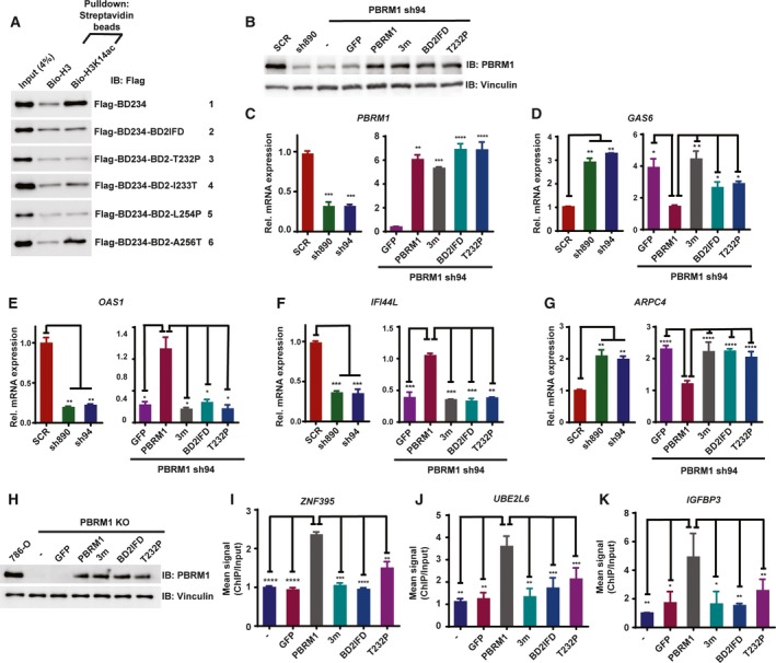
Polybromo-1 (PBRM1) is an important tumor suppressor in kidney cancer. It contains six tandem bromodomains (BDs), which are specialized structures that recognize acetyl-lysine residues. While BD2 has been found to bind acetylated histone H3 lysine 14 (H3K14ac), it is not known whether other BDs collaborate with BD2 to generate strong binding to H3K14ac, and the importance of H3K14ac recognition for the molecular and tumor suppressor function of PBRM1 is also unknown. We discovered that full-length PBRM1, but not its individual BDs, strongly binds H3K14ac. BDs 2, 4, and 5 were found to collaborate to facilitate strong binding to H3K14ac. Quantitative measurement of the interactions between purified BD proteins and H3K14ac or nonacetylated peptides confirmed the tight and specific association of the former. Interestingly, while the structural integrity of BD4 was found to be required for H3K14ac recognition, the conserved acetyl-lysine binding site of BD4 was not. Furthermore, simultaneous point mutations in BDs 2, 4, and 5 prevented recognition of H3K14ac, altered promoter binding and gene expression, and caused PBRM1 to relocalize to the cytoplasm. In contrast, tumor-derived point mutations in BD2 alone lowered PBRM1's affinity to H3K14ac and also disrupted promoter binding and gene expression without altering cellular localization. Finally, overexpression of PBRM1 variants containing point mutations in BDs 2, 4, and 5 or BD2 alone failed to suppress tumor growth in a xenograft model. Taken together, our study demonstrates that BDs 2, 4, and 5 of PBRM1 collaborate to generate high affinity to H3K14ac and tether PBRM1 to chromatin. Mutations in BD2 alone weaken these interactions, and this is sufficient to abolish its molecular and tumor suppressor functions.

Keywords: H3K14ac; PBRM1; bromodomain; kidney cancer; synergy.


Conflict of interest statement

The authors declare no conflict of interests.

Figures

Figure 1
Figure 1
BDs 2, 4, and 5 collaborate to strongly bind H3K14ac. (A) Flag‐PBRM1 was expressed in HEK293T cells. The EBC cell lysates were pulled down with indicated biotinylated peptides and analyzed with indicated antibodies (top). HEK293 lysates were treated the same way to examine the interaction between endogenous PBRM1 and biotinylated histone H3 peptides (bottom). (B) Full‐length PBRM1 or individual BDs of PBRM1 were expressed in HEK293T cells. Their binding to different biotinylated peptides was analyzed as described in (A). (C) Full‐length PBRM1 or PBRM1 with BDs sequentially truncated was expressed in HEK293T cells and analyzed as described above. (D) Full‐length PBRM1 or different combinations of BDs of PBRM1 were expressed in HEK293T cells and analyzed as described above. (E) The aligned protein sequences of BD 2 from different species, and the conserved YN residues that are critical for acetyl‐lysine recognition were labeled with a box. (F) Full‐length PBRM1 or different combinations of BDs of PBRM1, in the presence or absence of indicated mutations, were expressed in HEK293T cells and analyzed as described above. BD2* indicates N263A point mutation in BD2.
Figure 2
Figure 2
Quantitative measurement confirms that BD245 work together to create high affinity to H3K14ac. (A) The indicated GSTBD constructs of PBRM1 were expressed and purified from Escherichia coli, pulled down by the indicated peptides, and immunoblotted with anti‐GST antibody. (B) The bands in (A) were quantified with NIH ImageJ, and the ratios of bands associated with H3K14ac/H3 were calculated from three experiments. The P‐values were calculated using the two‐tailed Student's t‐test. *: P < 0.05; **: P < 0.01; ***: P < 0.001. N.S.: nonsignificant. (C) Top: The indicated GSTBD constructs of PBRM1 were expressed and purified from Escherichia coli. Similar amount of GST‐fusion protein was incubated with 3ug of nucleosome purified from HeLa cells, washed then analyzed with indicated antibodies. Arrow: the full‐length GSTBD12 protein. Bottom: The intensity of the pulled down H3 or H3K14ac bands was determined with ImageJ from three different experiments, and the ratios of H3/input and H3K14ac/input were calculated and plotted. The error bars represent standard error of the mean. (D) GSTBD fusion proteins (left) or cleaved and purified BD proteins (right) were pulled down with indicated peptides and analyzed with western blots. (E) BLI analysis of the interaction between PBRM1 BD proteins and the H3K14ac peptide. Binding activity was normalized to maximum response and is reported as relative binding. The association phase takes place from 0 to 900 s; the dissociation phase takes place from 901 to 2200 s. (F) Binding parameters of PBRM1 BD constructs and H3 peptide interactions. Apparent K d values (Kdapp) were obtained for the interaction between PBRM1 BD proteins and H3 peptides by fitting BLI data to a double exponential function. The H3:H3K14ac reports the preference of PBRM1 BD proteins to bind the H3K14ac peptide over the nonacetylated H3 peptide.
Figure 3
Figure 3
Concurrent point mutations in BDs 2, 4, and 5 abrogate PBRM1's recognition of H3K14ac. A) Sequence alignment of the six BDs of human PBRM1 and the BDs of BRG1 and BRM. *: conserved residues : or.: highly similar amino acids. The residues mutated in B–D were boxed. (B, C, and D) The indicated PBRM1 constructs were expressed in HEK293T cells, pulled down by the indicated peptides, and immunoblotted with anti‐Flag antibody. The * in the rows indicate point mutations.
Figure 4
Figure 4
Concurrent point mutations in BDs 2, 4, and 5 cause PBRM1 relocalization to the cytoplasm, while mutation in BD2 alone does not. (A) Wild‐type or mutant PBRM1 constructs were transiently expressed in HEK293T cells. The cells were fractionated into cytoplasmic, soluble nuclear, and chromatin‐bound fractions. The lysates were blotted with the indicated antibodies. The blots of IKKγ, IKKα, and Topo IIβ indicate the successful separation of cellular compartments. (B) Wild‐type or mutant PBRM1 constructs were expressed in HeLa cells. Immunofluorescence was performed using anti‐Flag antibody. The DNA is stained blue with DAPI, while the PBRM1 signal is stained green.
Figure 5
Figure 5
Tumor‐derived mutations mapped onto crystal structures of BDs. (A) Tumor‐derived mutations were mapped onto the available crystal structure of BD2 of PBRM1 (Filippakopoulos et al., 2010). (B) BD4* mutation was mapped onto the available crystal structure of BD4 of PBRM1 (Filippakopoulos et al., 2010). (C) The location of the critical N739 was mapped onto the available crystal structure of BD5 of PBRM1 (Filippakopoulos et al., 2010).
Figure 6
Figure 6
Tumor‐derived mutations in BD2 weaken PBRM1's recognition of H3K14ac, its ability to regulate gene expression, and its binding to promoters. (A) Wild‐type BD234 of PBRM1 or BD234 carrying tumor‐derived mutations were expressed in HEK293T cells. The lysates were pulled down with the indicated peptides and immunoblotted with anti‐Flag antibody. (B) 786‐O cells were infected to stably express the indicated shRNA and/or PBRM1 constructs. The lysates were blotted with indicated antibodies. The relative mRNA expression of (C) PBRM1, (D) GAS6, (E) OAS1, (F) IFI44L, and (G) ARPC4 over SCR‐expressing cells was examined with qRTPCR in the indicated cells from two experimental results. (H) PBRM1 constructs or GFP was transiently transfected into 786‐O cells with PBRM1 knocked out by CRISPR/Cas9. Anti‐PBRM1 ChIP was performed and the enrichment of promoter sequences for (I) ZNF‐395, (J) UBE2L6, or (K) IGFBP3 was measured by qPCR. The ChIP/input ratio for each promoter from the nontransfected cells was set as 1, and the relative enrichment was calculated by dividing the ChIP/input ratio from the transfected cells with that from the control cells from two experimental results. The error bars represent standard error of the mean. The P‐values were calculated using the two‐tailed Student's t‐test. *: P < 0.05; **: P < 0.01; ***: P < 0.001; ****: P < 0.0001.
Figure 7
Figure 7
H3K14ac recognition is critical to PBRM1's tumor suppressor function. Xenograft analyses were performed to compare tumor growth of 786‐O cells with PBRM1 stably suppressed by PBRM1‐sh94. (A) Representative tumors derived from cells expressing GFP or shRNA‐resistant wild‐type PBRM1; (B) quantification of tumor weights in A); C) representative tumors derived from cells expressing shRNA‐resistant wild‐type PBRM1 or PBRM1 with N263 in‐frame deletion; (D) quantification of tumor weights in (C); (E) representative tumors derived from cells expressing shRNA‐resistant wild‐type PBRM1 or PBRM1 with mutations in BDs 2, 4, and 5; (F) quantification of tumor weights in (E). The error bar depicts standard error of the mean, and P‐values were calculated using the two‐tailed Student's t‐test. n indicates the number of mice used in each experiment.

References

    1. Benusiglio PR, Couve S, Gilbert‐Dussardier B, Deveaux S, Le Jeune H, Da Costa M, Fromont G, Memeteau F, Yacoub M, Coupier I et al (2015) A germline mutation in PBRM1 predisposes to renal cell carcinoma. J Med Genet 52, 426–430. - PubMed
    1. Cancer Genome Atlas Research Network (2013) Comprehensive molecular characterization of clear cell renal cell carcinoma. Nature 499, 43–49. - PMC - PubMed
    1. Chandrasekaran R and Thompson M (2007) Polybromo‐1‐bromodomains bind histone H3 at specific acetyl‐lysine positions. Biochem Biophys Res Commun 355, 661–666. - PubMed
    1. Charlop‐Powers Z, Zeng L, Zhang Q and Zhou MM (2010) Structural insights into selective histone H3 recognition by the human Polybromo bromodomain 2. Cell Res 20, 529–538. - PMC - PubMed
    1. Choueiri TK and Motzer RJ (2017) Systemic Therapy for Metastatic Renal‐Cell Carcinoma. N Engl J Med 376, 354–366. - PubMed

Publication types

MeSH terms