. 2019 Jan 2;139(1):78-96.
doi: 10.1161/CIRCULATIONAHA.118.034588.

Uremic Toxin Indoxyl Sulfate Promotes Proinflammatory Macrophage Activation Via the Interplay of OATP2B1 and Dll4-Notch Signaling

Affiliations

Uremic Toxin Indoxyl Sulfate Promotes Proinflammatory Macrophage Activation Via the Interplay of OATP2B1 and Dll4-Notch Signaling

Toshiaki Nakano et al. Circulation. .

Abstract

Background: Chronic kidney disease (CKD) increases cardiovascular risk. Underlying mechanisms, however, remain obscure. The uremic toxin indoxyl sulfate is an independent cardiovascular risk factor in CKD. We explored the potential impact of indoxyl sulfate on proinflammatory activation of macrophages and its underlying mechanisms.

Methods: We examined in vitro the effects of clinically relevant concentrations of indoxyl sulfate on proinflammatory responses of macrophages and the roles of organic anion transporters and organic anion transporting polypeptides (OATPs). A systems approach, involving unbiased global proteomics, bioinformatics, and network analysis, then explored potential key pathways. To address the role of Delta-like 4 (Dll4) in indoxyl sulfate-induced macrophage activation and atherogenesis in CKD in vivo, we used 5/6 nephrectomy and Dll4 antibody in low-density lipoprotein receptor-deficient (Ldlr-/-) mice. To further determine the relative contribution of OATP2B1 or Dll4 to proinflammatory activation of macrophages and atherogenesis in vivo, we used siRNA delivered by macrophage-targeted lipid nanoparticles in mice.

Results: We found that indoxyl sulfate-induced proinflammatory macrophage activation is mediated by its uptake through transporters, including OATP2B1, encoded by the SLCO2B1 gene. The global proteomics identified potential mechanisms, including Notch signaling and the ubiquitin-proteasome pathway, that mediate indoxyl sulfate-triggered proinflammatory macrophage activation. We chose the Notch pathway as an example of key candidates for validation of our target discovery platform and for further mechanistic studies. As predicted computationally, indoxyl sulfate triggered Notch signaling, which was preceded by the rapid induction of Dll4 protein. Dll4 induction may result from inhibition of the ubiquitin-proteasome pathway, via the deubiquitinating enzyme USP5. In mice, macrophage-targeted OATP2B1/Slco2b1 silencing and Dll4 antibody inhibited proinflammatory activation of peritoneal macrophages induced by indoxyl sulfate. In low-density lipoprotein receptor-deficient mice, Dll4 antibody abolished atherosclerotic lesion development accelerated in Ldlr-/- mice. Moreover, coadministration of indoxyl sulfate and OATP2B1/Slco2b1 or Dll4 siRNA encapsulated in macrophage-targeted lipid nanoparticles in Ldlr-/- mice suppressed lesion development.

Conclusions: These results suggest that novel crosstalk between OATP2B1 and Dll4-Notch signaling in macrophages mediates indoxyl sulfate-induced vascular inflammation in CKD.

Keywords: Notch signaling; atherosclerosis; chronic kidney diseases; indoxyl sulfate; inflammation; macrophage.


Figures
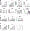
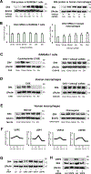
Figure 1.
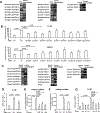
Figure 1.. Indoxyl sulfate induces pro-inflammatory cytokine expression in human primary macrophages derived from peripheral blood mononuclear cells (PMBC).
(A) mRNA expression of IL-1β, TNF-α, MCP-1 MRC1, AMAC1, IL-10 and CD163 was measured in human primary macrophages after stimulation with indoxyl sulfate for 3 hours (9 different PBMC donors). (B) IL-1β levels in supernatants of human primary macrophages were measured by ELISA after stimulation with indoxyl sulfate for 72 hours (9 donors). (C) mRNA expression of IL-1β, TNF-α and MCP-1 was measured in human primary macrophages after stimulation with tryptophan for 3 hours (6 donors). (D) mRNA expression of IL-1β, TNF-α, MCP-1 was measured in human primary macrophages after stimulation with indoxyl sulfate in medium with 4% albumin for 3 hours (8 donors). P value was calculated by one-way ANOVA followed by Tukey’s test, based on a comparison with 0 mM indoxyl sulfate. Error bars indicate ± SEM.
Figure 2.
Figure 2.. Macrophages express organic anion transporters (OATs) and organic anion transporting polypeptides (OATPs).
(A) mRNA expression of OATs / OATPs was examined by PCR in PBMC-derived human primary macrophages. Protein names are noted in brackets. (B) mRNA expression was measured in human primary macrophages after suppression by siRNA for 48 hours and stimulation with 0.5 mmol/L indoxyl sulfate (IS) for 3 hours (9 PBMC donors). (C) mRNA expression of OATs / OATPs was examined by PCR in mouse peritoneal macrophages. Protein names are noted in brackets. (D) mRNA expression was measured in mouse peritoneal macrophages after suppression by siRNA for 48 hours and stimulation with 0.5 mmol/L IS for 3 hours (n=6). (E) Cellular uptake of indoxyl sulfate (IS) in human primary macrophages was measured by HPLC in the absence or presence of the OAT/OATP inhibitor 10 mmol/L probenecid (PRO), 100 μmol/L rifampicin (RIF) or 10 μmol/L cyclosporin A (CyA) (6 PBMC donors). (F) Cellular uptake of indoxyl sulfate (IS) in human primary macrophages was measured by HPLC in the presence of SLCO2B1 siRNA or control siRNA (6 donors). (G) mRNA expression of IL-1β was measured after pretreatment with the OAT/OATP inhibitor 10 mmol/L probenecid (PRO), 100 μmol/L rifampicin (RIF) and 10 μmol/L cyclosporin A (CyA) for 30 minutes, and stimulation with 0.5 mmol/L IS for 3 hours (8 donors). P value was calculated by unpaired Student’s t-test or one-way ANOVA followed by Tukey’s test. Error bars indicate ± SEM.
Figure 3.
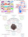
Figure 3.. Global proteomics, pathway network analysis and network analysis of the mouse macrophage cell line RAW264.7 stimulated with indoxyl sulfate.
(A) Model-based cluster analysis of indoxyl sulfate induced protein kinetics. The dashed red line indicates the theoretical trace for unchanged protein abundances. Red rectangles highlight early (30 min) induction clusters. (B) The network of significantly enriched pathways (FDR < 0.05) of the Cluster 9 proteins. Node size indicates the significance of each pathway in –log(q-value) and the edge thickness represents the overlap between the genes of the two pathways they connect, measured in terms of the Jaccard index (see Methods). Groups of pathways, which share genes and therefore represent common biological processes, are highlighted with red rectangles. (C) The network closeness of Cluster 9 first neighbors to various cardiovascular, malignant, metabolic and auto-immune diseases. The center network represents the first neighbor network of Cluster 9 consisting of the Cluster 9 proteins with FC>1.5 between 0 and 30 minutes (red nodes) and their first neighbors in the PPI network (grey nodes). The diseases inside the circle are those that are significantly close to the first neighbor network of Cluster 9 (empirical p-value <= 0.05). The diseases outside the circle are those that are not significantly close to the first neighbor network of Cluster 9 (empirical p-value > 0.05). Disease modules are built by mapping the respective disease-related genes onto the PPI network.
Figure 4.
Figure 4.. Indoxyl sulfate activates Dll4-Notch signaling.
(A) mRNA expression of Notch pathway components (Dll1, Dll4, Notch1) and protortypical target genes (Hes1, Hey1 and Hey2) was measured in human primary macrophages after stimulation with indoxyl sulfate for 3 hours (9 PBMC donors). (B) The expression of Dll4 and the accumulation of Notch1 intracellular domain (ICD) was measured in human primary macrophages by Western blotting after stimulation with 0.5 mmol/L indoxyl sulfate. These blots from one donor represent 3 different macrophage donors. (C) Flow cytometry analyses of Dll4-positive cells in human primary macrophages after stimulation with indoxyl sulfate (5 donors). P value was calculated by one-way ANOVA followed by Tukey’s test, based on a comparison with 0 minute (min). Error bars indicate ± SEM. (D) Double immunostaining (yellow) localized Dll4 (red) in CD68-positive (green) macrophages in human primary macrophages stimulated with 1 mmol/L indoxyl sulfate for 1 hour. Nuclei are visualized with DAPI. Scale bar = 50 μm. These data are representative of 3 different macrophage donors that produced similar results. (E) SPADE clustering of CyTOF data of human macrophages. Each SPADE tree is a color heat map showing relative Dll4, TNF-a or CD163 immunostaining of “nodes” or clusters of human macrophages after incubation with or without 1mmol/L indoxyl sulfate for 48 hours. The SPADE heatmap shows heterogenous expression patterns of Dll4, TNF-a and CD163 in each condition.
Figure 5.
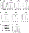
Figure 5.. Indoxyl sulfate-induced pro-inflammatory cytokine expression is mediated by SLCO2B1-Dll4-Notch signaling in human macrophages.
(A) mRNA expression was measured in human primary macrophages after pretreatment with Dll4 siRNA or control siRNA (ctrl) for 48 hours and stimulation with IS for 3 hours (8 PBMC donors). (B) mRNA expression was measured in human primary macrophages after pretreatment with Dll4 antibody (Ab) or control IgG for 24 hours and stimulation with IS for 3 hours (8 donors). (C) The expression of Dll4 was measured by Western blotting after suppression by siRNA for 48 hours and stimulation with 1.0 mmol/L IS for 1 hour. These blots from one donor are representative of the 3 different macrophage donors. (D) mRNA expression of the prototypical Notch target Hes1 was measured in human primary macrophages after suppression by siRNA for 48 hours and stimulation with 0.5 mM IS for 3 hours (6 donors). P value was calculated by unpaired Student’s t-test or one-way ANOVA followed by Tukey’s test. Error bars indicate ± SEM.
Figure 6.
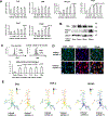
Figure 6.. Indoxyl sulfate suppresses the degradation of Dll4 protein in macrophages.
(A) The expression of Dll4 protein was measured in RAW264.7 cells (left) and human primary macrophages (right) by Western blotting after stimulation with 1.0 mmol/L indoxyl sulfate. (B) mRNA expression of Dll4 was measured in RAW264.7 cells (left, n=4 experiments) and human primary macrophages (right, n = 8 donors) after stimulation with 1mmol/L indoxyl sulfate. (C) Dll4 protein was measured in RAW264.7 cells treated with cycloheximide (CHX) with or without indoxyl sulfate (1 mmol/L). (D) Dll4 protein was measured in human primary macrophages treated with CHX with or without indoxyl sulfate (IS) (1 mmol/L). (E) Dll4 protein was measured in human primary macrophages pretreated with proteasome inhibitor MG132 (left) or lysosomal inhibitor chloroquine (right). (F) TMT-derived indoxyl sulfate-induced protein abundance profiles for USP5, 7, 14 and 48 in RAW264.7 cells.(G, H) Dll4 protein was measured in human primary macrophages by Western blotting after siRNA treatment for 48 hours and stimulation with 1.0 mmol/L IS for 1 hour. All Western blotting experiments are representative of either 3 different RAW264.7 experiments or 3 human PBMC donors that produced similar results.
Figure 7.
Figure 7.. The effects of Dll4 blockade on atherogenesis in CKD mice.
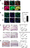
(A) In the aorta of CKD mice fed a HFD for 14 weeks, double immunostaining (yellow) localized Notch1 ICD (green) in Mac3-positive (red) macrophages. Fewer cells were double-positive in sham-operated mice. Nuclei are visualized with DAPI. The data represent 3 mice/group. Scale bar = 20 μm. (B) Double immunostaining (yellow) localized Dll4 (red) in Mac3-positive (green) macrophages in atherosclerotic plaque. Nuclei are visualized with DAPI. Scale bar = 20 μm. The graph shows the percentage of Dll4+ macrophages to total macrophages (n=7 and 5). The average of 3 high power fields was used for the analysis. (C) H&E staining on longitudinal sections of the aortic arch (each n=5 to 9). (D) Staining for Mac3 (macrophages) in the aorta (each n=5 to 9). Scale bar = 100 μm. P value was calculated by unpaired Student’s t-test or two-way ANOVA followed by the Bonferroni test. Error bars indicate ± SEM.
Figure 8.
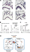
Figure 8.. The effects of macrophage-targeted Dll4 or Slco2b1 blockade on atherogenesis in mice.
(A) In the aorta of C57BL/6 mice fed a HFD for 12 weeks. After 8 weeks, indoxyl sulfate of 100 mg/kg/day or PBS was intraperitoneally injected to C57BL/6 mice for 4 weeks, and macrophage-targeted lipid nanoparticles (C12–200) containing control siRNA (Ctrl), Dll4 siRNA or Slco2b1 siRNA were also injected via tail vein twice a week for 4 weeks. H&E staining on longitudinal sections of the aortic arch. (B) Plaque size in the aortic arch and stenosis rate of brachiocephalic artery (each n=5 to 9). (C) The expression of IL1β mRNA in aorta of mice (each n=5 to 9). P value was calculated by one-way ANOVA followed by Tukey’s test. Error bars indicate ± SEM. (D)The potential mechanism for pro-inflammatory activation of macrophages in CKD via crosstalk between OATP2B1 and the Dll4-Notch axis. Macrophages take up indoxyl sulfate via transporters such as OATP2B1. Dll4 protein usually undergoes endocytosis and recycling. Indoxyl sulfate may inhibits Dll4 degradation via USP5 and increase its recycling. The enhanced interaction of Dll4 and a receptor (e.g., Notch1) may thus accelerate Notch signaling, inducing the expression of pro-inflammatory genes. Although our in vitro and in vivo data suggest the role of the Dll4-Notch pathway in indoxyl sulfate macrophage activation, other mechanisms may also contribute.

Comment in

  • Uremic Toxins Activate Macrophages.
    Hoyer FF, Nahrendorf M. Hoyer FF, et al. Circulation. 2019 Jan 2;139(1):97-100. doi: 10.1161/CIRCULATIONAHA.118.037308. Circulation. 2019. PMID: 30592654 Free PMC article. No abstract available.

References

    1. Tonelli M, Karumanchi SA, Thadhani R. Epidemiology and Mechanisms of Uremia-Related Cardiovascular Disease. Circulation. 2016;133:518–536. - PubMed
    1. Drueke TB, Massy ZA. Atherosclerosis in CKD: differences from the general population. Nat Rev Nephrol. 2010;6:723–735. - PubMed
    1. Aikawa E, Aikawa M, Libby P, Figueiredo JL, Rusanescu G, Iwamoto Y, Fukuda D, Kohler RH, Shi GP, Jaffer FA, Weissleder R. Arterial and aortic valve calcification abolished by elastolytic cathepsin S deficiency in chronic renal disease. Circulation. 2009;119:1785–1794. - PMC - PubMed
    1. Vanholder R, De Smet R, Glorieux G, Argiles A, Baurmeister U, Brunet P, Clark W, Cohen G, De Deyn PP, Deppisch R, Descamps-Latscha B, Henle T, Jorres A, Lemke HD, Massy ZA, Passlick-Deetjen J, Rodriguez M, Stegmayr B, Stenvinkel P, Tetta C, Wanner C, Zidek W, European Uremic Toxin Work G. Review on uremic toxins: classification, concentration, and interindividual variability. Kidney Int. 2003;63:1934–1943. - PubMed
    1. Barreto FC, Barreto DV, Liabeuf S, Meert N, Glorieux G, Temmar M, Choukroun G, Vanholder R, Massy ZA, European Uremic Toxin Work G. Serum indoxyl sulfate is associated with vascular disease and mortality in chronic kidney disease patients. Clin J Am Soc Nephrol. 2009;4:1551–1558. - PMC - PubMed

Publication types

MeSH terms