. 2019 Feb 5;139(6):799-811.
doi: 10.1161/CIRCULATIONAHA.118.034624.

A Premature Termination Codon Mutation in MYBPC3 Causes Hypertrophic Cardiomyopathy via Chronic Activation of Nonsense-Mediated Decay

Affiliations

A Premature Termination Codon Mutation in MYBPC3 Causes Hypertrophic Cardiomyopathy via Chronic Activation of Nonsense-Mediated Decay

Timon Seeger et al. Circulation. .

Abstract

Background: Hypertrophic cardiomyopathy (HCM) is frequently caused by mutations in myosin-binding protein C3 ( MYBPC3) resulting in a premature termination codon (PTC). The underlying mechanisms of how PTC mutations in MYBPC3 lead to the onset and progression of HCM are poorly understood. This study's aim was to investigate the molecular mechanisms underlying the pathogenesis of HCM associated with MYBPC3 PTC mutations by utilizing human isogenic induced pluripotent stem cell-derived cardiomyocytes (iPSC-CMs).

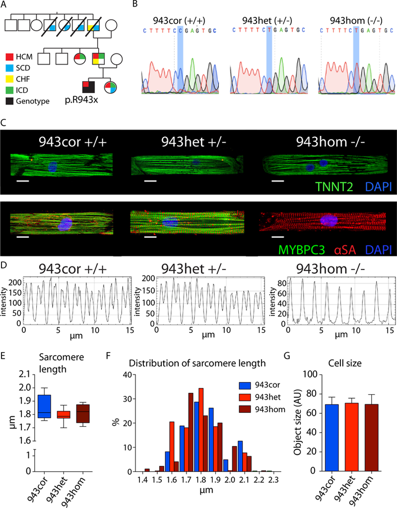
Methods: Isogenic iPSC lines were generated from HCM patients harboring MYBPC3 PTC mutations (p.R943x; p.R1073P_Fsx4) using genome editing. Comprehensive phenotypic and transcriptome analyses were performed in the iPSC-CMs.

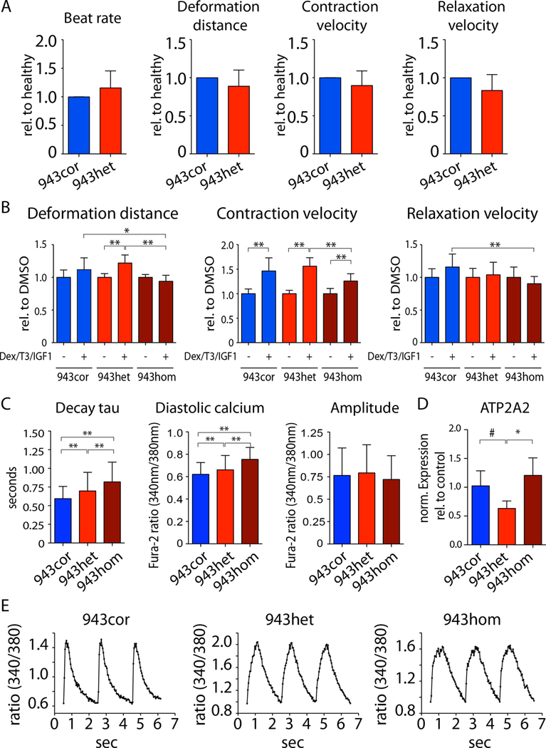
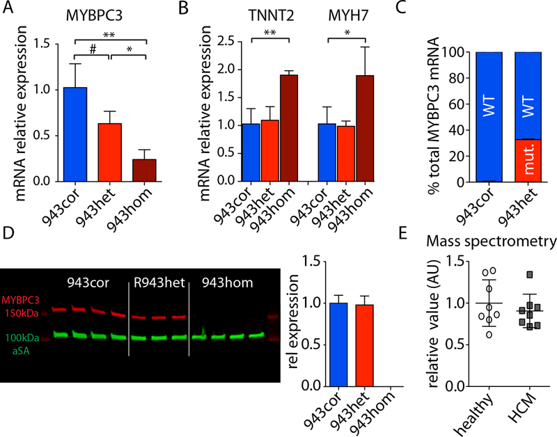
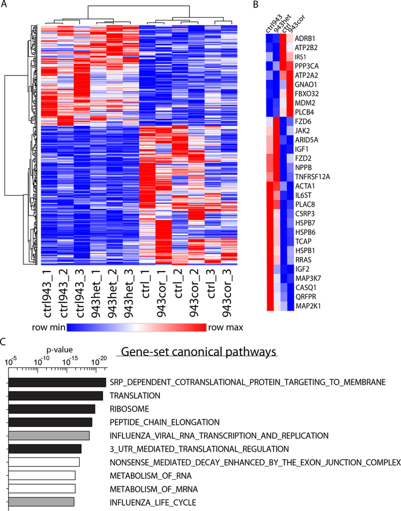
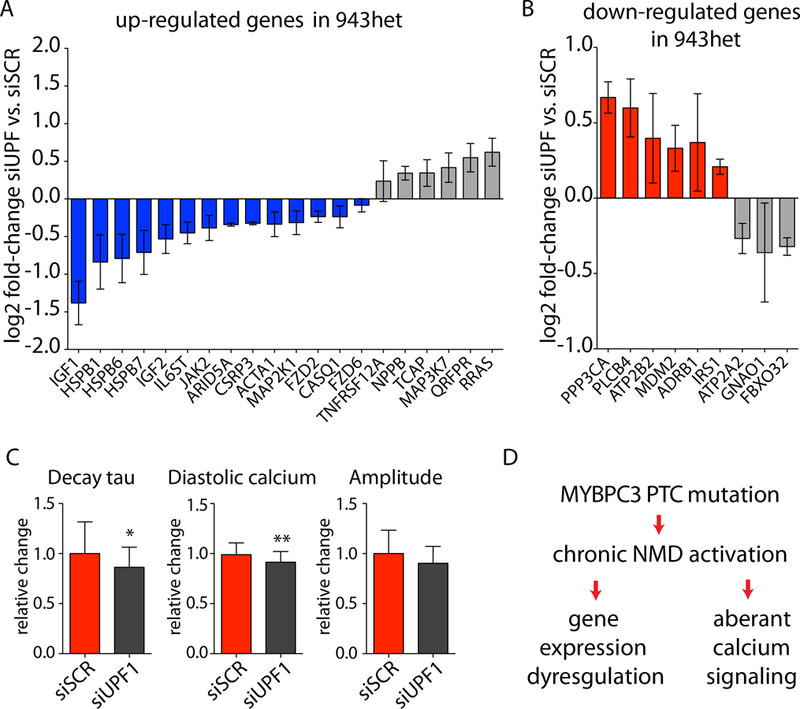
Results: We observed aberrant calcium handling properties with prolonged decay kinetics and elevated diastolic calcium levels in the absence of structural abnormalities or contracile dysfunction in HCM iPSC-CMs as compared to isogenic controls. The mRNA expression levels of MYBPC3 were significantly reduced in mutant iPSC-CMs, but the protein levels were comparable among isogenic iPSC-CMs, suggesting that haploinsufficiency of MYBPC3 does not contribute to the pathogenesis of HCM in vitro. Furthermore, truncated MYBPC3 peptides were not detected. At the molecular level, the nonsense-mediated decay pathway was activated, and a set of genes involved in major cardiac signaling pathways was dysregulated in HCM iPSC-CMs, indicating an HCM gene signature in vitro. Specific inhibition of the nonsense-mediated decay pathway in mutant iPSC-CMs resulted in reversal of the molecular phenotype and normalization of calcium-handling abnormalities.

Conclusions: iPSC-CMs carrying MYBPC3 PTC mutations displayed aberrant calcium signaling and molecular dysregulations in the absence of significant haploinsufficiency of MYBPC3 protein. Here we provided the first evidence of the direct connection between the chronically activated nonsense-mediated decay pathway and HCM disease development.

Keywords: cardiomyocytes; hypertrophic cardiomyopathy.


Figures

Figure 1:
Figure 1:. Genome editing and structural phenotype of MYBPC3 HCM iPSC-CMs.
(A) Pedigree of the affected family. The proband was diagnosed with HCM and a MYBPC3 truncation mutation (p.R943x) was identified by genetic screening. (B) Sanger sequencing of patient corrected (943cor), patient (943het), and patient homozygous (943hom) iPSCs. (C) Micropatterned iPSC-CMs stained with Troponin (green) and DAPI (top row), and MYBPC3 (green), alpha sarcomeric actin (αSA; red), and DAPI (bottom row; scale bar 10 µm). (D) Representative plots of fluorescence intensity longitudinally throughout myofibers analyzed using plot profile (ImageJ), showing MYBPC3 expression in micro-patterned iPSC-CMs (943cor, 943het) and αSA (943hom). (E) Quantification of sarcomeric length based on αSA stained micro-patterned iPSC-CMs (n=10, 11, and 4 cells, respectively). (F) Distribution of the obtained sarcomere length measurements in 943cor (blue), 943het (red), and 943hom (dark). (G) Cell size measurement of unpatterned iPSC-CMs (n=4–6 differentiation batches with 20–130 cells each, respectively). HCM - hypertrophic cardiomyopathy; SCD - sudden cardiac death; CHF - congestive heart failure; ICD - implantable cardioverter defibrillator; AU - arbitrary units.
Figure 2:
Figure 2:. Functional evaluation of HCM iPSC-CMs.
(A) Contraction analysis of iPSC-CM monolayers using a vector-based imaging approach. The relative differences of disease (943het) vs. healthy (943cor) were calculated for each respective experiment (n=5). (B) iPSC-CMs from 943cor, 943het, and 943hom were treated with dexamethasone (Dex), triiodothyronine (T3), and insulin like growth factor 1 (IGF1) for 72 hours prior to contractility analysis (n=3 batches). Analysis was performed using vector-based imaging approach. (C) Analysis of calcium handling properties in isogenic iPSC-CMs (943cor, 943het, 943hom) using ratiometric measurements of fluorescence after Fura-2 staining under continuous field potential stimulation (n=4/4/6 differentiation batches each, 40 cells per batch). (D) Relative expression of ATP2A2 in iPSC-CMs (943cor, 943het, 943hom; n=3–6 differentiation batches each). (E) Representative raw traces of intracellular calcium signaling. # p<0.1, * p<0.05; ** p<0.01.
Figure 3:
Figure 3:. MYBPC3 expression in HCM iPSC-CMs.
(A,B) Total MYBPC3, troponin T (TNNT2), and myosin heavy chain 7 (MYH7) mRNA expression in isogenic iPSC-CMs measured by qPCR (943cor, 943het, 943hom; n=3–4, respectively). (C) Fraction of mutant mRNA (mut.) and wild type (WT) within total MYBPC3 mRNA accounts for 32.7% ± 0.6% in 943het iPSC-CMs (quantification using RNA-seq data; 943cor and 943het; n=3, respectively). (D) Western blot analysis of MYBPC3 protein levels and quantification of MYBPC3 protein expression normalized to alpha sarcomeric actin (αSA) in isogenic iPSC-CMs (using Santa Cruz E7 anti-MYBPC3; 943cor and 943het). (E) Mass spectrometry analysis of MYBPC3 protein expression in healthy (943cor, ctrl) and HCM (943het, ctrl943) iPSC-CMs (n=4 differentiation batches, respectively). # p<0.1; * p<0.05; ** p<0.01.
Figure 4:
Figure 4:. RNA-sequencing reveals a molecular HCM phenotype.
(A) Heatmap of the 298 significantly differentially regulated genes comparing HCM (943het, ctrl943) and healthy (943cor, ctrl) iPSC-CMs (fold-change ± 1.4; q-value <0.05; n=3 differentiation batches each iPSC line). 125 genes were upregulated and 173 genes were downregulated in HCM compared to healthy iPSC-CMs. (B) Within the top significant genes, a set of 30 genes were identified to be involved in cardiac hypertrophy, anabolic processes, and calcium signaling. (C) Top 10 canonical pathways activated in HCM iPSC-CMs identified by gene-set enrichment analysis (GSEA) of the genes. Pathways involved in translation are highlighted in black and pathways involved in RNA metabolism are highlighted in white.
Figure 5:
Figure 5:. Modulation of nonsense-mediated decay pathway reverses HCM phenotype.
Regulation of genes contributing to the molecular HCM phenotype by siUPF1 treatment compared to scramble (siSCR) control in 943het iPSC-CMs. (A) Genes upregulated in HCM (943het, ctrl) being reversed by NMD inhibition are highlighted in blue and (B) genes downregulated in HCM being reversed by NMD inhibition are highlighted in red (943het iPSC-CMs; n=3 differentiation batches; data are presented as mean log2 fold-change of siUPF1 vs. siSCR control ± SEM). (C) NMD modulation by UPF1 inhibition resulted in a reversion of calcium handling abnormalities in 943het iPSC-CMs. (D) Proposed mechanism of NMD activation by PTC carrying MYBPC3 mRNA resulting in a molecular HCM phenotype and abnormal calcium signaling. Short-term inhibition of UPF1 results in a partial reversion of the HCM phenotypes supporting a direct link between NMD activation and HCM. * p<0.05; ** p<0.01.

Comment in

References

    1. Maron BJ, Ommen SR, Semsarian C, Spirito P, Olivotto I, Maron MS. Hypertrophic Cardiomyopathy: Present and future, with translation into contemporary cardiovascular medicine. JACC 2014;64:83–99. - PubMed
    1. Maron BJ, Maron MS, Semsarian C. Genetics of hypertrophic cardiomyopathy After 20 Years. JACC 2012;60:705–715. - PubMed
    1. Marston S, Copeland O, Jacques A, Livesey K, Tsang V, McKenna WJ, Jalilzadeh S, Carballo S, Redwood C, Watkins H. Evidence from human myectomy samples that MYBPC3 mutations cause hypertrophic cardiomyopathy through haploinsufficiency. Circ Res 2009;105:219–222. - PubMed
    1. Vignier N, Schlossarek S, Fraysse B, Mearini G, Kramer E, Pointu H, Mougenot N, Guiard J, Reimer R, Hohenberg H, Schwartz K, Vernet M, Eschenhagen T, Carrier L. Nonsense-mediated mRNA decay and ubiquitin-proteasome system regulate cardiac myosin-binding protein C mutant levels in cardiomyopathic Mice. Circ Res 2009;105:239–248. - PubMed
    1. Karakikes I, Ameen M, Termglinchan V, Wu JC. Human induced pluripotent stem cell-derived cardiomyocytes: insights into molecular, cellular, and functional phenotypes. Circ Res 2015;117:80–88. - PMC - PubMed

Publication types