. 2019 Feb 12;139(7):918-931.
doi: 10.1161/CIRCULATIONAHA.118.033942.

Platelet Serotonin Aggravates Myocardial Ischemia/Reperfusion Injury via Neutrophil Degranulation

Affiliations

Platelet Serotonin Aggravates Myocardial Ischemia/Reperfusion Injury via Neutrophil Degranulation

Maximilian Mauler et al. Circulation. .

Abstract

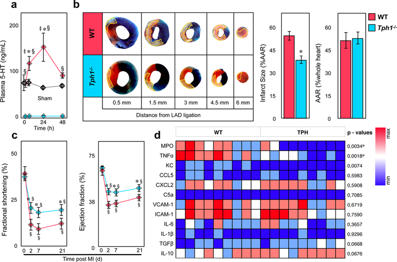
Background: Platelets store large amounts of serotonin that they release during thrombus formation or acute inflammation. This facilitates hemostasis and modulates the inflammatory response.

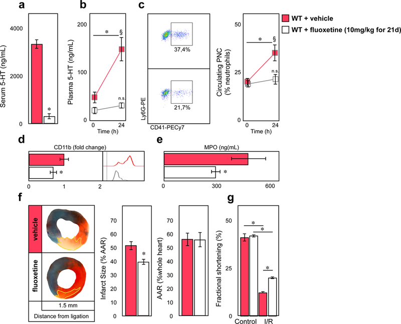
Methods: Infarct size, heart function, and inflammatory cell composition were analyzed in mouse models of myocardial reperfusion injury with genetic and pharmacological depletion of platelet serotonin. These studies were complemented by in vitro serotonin stimulation assays of platelets and leukocytes in mice and men, and by measuring plasma serotonin levels and leukocyte activation in patients with acute coronary syndrome.

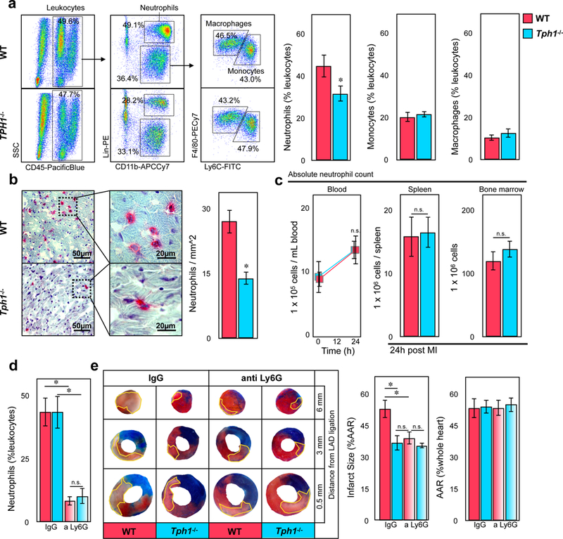
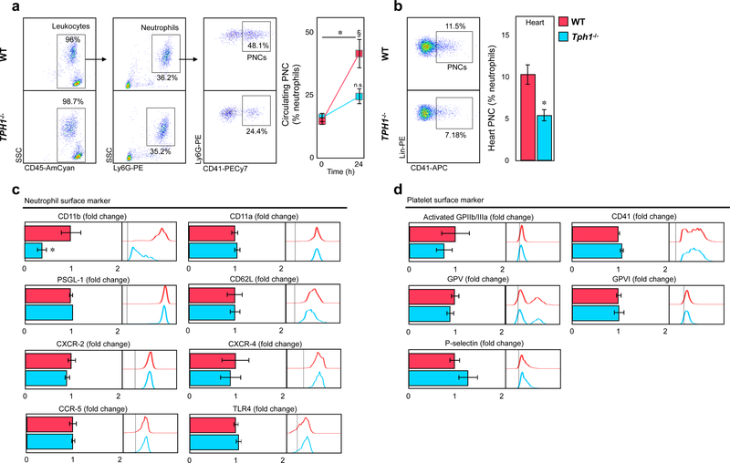
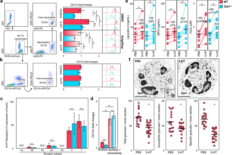
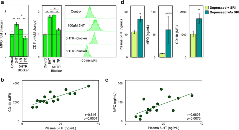
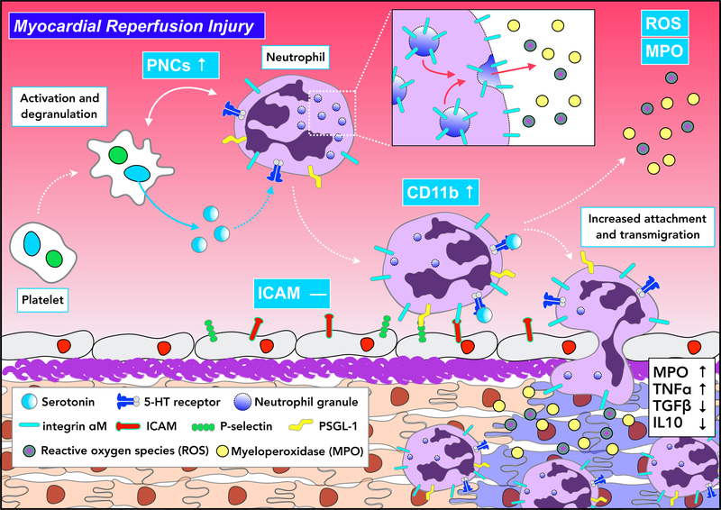
Results: Platelet-derived serotonin induced neutrophil degranulation with release of myeloperoxidase and hydrogen peroxide (H2O2) and increased expression of membrane-bound leukocyte adhesion molecule CD11b, leading to enhanced inflammation in the infarct area and reduced myocardial salvage. In patients hospitalized with acute coronary syndrome, plasmatic serotonin levels correlated with CD11b expression on neutrophils and myeloperoxidase plasma levels. Long-term serotonin reuptake inhibition-reported to protect patients with depression from cardiovascular events-resulted in the depletion of platelet serotonin stores in mice. These mice displayed a reduction in neutrophil degranulation and preserved cardiac function. In line, patients with depression using serotonin reuptake inhibition, presented with suppressed levels of CD11b surface expression on neutrophils and lower myeloperoxidase levels in blood.

Conclusions: Taken together, we identify serotonin as a potent therapeutic target in neutrophil-dependent thromboinflammation during myocardial reperfusion injury.

Keywords: blood platelets; inflammation; integrins; neutrophils; reactive oxygen species; reperfusion injury; serotonin.


Conflict of interest statement

Disclosures: The authors declare that there are no conflicts of interest to disclose.

Figures

Figure 1
Figure 1
Myocardial reperfusion injury is dampened in Tph1−/− mice. (a) Plasma serotonin levels in WT (red) and Tph1−/− (blue) before, 6, 24, and 48 hours after myocardial infarction with reperfusion. Values in sham (thoracotomy was performed without ligation of the LAD) operated WT mice are shown in grey. Results are presented as mean ± SEM, p<0.05 denote significant changes compared to time point 0 hours within groups, § p<0.05 denote significant changes between WT and Tph1−/− mice, and ‡ p<0.05 between WT and Sham, two-way ANOVA with Bonferroni’s multiple comparisons testing, n≥6 independent animals per group and time point. (b) Representative images of heart sections of WT and Tph1−/− mice after 30 minutes of LAD ligation with 24 hours of reperfusion at increasing distance from the site of ligation. The infarct area (white tissue) is circumscribed in yellow and quantified as percentage of area at risk (AAR; non-blue tissue). The AAR is presented as percentage of the entire heart section. Viable tissue within the AAR was stained in red. Results are presented as mean ± SEM, p<0.05, Student´s t-test, n≥10 per group, WT (red) and Tph1−/− (blue). (c) Time course of fractional shortening (FS) and left ventricular ejection fraction of WT and Tph1−/− mice recorded under baseline (d0) conditions and repetitively over 3 weeks (at day 2, 7, and 21) following myocardial infarction with reperfusion. Results are presented as mean ± SEM, ✽,§p<0.05, denotes significant changes between different genotypes; § denotes significance compared to d0 within groups; Two-way repeated measures ANOVA with Bonferroni’s multiple comparison testing, n=8 per group, WT (red) and Tph1−/− (blue). (d) Expression of pro- and anti-inflammatory target genes in homogenized heart tissue after I/R injury. Results are presented in a heat map depicting 2−▵Ct values, n=9 per group, each column represents one WT or Tph1−/− mouse. P values were derived from comparison of respective 2−▵▵Ct values by unpaired t-tests, and asterisks denote significant differences according to correction for multiple comparisons using the Holm-Sidak method.
Figure 2
Figure 2
Reduced neutrophil accumulation in hearts of Tph1−/− mice following myocardial I/R injury. (a) Representative flow cytometric dot plots of digested cardiac tissues of WT and Tph1−/− mice 24 hours after myocardial ischemia and subsequent reperfusion (left panels). Quantification of neutrophils, monocytes, and macrophages in cardiac tissue of WT (red) and Tph1−/− (blue) mice 24 hours after myocardial I/R injury (right panels). Results are presented as mean percent of CD45.2+ leukocytes ± SEM, p<0.05, Student´s t-test, n≥7 per group. (b) Representative immunohistology of myocardial infarct tissue. Ly6G+ neutrophils are stained in red (left panels) and quantified per mm tissue section of WT (red) and Tph1−/− (blue) mice. Results are presented as mean ± SEM, p<0.05, Student´s t-test, n≥9 per group. (c) Neutrophil counts of WT (red) and Tph1−/− (blue) mice in blood before, and in blood, spleen and bone marrow after myocardial I/R injury. Results are presented as mean ± SEM, n.s., two-way ANOVA with Bonferroni’s multiple comparison testing was used for blood samples, Student´s t-test was used for spleen and bone marrow, n=6 independent animals per group and time point. (d) Quantification of blood neutrophils following depletion with anti-Ly6G versus IgG control. Results are presented as mean ± SEM, p<0.05, two-way ANOVA with Bonferroni’s multiple comparison testing, n=6 per group, WT (red) and Tph1−/− (blue). (e) Representative images of heart sections of WT and Tph1−/− mice after 30 minutes of LAD ligation followed by 24 hours of reperfusion at increasing distance from the site of ligation. The infarct area (white tissue) is circumscribed in yellow and quantified as percentage of area at risk (AAR; non-blue tissue). The AAR is presented as percentage of the entire heart section. Viable tissue within the AAR was stained in red. Results are presented as mean ± SEM, p<0.05, two-way ANOVA with Bonferroni’s multiple comparison testing, n=6 per group, WT (red) and Tph1−/− (blue)
Figure 3
Figure 3
Platelet neutrophil complex formation is reduced in Tph1−/− mice after reperfusion injury. (a) Representative dot plots displaying platelet neutrophil complexes (PNCs) in blood of WT and Tph1−/− mice (left panel). PNCs were quantified before and after 24h of reperfusion (right panel). Results are presented as mean ± SEM, p<0.05 comparing time points, § p<0,05 comparing groups, two-way ANOVA with Bonferroni’s multiple comparison testing, n≥10 independent animals per group and time point, WT (red) and Tph1−/− (blue).(b) Representative dot plots and quantification of PNCs within the AAR of WT (red) and Tph1−/− (blue) mice at 24 hours after myocardial ischemia with reperfusion. Results are presented as mean ± SEM, p<0.05, Student´s t-test, n≥7 per group. (c) Quantification of CD11b, CD11a, CXCR-2, CD62L and PSGL-1 surface expression in PNC of WT (red) and Tph1−/− (blue) mice 24 hours after myocardial I/R injury. Results are presented as normalized mean ± SEM, p<0.05, Student´s t-test, n=10 per group. (d) Activated GPIIb/IIIa, GPV, GPVI, and P-selectin platelet marker expression in PNC of WT and Tph1−/− mice 24 hours after myocardial I/R injury. Results are presented as normalized mean ± SEM, n.s., Student´s t-test, n=10 per group. Bar graphs are accompanied by representative histograms of mean fluorescence intensities (MFI) of respective markers.
Figure 4
Figure 4
Serotonin mediates degranulation of neutrophils in vitro. (a) Representative dot plots displaying uncomplexed (free) neutrophils (SSChigh, Ly6G+, CD41) in blood (left panel). Quantification of neutrophil CD11b expression in WT (red) and Tph1−/− (blue) mice upon stimulation with PBS (control) or 100µM serotonin co-incubated with DMSO or GolgiStop/Plug reagent (right panel). Results are presented as mean ± SEM, p<0.05, two-way ANOVA with Bonferroni test, n≥12 per group (b) Dot plots of uncomplexed (free) monocytes (SSClow, CD11b+, CD41) in blood (left panel) and quantification of CD11b expression following serotonin or PBS treatment (right panel). Results are presented as mean ± SEM, p<0.05, two-way ANOVA with Bonferroni test, n≥12 per group, WT (red) and Tph1−/− (blue). (c) Expression of different serotonin receptors in isolated neutrophils in WT (red) and Tph1−/− (blue) mice. Results are presented as normalized mean ± SEM, n.s., Student´s t-test, n=5 per group. (d) Intracellular expression of CD11b in neutrophils compared to matched surface expression in blood of WT (red) and Tph1−/− (blue) mice. Results are presented as mean ± SEM normalized to WT surface staining, p<0.05 denotes significant changes between surface and intracellular staining, two-way ANOVA for repeated measures with Bonferroni’s multiple comparison testing, n=5 per group. (e) Plasma concentration of lipocalin-2,MPO, and H2O2 in WT (red) and Tph1−/− (blue) blood after stimulation with either serotonin or PBS. Results are presented as normalized mean ± SEM, p<0.05, Paired Student´s t-test, n=9 per group. (f) Electron microscopic images of neutrophils after treatment with serotonin or PBS (top panel). Quantification of azurophilic granules (AG) and specific granules (SG) per cross section after stimulation with serotonin (lower panels). Results are presented as mean ± SEM, p<0.05, Student´s t-test, n≥15 per treatment.
Figure 5
Figure 5
Pharmacologic platelet serotonin depletion protects from myocardial ischemia reperfusion injury. (a) Three week administration of fluoxetine (Flx) depletes platelet dense granule serotonin storage (indirectly assessed by freshly provoked serum). Results are presented as mean ± SEM, p<0.05, Student´s t-test, n=7 per vehicle (red) or Flx (white) treatment. (b) Plasma serotonin levels in vehicle (red) and Flx (white) before, and 24 hours after myocardial infarction with reperfusion. Results are presented as mean ± SEM, p<0.05 comparing time points within groups, § p<0.05 comparing groups, two-way ANOVA with Bonferroni’s multiple comparison testing, n=7 independent animals per treatment and time point. (c) Representative dot plots of circulating PNCs in vehicle (red) and Flx (white) treated mice (left panel) and quantification before and after 24 hours following myocardial I/R injury. Results are presented as mean ± SEM, p<0.05 comparing time points within groups, § p<0.05 comparing groups, two-way ANOVA with Bonferroni’s multiple comparison testing, n=7 independent animals per treatment and time point. (d) Quantification of CD11b surface expression in PNC and (e) plasma MPO levels of vehicle (red) and Flx (white) treated mice 24 hours after myocardial ischemia and reperfusion. Results are presented as normalized mean ± SEM, p<0.05, Student´s t-test, n=7 per group. (f) Representative images of heart sections of vehicle and fluoxetine treated mice after 30 minutes of LAD ligation with 24 hours of reperfusion. Infarcted areal (white tissue) is circumscribed in yellow and quantified as percentage of area at risk (AAR; non-blue tissue). The AAR is presented as percentage of the entire heart section. Viable tissue within the AAR was tained in red. Results are presented as mean ± SEM, p<0.05, Student´s t-test, n=7 per vehicle (red) and Flx (white) treatment. (g) LV fractional shortening in vehicle (red) and Flx (white) treated mice following I/R injury. Results are presented as mean ± SEM, p<0.05, two-way ANOVA with Bonferroni test, n=7 per group.
Figure 6
Figure 6
Human neutrophil degranulation is regulated by serotonin. (a) Supernatant MPO concentration and surface CD11b expression on human neutrophils after stimulation with 100µM serotonin or PBS with and without addition of specific serotonin receptor antagonist (5HTR4 and 5HTR7). Representative histograms of changes in CD11b expression are shown on the right. Results are presented as mean ± SEM, p<0.05, One-way ANOVA with Tukey t-test, n=4 per treatment. (b) Correlation of plasma serotonin concentration and CD11b (p<0.0001) on blood neutrophils and (c) plasma MPO concentration of patients with acute coronary syndrome. Each point represents an individual patient, r: Pearson correlation coefficient. (d) Quantification of plasma serotonin and MPO levels and CD11b expression in blood of depressed patients treated with (lime green) or without (blue) antidepressants. p<0.05, Student´s t-test, n=10 per group
Figure 7
Figure 7
Proposed model of platelet serotonin-neutrophil interactions during myocardial reperfusion injury. Serotonin released by platelets triggers the degranulation of neutrophils, subsequently leading to increased CD11b surface expression. This enhances circulating PNC levels and supports the accumulation of pro-inflammatory cytokine-releasing neutrophils within the injured myocardium.

Comment in

References

    1. Yellon DM and Hausenloy DJ. Myocardial reperfusion injury. N Engl J Med 2007;357:1121–1135. - PubMed
    1. Hausenloy DJ and Yellon DM. Myocardial ischemia-reperfusion injury: a neglected therapeutic target. J Clin Invest 2013;123:92–100. - PMC - PubMed
    1. Hausenloy DJ, Botker HE, Engstrom T, Erlinge D, Heusch G, Ibanez B, Kloner RA, Ovize M, Yellon DM and Garcia-Dorado D. Targeting reperfusion injury in patients with ST-segment elevation myocardial infarction: trials and tribulations. Eur Heart J 2017;38:935–941. - PMC - PubMed
    1. Heusch G and Gersh BJ. The pathophysiology of acute myocardial infarction and strategies of protection beyond reperfusion: a continual challenge. Eur Heart J 2017;38:774–784. - PubMed
    1. Vinten-Johansen J Involvement of neutrophils in the pathogenesis of lethal myocardial reperfusion injury. Cardiovascular research 2004;61:481–497. - PubMed

Publication types

MeSH terms