. 2019 Apr 2;139(14):1710-1724.
doi: 10.1161/CIRCULATIONAHA.118.037642.

Loss of Endothelium-Derived Wnt5a Is Associated With Reduced Pericyte Recruitment and Small Vessel Loss in Pulmonary Arterial Hypertension

Affiliations

Loss of Endothelium-Derived Wnt5a Is Associated With Reduced Pericyte Recruitment and Small Vessel Loss in Pulmonary Arterial Hypertension

Ke Yuan et al. Circulation. .

Abstract

Background: Pulmonary arterial hypertension (PAH) is a life-threatening disorder of the pulmonary circulation associated with loss and impaired regeneration of microvessels. Reduced pericyte coverage of pulmonary microvessels is a pathological feature of PAH and is caused partly by the inability of pericytes to respond to signaling cues from neighboring pulmonary microvascular endothelial cells (PMVECs). We have shown that activation of the Wnt/planar cell polarity pathway is required for pericyte recruitment, but whether production and release of specific Wnt ligands by PMVECs are responsible for Wnt/planar cell polarity activation in pericytes is unknown.

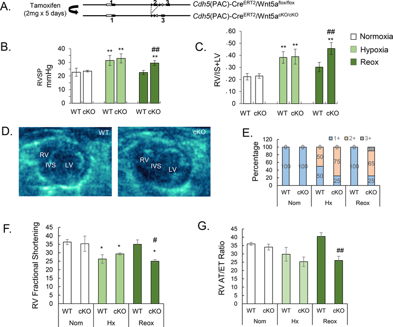
Methods: Isolation of pericytes and PMVECs from healthy donor and PAH lungs was carried out with 3G5 or CD31 antibody-conjugated magnetic beads. Wnt expression profile of PMVECs was documented via quantitative polymerase chain reaction with a Wnt primer library. Exosome purification from PMVEC media was carried out with the ExoTIC device. Hemodynamic profile, right ventricular function, and pulmonary vascular morphometry were obtained in a conditional endothelium-specific Wnt5a knockout ( Wnt5aECKO) mouse model under normoxia, chronic hypoxia, and hypoxia recovery.

Results: Quantification of Wnt ligand expression in healthy PMVECs cocultured with pericytes demonstrated a 35-fold increase in Wnt5a, a known Wnt/planar cell polarity ligand. This Wnt5a spike was not seen in PAH PMVECs, which correlated with an inability to recruit pericytes in Matrigel coculture assays. Exosomes purified from media demonstrated an increase in Wnt5a content when healthy PMVECs were cocultured with pericytes, a finding that was not observed in exosomes of PAH PMVECs. Furthermore, the addition of either recombinant Wnt5a or purified healthy PMVEC exosomes increased pericyte recruitment to PAH PMVECs in coculture studies. Although no differences were noted in normoxia and chronic hypoxia, Wnt5aECKO mice demonstrated persistent pulmonary hypertension and right ventricular failure 4 weeks after recovery from chronic hypoxia, which correlated with significant reduction, muscularization, and decreased pericyte coverage of microvessels.

Conclusions: We identify Wnt5a as a key mediator for the establishment of pulmonary endothelium-pericyte interactions, and its loss could contribute to PAH by reducing the viability of newly formed vessels. We speculate that therapies that mimic or restore Wnt5a production could help prevent loss of small vessels in PAH.

Keywords: Wnt5a; endothelial cells; exosomes; pericytes; pulmonary hypertension.


Figures

Figure 1.
Figure 1.. PAH PMVECs fail to recruit healthy pulmonary pericytes (Pc).
(A) Matrigel assays of PMVEC from healthy donors (A, top) or from PAH patients alone (A, bottom) or in the presence of healthy donor Pc. Inset: IF of boxed areas. PMVEC are stained with PKH67 (green) and Pc with PKH26 (red). (B) Total tube length (left), total branching points (middle), and number of loops (right) with healthy donor or PAH PMVECs along or with pericytes from healthy subjects. (C) Boyden chamber comparing translocation of Pc in the presence of healthy donor or PAH PMVECs. After 6 hours, pericytes at the bottom of the inserts were fixed and stained with hematoxylin and eosin. The mean number of pericytes from four random 20x fields was used for comparison. (D) Representative images of wound-healing co-culture assays in which PMVECs are on the left and pericytes on the right. Bottom row shows enlargements of the boxes in the top row. Cell polarity was assessed by pericentrin (red dots, indicates direction of cell movement), a marker of the microtubule organization center (MTOC). F-actin was stained in Alexa Fluor 488 phalloidin and DAPI was in blue. Red arrowheads indicate cells pointing toward EC layer whereas white arrowheads indicate cells pointing the opposite direction. The white lines indicate 0 hours; dashed lines, 6 hours. Quantification of cell distance (bottom left) and percentage of polarized cells (bottom right) after 6 hours. Data are expressed as means ± SEM of three experiments. ∗P < 0.05 ∗∗P < 0.01, ∗∗∗P < 0.001 versus PMVEC alone (one-way analysis of variance with Dunnett post-test (B,C)) or versus control (unpaired t-test (D)). Scale bars = 100 μm.
Figure 2.
Figure 2.. Expression of Wnt5a fails to increase in PAH PMVECs co-cultured with healthy donor pericytes.
(A) SYBR Green qPCR analysis for Wnt/PCP-related components in PAH PMVEC or healthy donor co-culture with healthy Pc. The expression of each gene is shown relative to that in monoculture of healthy and PAH PMVECs. Data presented is the result of three independent studies. (B) Representative Western immunoblot (WB) images of Wnt5a in PAH PMVEC in mono or co-culture versus healthy donors. ∗∗P < 0.01 versus monoculture control (one-way analysis of variance with Dunnett’s post-test). OD, optical density. Densitometry was performed against a-tubulin. (C) IF of lung sections from human healthy donor and PAH patients stained for CD31 (green) and anti-human Wnt5a (red). (D) IF of human healthy vessel and PAH vascular lesions stained with 3G5 (red) for pericytes. CD31 is green and DAPI is blue. Graph indicates average number of 3G5 positive cells associated with microvessels by counting the ratio of number of 3G5+ cells overlapped with CD31+ cells in each group to the total number of CD31+ cells. White arrows indicate 3G5+CD31+ whereas white arrowheads indicate only 3G5+ staining. ∗∗P < 0.01 using the unpaired t-test. (E) Representative WB images of Wnt5a in isolated PMVECs from three donors versus three patients with PAH. Densitometry was performed against a-tubulin. ∗∗∗P < 0.001 using the unpaired t-test. Scale bar = 50 μm.
Figure 3.
Figure 3.. Knockdown of Wnt5a in healthy donor PMVECs reduces pericyte recruitment but can be partially restored with recombinant Wnt5a (rWnt5a).
(A) WB for Wnt5a in total cell lysate from PMVEC transfected with either nontargeting (siCtrl) or Wnt5a-specific (siWnt5a) siRNAs. Densitometry was performed against a-tubulin. ∗∗P < 0.01 versus siCtrl using the unpaired t-test. (B) Boyden chamber assay comparing translocation of Pc in the presence of PMVECs with siCtrl or siWnt5a or rWnt5a. (C) Representative images of wound-healing co-culture assays. Enlargements of the boxes are shown in right panels. Cell polarity was assessed by pericentrin. The red lines indicate 0 hours; dashed lines, 6 hours. Quantification of distance migrated (D) and percentage of polarized cells (E) was performed after 6 hours. ∗P < 0.05 versus siCtrl using the unpaired t-test. (F) Matrigel assays of healthy PMVEC with siCtrl, siWnt5a or rWnt5a in the presence of healthy Pc. Images in the lower panel are tubular-like structures formed by PMVEC (PKH67, green) with or without the presence of pericytes (Pc) (PKH26, red). Total tube length (left), total branching points (middle), and number of loops (right) of PMVEC siCtrl, siWnt5a or rWnt5a with healthy pericytes from healthy subjects. ∗∗∗P < 0.001 versus siCtrl (one-way analysis of variance with Dunnett post-test); # P< 0.05, ##P<0.01, ###P<0.001 (one-way analysis of variance with Bonferroni post-test vs. siWnt5a +Pc). Scale bar = 100μm.
Figure 4.
Figure 4.. Wnt5a is secreted in exosomes by PMVECs.
(A) IF for Wnt5a (green) in healthy donor or PAH PMVECs alone or co-cultured with pericytes in Boyden chambers. (B) TEM of one exosome (upper) and a CD63 immunogold labeled exosome (lower) isolated from co-culture. Scale bar=100nm. (C and D) WB for Wnt5a in total cell exosome lysate (CD81 as an internal control) from healthy donor (C) and PAH PMVEC (D) in mono and co-culture. ***P < 0.001 versus monoculture using the unpaired t-test. (E) Wound healing assay of PMVEC transfected with either nontargeting (siCtrl) or Wnt5a-specific (siWnt5a) siRNAs treated with exosomes isolated from healthy co-culture media. Cell polarity was assessed by pericentrin. F-actin was stained in Alexa Fluor 488 phalloidin and DAPI was in blue. ∗∗P < 0.01 versus Ctrl (one-way analysis of variance with Dunnett’s post-test); ## P< 0.01, ### P<0.001 (one-way analysis of variance with Bonferroni post-test vs. PAH PMVECs + Pc). Scale bar= 100um.
Figure 5.
Figure 5.. Persistent elevation of right ventricular systolic pressure (RVSP) and RV failure in Wnt5aECKO mice after recovery from hypoxia (Reox).
(A) Diagram showing the strategy for generation of Wnt5aECKO mice. (B) RVSP and (C) Fulton Index of wild type (WT) and Wnt5aECKO (cKO) in normoxia, hypoxia and recovery (Reox). (D) Echo images of WT and Wnt5aECKO in recovery. right (RV) and left ventricles (LV), interventricular septum (IVS). (E) The percentage of RV enlargement in echo images was scored as follows: 1+ normal (RV is invisible), 2+ some degree impaired (visible), 3+ RV hypertrophy (obvious enlargement). RV Fractional Shortening (F) and RV acceleration time over ejection time (G) in normoxia, hypoxia and recovery (reox). Data are expressed as means ± SEM of at least six random images. N=11 for each group. ∗ P<0.05, ∗∗P<0.01 (one-way analysis of variance with Dunnett’s post-test). # P< 0.05, ##P<0.01, (one-way analysis of variance with Bonferroni post-test vs. WT Reox).
Figure 6.
Figure 6.. Wnt5aECKO mice have persistent loss and muscularization of microvessels in recovery.
(A) Quantification of number of vessels per 100 alveoli and (B) number of muscularized in microvessels (diameter < 50um). Data are expressed as means ± SEM of at least six random images; each group has 11 different mice. (C) MicroCt imaging and enlarged images of distal vessels from Reox WT and cKO mice. Quantification of tertiary blood vessel length (um) and number of distal vessels on the right. (D) IF of OCT embedded lung sections from Reox WT (upper) and cKO (lower) mice. Smooth muscle layer was stained with a-SMA (red), pericytes were stained with NG2 (magenta) and endothelium was stained with CD31 (green). Scale bar =25um (E) Quantification of NG2 positive cell pericyte coverage in vessels from WT and cKO mice in recovery group. (F) IF of OCT embedded lung sections from Reox WT and cKO mice which stained for CD31 (green), 3G5(red) and cPARP. White arrow indicates 3G5 overlap with cPARP. ∗∗P < 0.01, unpaired t-test. ∗∗∗P<0.001 (one-way analysis of variance with Dunnett’s post-test); ##P<0.01, (one-way analysis of variance with Bonferroni post-test vs. WT Reox). Scale bar =10um.
Figure 7.
Figure 7.. Proposed model.
In the setting of vascular injury, PMVECs produce Wnt5a packaged in exosomes to recruit pericytes to associate with blood vessels, thereby preserving the number of functional vessels within the microcirculation (A). A lack of Wnt5a production significantly impair establishment of endothelial-pericyte interactions thus resulting in loss of small vessels leading to PAH (B). MVE=microvesicles.

References

    1. Ribatti D, Nico B and Crivellato E. The role of pericytes in angiogenesis. Int J Dev Bio. 2011;55:261–268. - PubMed
    1. Yamagishi S and Imaizumi T. Pericyte biology and diseases. Int J Tissue React 2005;27:125–135. - PubMed
    1. Diaz-Flores L, Gutierrez R, Madrid JF, Varela H, Valladares F, Acosta E, Martin-Vasallo P and Diaz-Flores L Jr. Pericytes. Morphofunction, interactions and pathology in a quiescent and activated mesenchymal cell niche. Histol Histopathol. 2009;24:909–969. - PubMed
    1. Kapanci Y, Ribaux C, Chaponnier C and Gabbiani G. Cytoskeletal features of alveolar myofibroblasts and pericytes in normal human and rat lung. J Histochem Cytochem. 1992;40:1955–1963. - PubMed
    1. Teichert-Kuliszewska K, Kutryk MJ, Kuliszewski MA, Karoubi G, Courtman DW, Zucco L, Granton J and Stewart DJ. Bone morphogenetic protein receptor-2 signaling promotes pulmonary arterial endothelial cell survival: implications for loss-of-function mutations in the pathogenesis of pulmonary hypertension. Circ Res. 2006;98:209–217. - PubMed

Publication types

MeSH terms