Review
. 2018 Dec 24;11(1):6.
doi: 10.3390/pharmaceutics11010006.

Electrospinning of Cyclodextrin Functional Nanofibers for Drug Delivery Applications

Affiliations
Review

Electrospinning of Cyclodextrin Functional Nanofibers for Drug Delivery Applications

Fuat Topuz et al. Pharmaceutics. .

Abstract

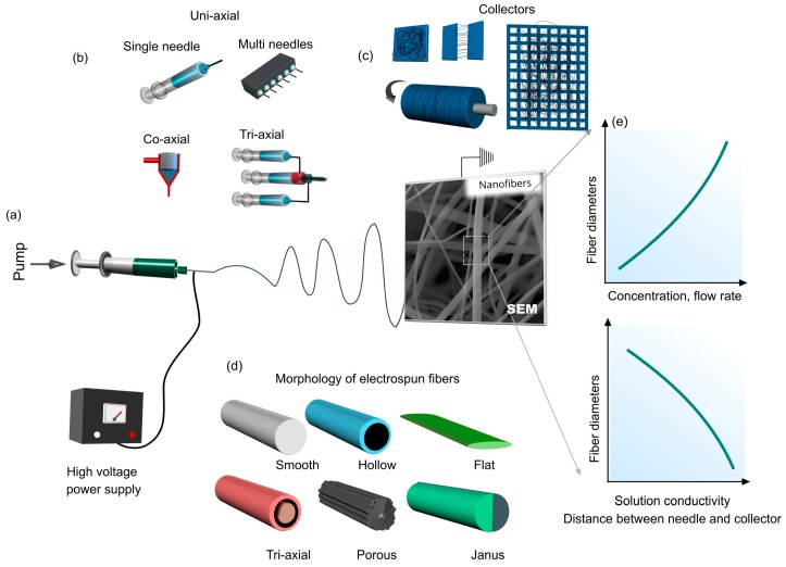
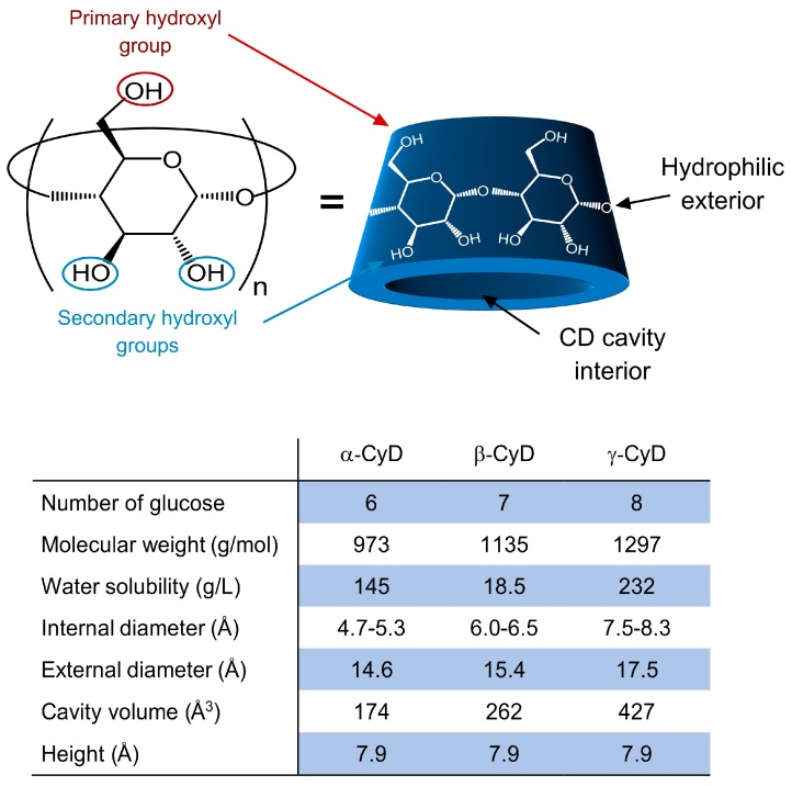
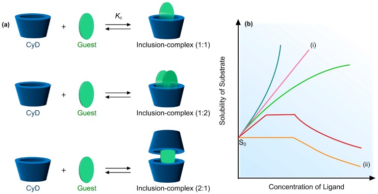
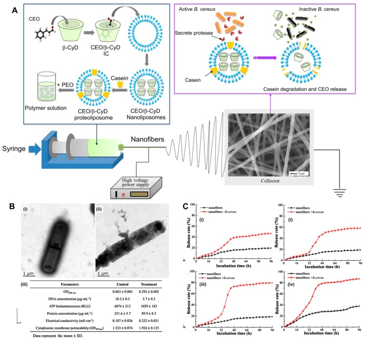
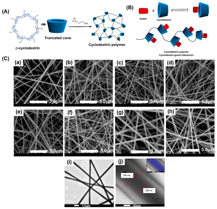
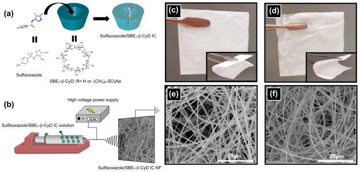
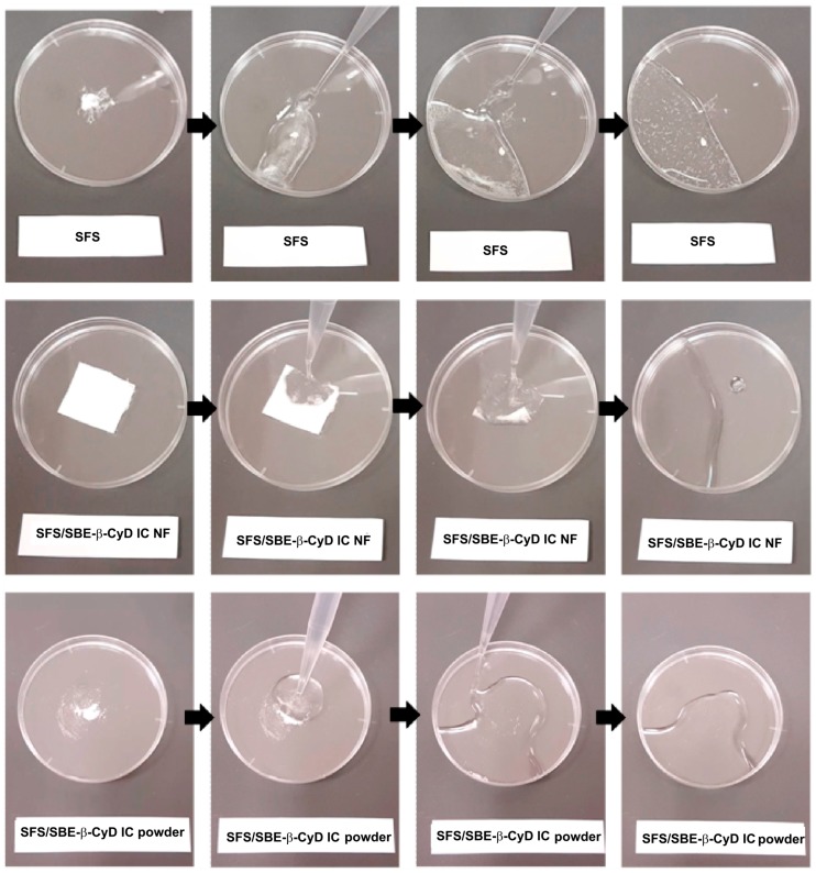
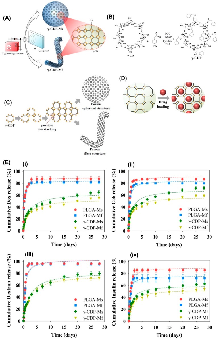
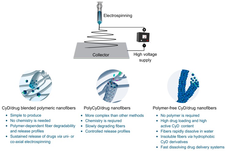
Electrospun nanofibers have sparked tremendous attention in drug delivery since they can offer high specific surface area, tailored release of drugs, controlled surface chemistry for preferred protein adsorption, and tunable porosity. Several functional motifs were incorporated into electrospun nanofibers to greatly expand their drug loading capacity or to provide the sustained release of the embedded drug molecules. In this regard, cyclodextrins (CyD) are considered as ideal drug carrier molecules as they are natural, edible, and biocompatible compounds with a truncated cone-shape with a relatively hydrophobic cavity interior for complexation with hydrophobic drugs and a hydrophilic exterior to increase the water-solubility of drugs. Further, the formation of CyD-drug inclusion complexes can protect drug molecules from physiological degradation, or elimination and thus increases the stability and bioavailability of drugs, of which the release takes place with time, accompanied by fiber degradation. In this review, we summarize studies related to CyD-functional electrospun nanofibers for drug delivery applications. The review begins with an introductory description of electrospinning; the structure, properties, and toxicology of CyD; and CyD-drug complexation. Thereafter, the release of various drug molecules from CyD-functional electrospun nanofibers is provided in subsequent sections. The review concludes with a summary and outlook on material strategies.

Keywords: antibacterial; antibiotics; cyclodextrin; cyclodextrin-inclusion complexes; drug delivery; electrospinning; electrospun nanofibers; essential oils; nanofibers; poly-cyclodextrin.


Conflict of interest statement

The authors have no conflicts of interest to declare.

Figures

Figure 1
Figure 1
An electrospinning setup with important parameters is shown. (a) A cartoon scheme of an electrospinning system with the scanning electron micrograph of electrospun fibers, (b) common spinneret systems used in electrospinning, (c) collector types, (d) the morphology of electrospun fibers, and (e) diagrams showing the influence of electrospinning process parameters and solution properties on the electrospun fibers.
Figure 2
Figure 2
The chemical structure and the representative cartoon illustration of a native cyclodextrin (CyD) molecule in the 3D form. The general characteristics of CyD are given in the inset table [49].
Figure 3
Figure 3
(a) The inclusion complex formation between CyD and guest molecules at various stoichiometries. (b) The plot shows a phase solubility of guest molecules; (i) represents the formation of soluble inclusion-complex (IC), and (ii) denotes the formation of IC with limited solubility.
Figure 4
Figure 4
(A) Cartoon schemes of the production of CEO/β-CyD proteoliposomes incorporated poly(ethylene oxide) (PEO) fibers and B. cereus proteinase triggered cinnamon essential oil (CEO) delivery from CEO/β-CyD proteoliposomes. (B) TEM images of B. cereus before (i) and after the treatment (ii) of CEO/β-CyD proteoliposomes. (iii) The respective analysis results on the release of B. cereus cell constituents and the cell membrane permeability before and after proteoliposomes treatment. (C) The release rate of CEO/β-CyD proteoliposomes nanofibers stored at different temperatures 4 °C (i), 12 °C (ii), 25 °C (iii), and 37 °C (iv) for 4 days. The figure was reproduced from [133] with the permission of Elsevier, 2017.
Figure 5
Figure 5
Cartoon schemes of (A) the synthesis pathway of CyD polymers and (B) their complexation with guest molecules. (C) scanning electron microscopy (SEM) and transmission electron microscopy (TEM) images of uni- and co-axial PMAA/polyCyD fibers: (a) SEM uniaxial–PMAA; (b) SEM uniaxial–PMAA + PROP; (c) SEM uniaxial PMAA/polyCyD (80:20); (d) SEM uniaxial PMAA/polyCyD (80:20) + PROP; (e) SEM uniaxial PMAA/polyCyD (60:40); (f) SEM uniaxial PMAA/polyCyD (60:40) + PROP; (g) SEM coaxial–shell (PMAA) and core (polyCyD); (h) SEM coaxial–shell (PMAA) and core (polyCyD + PROP); (i) TEM coaxial–shell (PMAA) and core (polyCyD + PROP); and (j) TEM coaxial–shell (PMAA) and core (polyCyD + PROP). The figure was reproduced from [151] with the permission of Elsevier, 2015.
Figure 6
Figure 6
(a) Cartoon illustration of inclusion-complexation between CyD and sulfisoxazole (SFS). The chemical structure of sulfisoxazole and SBE7-β-CyD with a schematic representation of sulfisoxazole, SBE7-β-CyD and their IC, (b) schematic representation of the electrospinning of SFS/SBE7-β-CyD-IC NF. Photographs of electrospun (c) SBE7-β-CyD nanofibers, (d) SFS/SBE7-β-CyD-IC nanofibers, and SEM images of (e) SBE7-β-CyD NF, (f) SFS/SBE7-β-CyD-IC nanofibers. The figure was reproduced from [179] with the permission of Elsevier, 2017.
Figure 7
Figure 7
Typical water-solubility of the drug loaded polymer-free CyD fibers. The representative photos of the SFS and SFS/ SBE7-β-CyD IC powder and SFS/ SBE7-β-CyD IC nanofibers on exposure to water. The figure was reproduced from [179] with the permission of Elsevier, 2017.
Figure 8
Figure 8
(A) A schematic representation of the electrospinning and electrospraying of γ-CyDPs. (B) The synthesis pathway of γ-CyDP. (C) Cartoon schemes of γ-CyDP-microspheres (Ms) or γ-CyDP microfibers (Mf) with porous structure and (D) their drug loading. (E) The cumulative molecule (i) Dox, (ii) Ce6, (iii) dextran, and (iv) insulin release (wt. %) from PLGA-Ms, PLGA-Mf, γ-CyDP-Ms, and γ-CyDP-Mf (n = 3). The figure was reproduced from [182] with the permission of Elsevier, 2018.
Figure 9
Figure 9
Cartoon illustration of CyD-functional electrospun nanofibers used for drug delivery applications.

References

    1. Zeng J., Xu X., Chen X., Liang Q., Bian X., Yang L., Jing X. Biodegradable electrospun fibers for drug delivery. J. Control. Release. 2003;92:227–231. doi: 10.1016/S0168-3659(03)00372-9. - DOI - PubMed
    1. Sill T.J., von Recum H.A. Electrospinning: Applications in drug delivery and tissue engineering. Biomaterials. 2008;29:1989–2006. doi: 10.1016/j.biomaterials.2008.01.011. - DOI - PubMed
    1. Yoo H.S., Kim T.G., Park T.G. Surface-functionalized electrospun nanofibers for tissue engineering and drug delivery. Adv. Drug Deliv. Rev. 2009;61:1033–1042. doi: 10.1016/j.addr.2009.07.007. - DOI - PubMed
    1. Kenawy E.-R., Bowlin G.L., Mansfield K., Layman J., Simpson D.G., Sanders E.H., Wnek G.E. Release of tetracycline hydrochloride from electrospun poly(ethylene-co-vinylacetate), poly(lactic acid), and a blend. J. Control. Release. 2002;81:57–64. doi: 10.1016/S0168-3659(02)00041-X. - DOI - PubMed
    1. Zeng J., Yang L., Liang Q., Zhang X., Guan H., Xu X., Chen X., Jing X. Influence of the drug compatibility with polymer solution on the release kinetics of electrospun fiber formulation. J. Control. Release. 2005;105:43–51. doi: 10.1016/j.jconrel.2005.02.024. - DOI - PubMed

LinkOut - more resources