

The Participation of Regulatory Lipids in Vacuole Homotypic Fusion
- PMID: 30587414
- PMCID: PMC6814398
- DOI: 10.1016/j.tibs.2018.12.003
The Participation of Regulatory Lipids in Vacuole Homotypic Fusion
Abstract
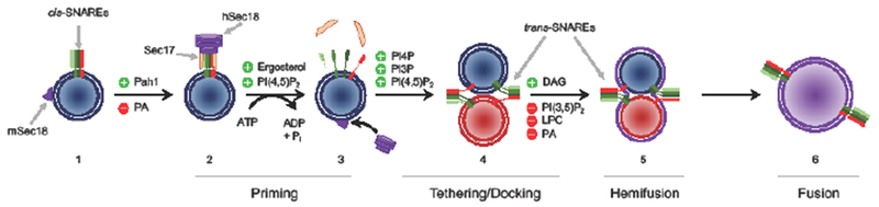
In eukaryotes, organelles and vesicles modulate their contents and identities through highly regulated membrane fusion events. Membrane trafficking and fusion are carried out through a series of stages that lead to the formation of SNARE complexes between cellular compartment membranes to trigger fusion. Although the protein catalysts of membrane fusion are well characterized, their response to their surrounding microenvironment, provided by the lipid composition of the membrane, remains to be fully understood. Membranes are composed of bulk lipids (e.g., phosphatidylcholine), as well as regulatory lipids that undergo constant modifications by kinases, phosphatases, and lipases. These lipids include phosphoinositides, diacylglycerol, phosphatidic acid, and cholesterol/ergosterol. Here we describe the roles of these lipids throughout the stages of yeast vacuole homotypic fusion.
Keywords: Dgk1; Diacylglycerol; Lipin1; Pah1; Sec18; phosphatidic acid.
Copyright © 2018 Elsevier Ltd. All rights reserved.
Figures
Similar articles
-
Ergosterol is required for the Sec18/ATP-dependent priming step of homotypic vacuole fusion.EMBO J. 2001 Aug 1;20(15):4035-40. doi: 10.1093/emboj/20.15.4035. EMBO J. 2001. PMID: 11483507 Free PMC article.
-
Enhanced membrane fusion in sterol-enriched vacuoles bypasses the Vrp1p requirement.Mol Biol Cell. 2004 Oct;15(10):4609-21. doi: 10.1091/mbc.e04-03-0194. Epub 2004 Jul 14. Mol Biol Cell. 2004. PMID: 15254266 Free PMC article.
-
Interdependent assembly of specific regulatory lipids and membrane fusion proteins into the vertex ring domain of docked vacuoles.J Cell Biol. 2004 Dec 20;167(6):1087-98. doi: 10.1083/jcb.200409068. J Cell Biol. 2004. PMID: 15611334 Free PMC article.
-
Lipid dynamics in exocytosis.Cell Mol Neurobiol. 2010 Nov;30(8):1335-42. doi: 10.1007/s10571-010-9577-x. Epub 2010 Nov 16. Cell Mol Neurobiol. 2010. PMID: 21080057 Free PMC article. Review.
-
[Lipids in the process of synaptic vesicle exo- and endocytosis].Ross Fiziol Zh Im I M Sechenova. 2010 Aug;96(8):753-65. Ross Fiziol Zh Im I M Sechenova. 2010. PMID: 20968061 Review. Russian.
Cited by
-
Use of Bio-Layer Interferometry (BLI) to Measure Binding Affinities of SNAREs and Phosphoinositides.Methods Mol Biol. 2025;2887:103-117. doi: 10.1007/978-1-0716-4314-3_7. Methods Mol Biol. 2025. PMID: 39806149
-
Human atlastins are sufficient to drive the fusion of liposomes with a physiological lipid composition.J Cell Biol. 2023 Apr 3;222(4):e202109090. doi: 10.1083/jcb.202109090. Epub 2023 Feb 9. J Cell Biol. 2023. PMID: 36757370 Free PMC article.
-
Vac8 Controls Vacuolar Membrane Dynamics during Different Autophagy Pathways in Saccharomyces cerevisiae.Cells. 2019 Jun 30;8(7):661. doi: 10.3390/cells8070661. Cells. 2019. PMID: 31262095 Free PMC article.
-
Sphingolipids containing very long-chain fatty acids regulate Ypt7 function during the tethering stage of vacuole fusion.J Biol Chem. 2024 Nov;300(11):107808. doi: 10.1016/j.jbc.2024.107808. Epub 2024 Sep 21. J Biol Chem. 2024. PMID: 39307308 Free PMC article.
-
Differential Roles of Lipin1 and Lipin2 in the Hepatitis C Virus Replication Cycle.Cells. 2019 Nov 18;8(11):1456. doi: 10.3390/cells8111456. Cells. 2019. PMID: 31752156 Free PMC article.
References
-
- Gowri PM, Sengupta S, Bertera S and Katzenellenbogen BS (2007) Lipin1 regulation by estrogen in uterus and liver: implications for diabetes and fertility. Endocrinology 148, 3685–3693 - PubMed
-
- Mlinar B, Ferk P, Pfeifer M, Gersak K and Marc J (2011) Lipin 1 gene polymorphisms in polycystic ovary syndrome. Horm Metab Res 43, 427–432 - PubMed
Publication types
MeSH terms
Substances
Grants and funding
LinkOut - more resources
Full Text Sources
Medical
Molecular Biology Databases
