MiR-1 Suppresses Proliferation of Osteosarcoma Cells by Up-regulating p21 via PAX3
- PMID: 30587501
- PMCID: PMC6348397
- DOI: 10.21873/cgp.20113
MiR-1 Suppresses Proliferation of Osteosarcoma Cells by Up-regulating p21 via PAX3
Abstract
Background/aim: miRNA-1(miR-1) is down-regulated in various cancer cells including osteosarcoma cells. This study was conducted to analyze the function of miR-1 in osteosarcoma cells.
Materials and methods: miR-1 expression in osteosarcoma cells was evaluated by qRT-PCR. Cell proliferation was evaluated after transfecting miR-1 by WST8 assay and FACS analysis, both in vitro and in vivo.
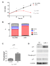
Results: Overexpression of miR-1 suppressed cell proliferation and induced cell-cycle arrest in the G0-G1 phase by increasing p21 levels via a p53-independent pathway. Overexpression of miR-1 down-regulated PAX3, a potential p21-regulating gene. Moreover, knockdown of PAX3 suppressed cell proliferation by increasing p21 levels, and induced arrest at the G0/G1 phase. Administration of miR-1 showed an in vivo antitumor effect.
Conclusion: Overexpression of miR-1 suppressed cell proliferation and induced arrest in the G0/G1 phase by increasing p21 levels via a p53-independent pathway through PAX3 suppression. These results indicate that miR-1 could be a therapeutic target for osteosarcoma.
Keywords: Osteosarcoma; PAX3; miR-1; p21; p53-independent pathway.
Copyright© 2019, International Institute of Anticancer Research (Dr. George J. Delinasios), All rights reserved.
Conflict of interest statement
No potential conflicts of interest were reported by the Authors.
Figures


Similar articles
-
Loss of MicroRNA-489-3p promotes osteosarcoma metastasis by activating PAX3-MET pathway.Mol Carcinog. 2017 Apr;56(4):1312-1321. doi: 10.1002/mc.22593. Epub 2016 Nov 29. Mol Carcinog. 2017. PMID: 27859625
-
MiR-223/Ect2/p21 signaling regulates osteosarcoma cell cycle progression and proliferation.Biomed Pharmacother. 2013 Jun;67(5):381-6. doi: 10.1016/j.biopha.2013.03.013. Epub 2013 Apr 3. Biomed Pharmacother. 2013. PMID: 23601845
-
MicroRNA-206 Reduces Osteosarcoma Cell Malignancy In Vitro by Targeting the PAX3-MET Axis.Yonsei Med J. 2019 Feb;60(2):163-173. doi: 10.3349/ymj.2019.60.2.163. Yonsei Med J. 2019. PMID: 30666838 Free PMC article.
-
[miR-365 inhibits proliferation and promotes apoptosis of SOSP9607 osteosarcoma cells].Xi Bao Yu Fen Zi Mian Yi Xue Za Zhi. 2016 Jan;32(1):44-8. Xi Bao Yu Fen Zi Mian Yi Xue Za Zhi. 2016. PMID: 26728377 Chinese.
-
Implication of the p53-Related miR-34c, -125b, and -203 in the Osteoblastic Differentiation and the Malignant Transformation of Bone Sarcomas.Cells. 2020 Mar 27;9(4):810. doi: 10.3390/cells9040810. Cells. 2020. PMID: 32230926 Free PMC article. Review.
Cited by
-
Analysis of a Preliminary microRNA Expression Signature in a Human Telangiectatic Osteogenic Sarcoma Cancer Cell Line.Int J Mol Sci. 2021 Jan 25;22(3):1163. doi: 10.3390/ijms22031163. Int J Mol Sci. 2021. PMID: 33503899 Free PMC article.
-
Molecular mechanism of microRNAs regulating apoptosis in osteosarcoma.Mol Biol Rep. 2022 Jul;49(7):6945-6956. doi: 10.1007/s11033-022-07344-x. Epub 2022 Apr 26. Mol Biol Rep. 2022. PMID: 35474050 Review.
-
Role of MicroRNAs in Human Osteosarcoma: Future Perspectives.Biomedicines. 2021 Apr 23;9(5):463. doi: 10.3390/biomedicines9050463. Biomedicines. 2021. PMID: 33922820 Free PMC article. Review.
-
The Role of Cell Cycle Regulators in Cell Survival-Dual Functions of Cyclin-Dependent Kinase 20 and p21Cip1/Waf1.Int J Mol Sci. 2020 Nov 12;21(22):8504. doi: 10.3390/ijms21228504. Int J Mol Sci. 2020. PMID: 33198081 Free PMC article. Review.
-
SLC25A10 performs an oncogenic role in human osteosarcoma.Oncol Lett. 2020 Oct;20(4):2. doi: 10.3892/ol.2020.11863. Epub 2020 Jul 14. Oncol Lett. 2020. PMID: 32774476 Free PMC article.
References
-
- Uribe-Botero G, Russell WO, Sutow WW, Martin RG. Primary osteosarcoma of bone. Clinicopathologic investigation of 243 cases, with necropsy studies in 54. Am J Clin Pathol. 1977;67(5):427–435. - PubMed
-
- Bielack SS, Kempf-Bielack B, Delling G, Exner GU, Flege S, Helmke K, Kotz R, Salzer-Kuntschik M, Werner M, Winkelmann W, Zoubek A, Jurgens H, Winkler K. Prognostic factors in high-grade osteosarcoma of the extremities or trunk: an analysis of 1,702 patients treated on neoadjuvant cooperative osteosarcoma study group protocols. J Clin Oncol. 2002;20(3):776–790. - PubMed
-
- Kempf-Bielack B, Bielack SS, Jurgens H, Branscheid D, Berdel WE, Exner GU, Gobel U, Helmke K, Jundt G, Kabisch H, Kevric M, Klingebiel T, Kotz R, Maas R, Schwarz R, Semik M, Treuner J, Zoubek A, Winkler K. Osteosarcoma relapse after combined modality therapy: an analysis of unselected patients in the Cooperative Osteosarcoma Study Group (COSS) J Clin Oncol. 2005;23(3):559–568. - PubMed
-
- Szuhai K, Cleton-Jansen AM, Hogendoorn PC, Bovee JV. Molecular pathology and its diagnostic use in bone tumors. Cancer Genet. 2012;205(5):193–204. - PubMed
MeSH terms
Substances
LinkOut - more resources
Full Text Sources
Medical
Research Materials
Miscellaneous
