. 2019 Apr;17(4):974-986.
doi: 10.1158/1541-7786.MCR-18-0504. Epub 2018 Dec 26.

Ovarian Cancer Cells Commonly Exhibit Defective STING Signaling Which Affects Sensitivity to Viral Oncolysis

Affiliations

Ovarian Cancer Cells Commonly Exhibit Defective STING Signaling Which Affects Sensitivity to Viral Oncolysis

Nina Marí Gual Pimenta de Queiroz et al. Mol Cancer Res. 2019 Apr.

Abstract

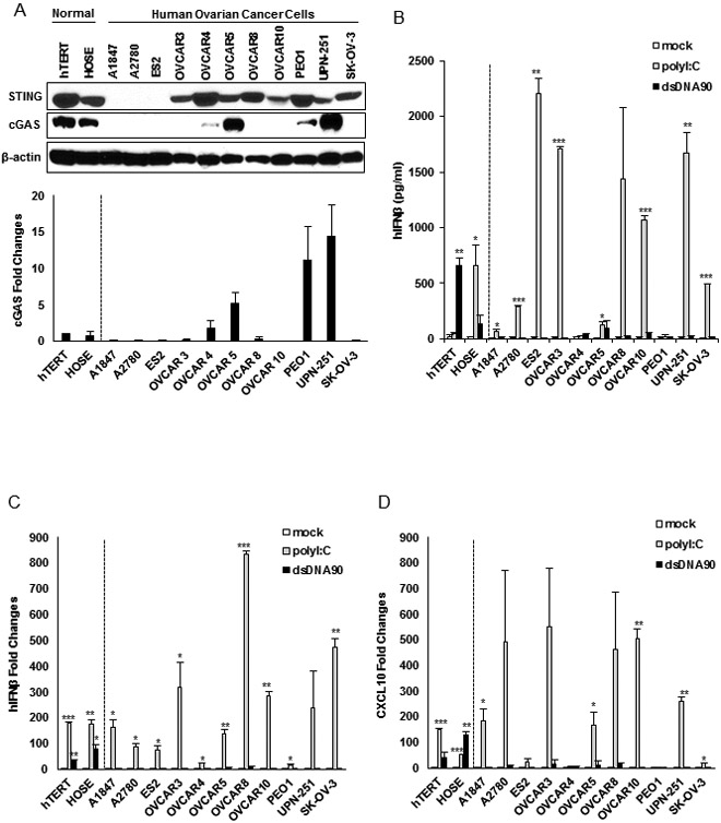
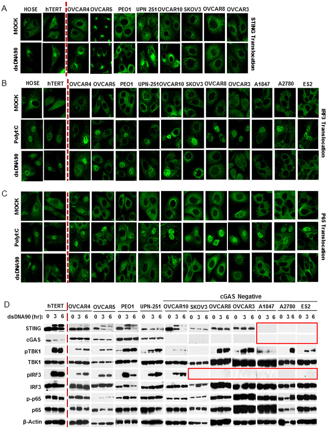
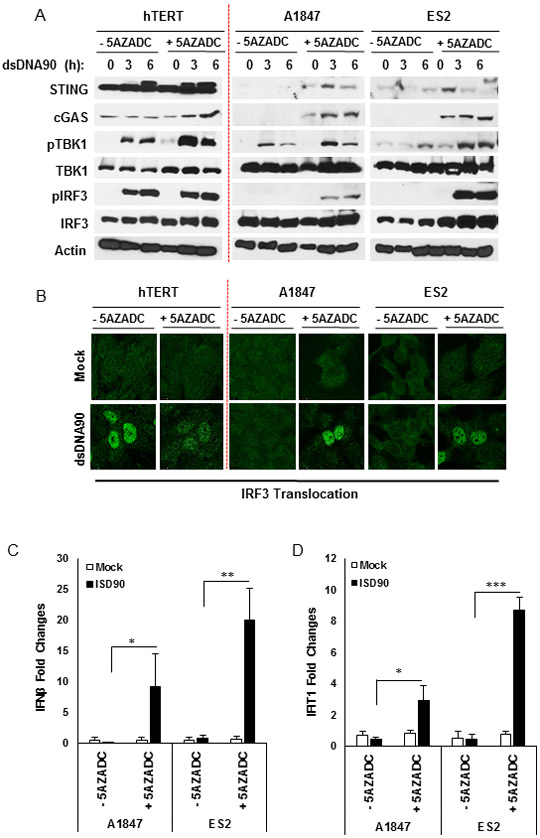
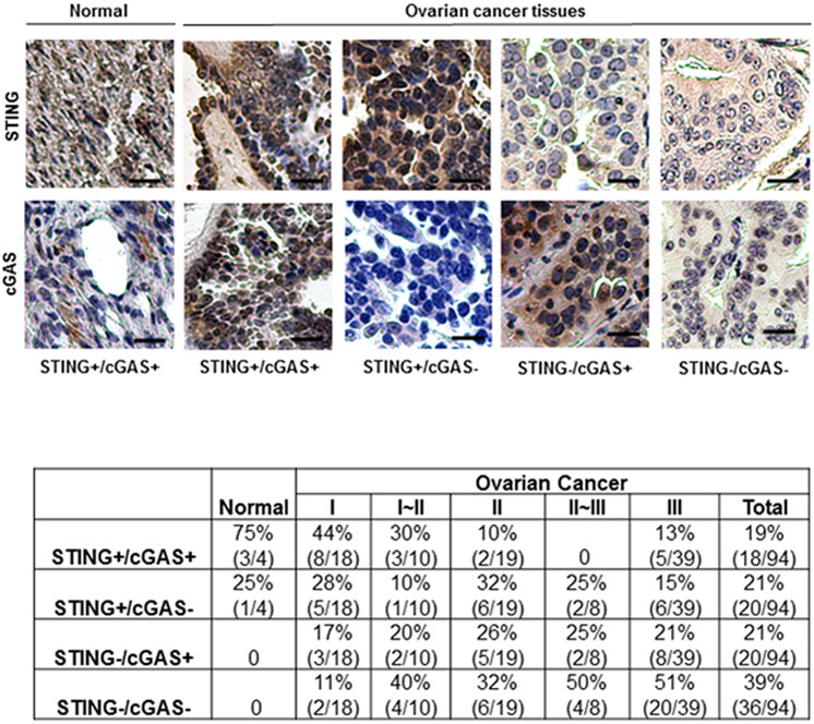
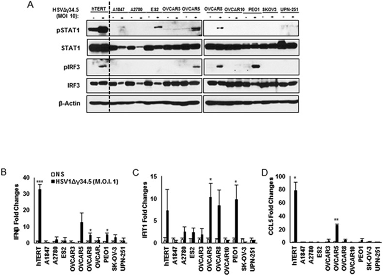
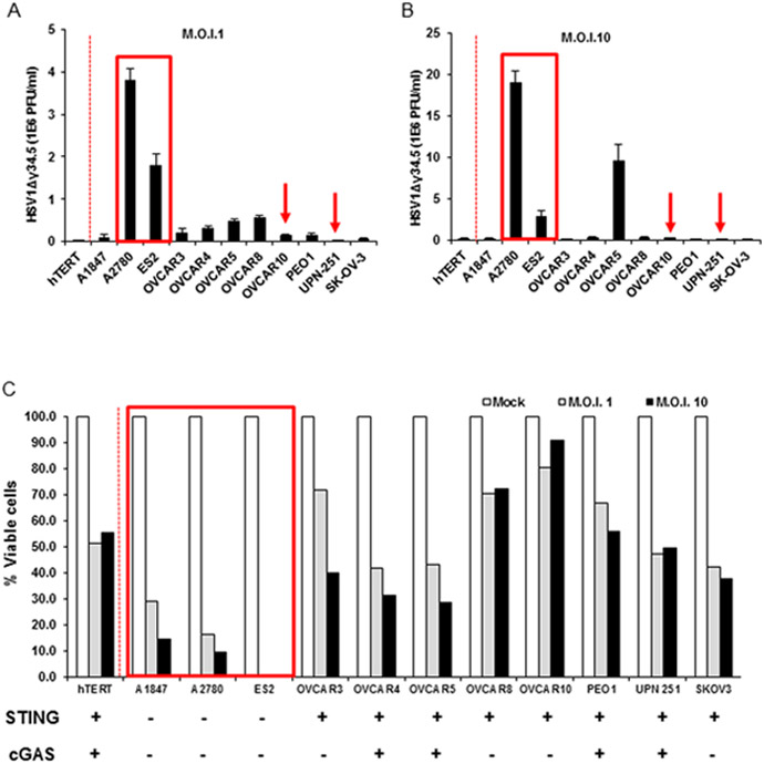
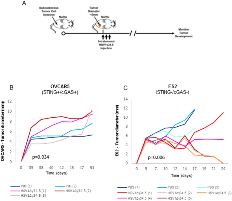
Ovarian cancer is the sixth most prevalent cancer in women and the most lethal of the gynecologic malignancies. Treatments have comprised the use of immunotherapeutic agents as well as oncolytic viruses, with varying results for reasons that remain to be clarified. To better understand the mechanisms that may help predict treatment outcome, we have evaluated innate immune signaling in select ovarian cancer cell lines, governed by the Stimulator of Interferon Genes (STING), which controls self or viral DNA-triggered cytokine production. Our results indicate that STING-dependent signaling is habitually defective in majority of ovarian cancer cells examined, frequently through the suppression of STING and/or the cyclic dinucleotide (CDN) enzyme Cyclic GMP-AMP synthase (cGAS) expression, by epigenetic processes. However, STING-independent, dsRNA-activated innate immune cytokine production, which require RIG-I/MDA5, were largely unaffected. Such defects enabled ovarian cancer cells to avoid DNA damage-mediated cytokine production, which would alert the immunosurveillance system. Loss of STING signaling also rendered ovarian cancer cells highly susceptible to viral oncolytic γ34.5 deleted-HSV1 (Herpes simplex virus) infection in vitro and in vivo. IMPLICATIONS: STING signaling evaluation in tumors may help predict disease outcome and possibly dictate the efficacy of oncoviral and other types of cancer therapies.


Conflict of interest statement

Disclosure of Potential Conflict of Interests: No potential conflicts of interest were disclosed.

Figures

Figure 1.
Figure 1.
STING-mediated dsDNA-induced innate immune activation is impaired in the majority of human ovarian cancer cell lines. A, Immunoblot of STING and cGAS in hTERT fibroblasts, normal human ovarian surface epithelial (HOSE) and a series of human ovarian cancer cell lines. cGAS expression was also analyzed by qPCR (bottom). B, ELISA analysis of human IFNβ production in the media of cells (same as in A) transfected with 3 μg/ml polyI:C or dsDNA90 or mock transfected for 16 hours. C, qPCR analysis of human IFNβ expression in cells (same as in A) transfected with 3 μg/ml polyI:C or dsDNA90 or mock transfected for 6 hours. D, qPCR analysis of human CXCL10 expression in cells (same as in C). Data are representative of at least two independent experiments. Error bars indicate SD. *p < 0.05, **p < 0.01, and ***p < 0.001; Student’s t test.
Figure 2.
Figure 2.
dsDNA-induced STING signaling pathway is defective in human ovarian cancer cell lines. A, Immunofluorescence microscopy analysis of STING translocation in normal and human ovarian cancer cell lines transfected with 3 μg/ml dsDNA90 or polyI:C or mock transfected for 3 hours. B, Immunofluorescence microscopy analysis of IRF3 translocation in normal and human ovarian cancer cell lines. C, Immunofluorescence microscopy analysis of p65 translocation in cells (same as in B). D, Immunoblot analysis of STING signal activation in cells (same as above) transfected with 3 μg/ml dsDNA90 for the indicated time periods.
Figure 3.
Figure 3.
DNA demethylation recapitulated STING and cGAS expression in human ovarian cancer cell lines. A, Immunoblot analysis of STING signal activation in ovarian cancer cells mock or treated with 10 μM 5-aza-2’-deoxycytidine (5AZADC) for 4 days, followed by dsDNA90 transfection at 3 μg/ml for the indicated time periods. B, Immunofluorescence microscopy analysis of IRF3 translocation in ovarian cancer cells treated with 5AZADC (same as above) followed by dsDNA90 transfection at 3 μg/ml for 3 hours. C, Ovarian cancer cells mock treated or treated with 10 μM 5AZADC for 4 days, followed by dsDNA90 transfection at 3 μg/ml for 6 hours. human IFN-β expression was analyzed by qPCR. D, qPCR analysis of human IFIT1 expression in ovarian cancer cells (same as in C). Data are representative of at least two independent experiments. Error bars indicate SD. *p < 0.05, **p < 0.01, and ***p < 0.001; Student’s t test.
Figure 4.
Figure 4.
STING and cGAS expression were suppressed in high percentage of human ovarian cancer. IHC analysis of STING and cGAS in 98 human ovarian TMA containing normal/benign and cancer tissues. Representative images of normal human epidermal and human ovarian cancer tissues stained for STING and cGAS. Images are shown at 200x. Bar size, 50 μm. STING and cGAS expression status is summarized and shown in the bottom.
Figure 5.
Figure 5.
DNA virus infection (HSV1Δγ34.5) does not activate tipe I IFN response in STING/cGAS-deficient cells. A, Normal and human ovarian cancer cell lines were infected with HSV1Δγ34.5 at M.O.I. 10 for 1 hour and analysed 4 hours post infection by immunoblot (pSTAT1, STAT1, pIRF3, IRF3 and β-actin). Same cells as above were infected with HSV1Δγ34.5 at M.O.I. 1 for 1 hour and samples collected 6 hours post infection to check RNA expression by qPCR for IFNβ (B), IFIT1 (C) and CCL5 (D). Data are representative of at least two independent experiments. Error bars indicate SD. *p < 0.05, **p < 0.01, and ***p < 0.001; Student’s t test.
Figure 6.
Figure 6.
STING signal defect leads ovarian cancer cells more susceptible to HSV1Δγ34.5. Normal human hTERT cells and human ovarian cancer cell lines were infected with HSV1Δγ34.5 at the M.O.I. 1 (A) or M.O.I. 10 (B) for 1 hour and titration of HSV1Δγ34.5 was analyzed by standard plaque assay in Vero cells 24 hours later. (C) Cells (same as in A) were infected with HSV1Δγ34.5 at M.O.I. 1 and 10 for 1 hour, and cell viability was analyzed by trypan blue staining 24 hours later. Data are representative of at least two independent experiments.
Figure 7.
Figure 7.
Increased HSV1Δγ34.5 oncolytic effect was observed in ovarian cancer cells with impaired STING signal in vivo. A, Scheme of HSV1Δγ34.5 treatment on a xenograft tumor in nude mice. B-C, The indicated xenograft tumors were generated in the right flank of nude BALB/c mice. When tumors reached 5 mm in diameter, they were injected every other day, for a total of three times, with 1E8 PFU HSV1Δγ34.5 in 50 μl PBS or 50 μl PBS only, and tumor growth was measured every other day. Statistical analysis was carried out comparing the two treatment groups at the last time point using the unpaired Student’s t test. p values are as indicated.

References

    1. Cannistra SA. Cancer of the ovary. N Engl J Med 2004;351(24):2519–29 doi 10.1056/NEJMra041842. - DOI - PubMed
    1. Jemal A, Siegel R, Ward E, Hao Y, Xu J, Murray T, et al. Cancer statistics, 2008. CA Cancer J Clin 2008;58(2):71–96 doi 10.3322/CA.2007.0010. - DOI - PubMed
    1. Li S, Tong J, Rahman MM, Shepherd TG, McFadden G. Oncolytic virotherapy for ovarian cancer. Oncolytic Virother 2012;1:1–21. - PMC - PubMed
    1. Lengyel E Ovarian cancer development and metastasis. Am J Pathol 2010;177(3):1053–64 doi 10.2353/ajpath.2010.100105. - DOI - PMC - PubMed
    1. Naora H, Montell DJ. Ovarian cancer metastasis: integrating insights from disparate model organisms. Nat Rev Cancer 2005;5(5):355–66 doi 10.1038/nrc1611. - DOI - PubMed

Publication types