. 2019 Feb 13;39(7):1150-1168.
doi: 10.1523/JNEUROSCI.1401-18.2018. Epub 2018 Dec 26.

Genetic Analysis of the Organization, Development, and Plasticity of Corneal Innervation in Mice

Affiliations

Genetic Analysis of the Organization, Development, and Plasticity of Corneal Innervation in Mice

Nacim Bouheraoua et al. J Neurosci. .

Abstract

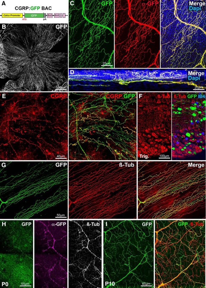
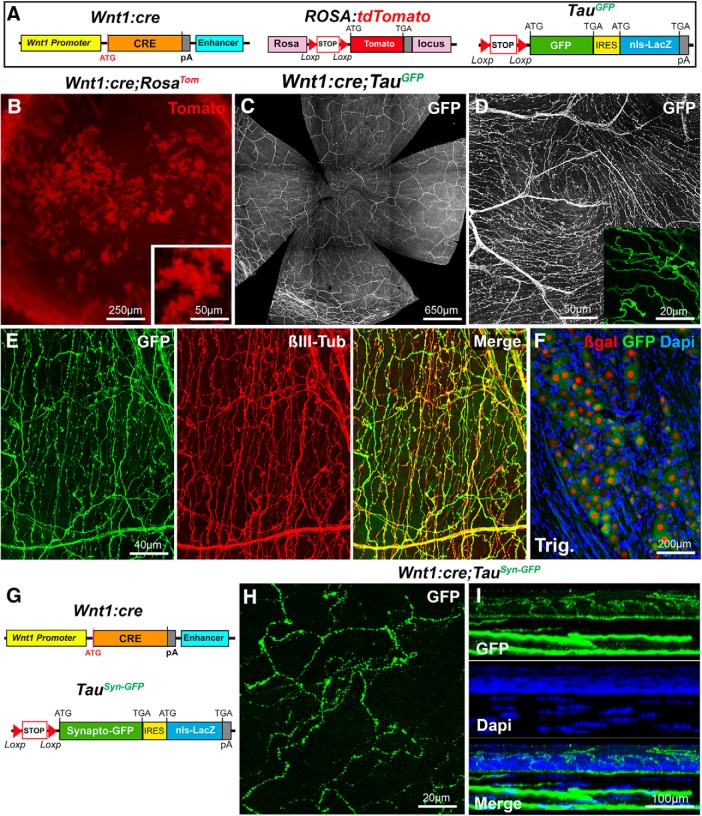
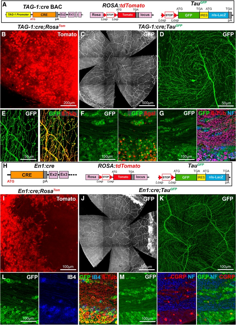
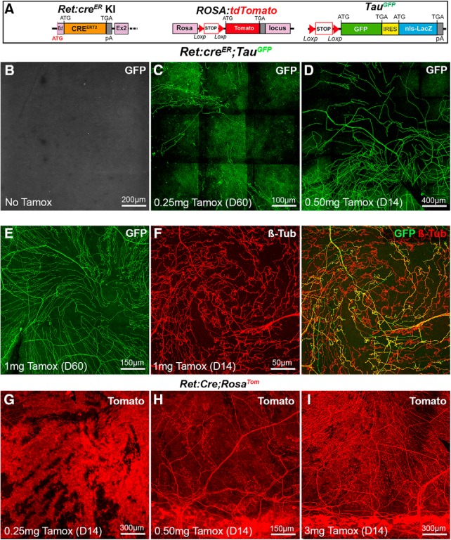
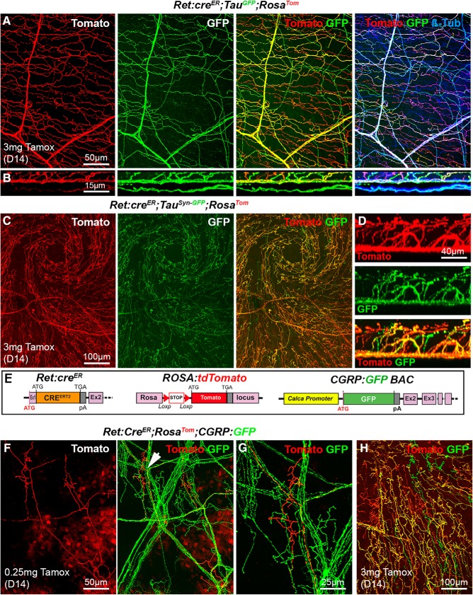
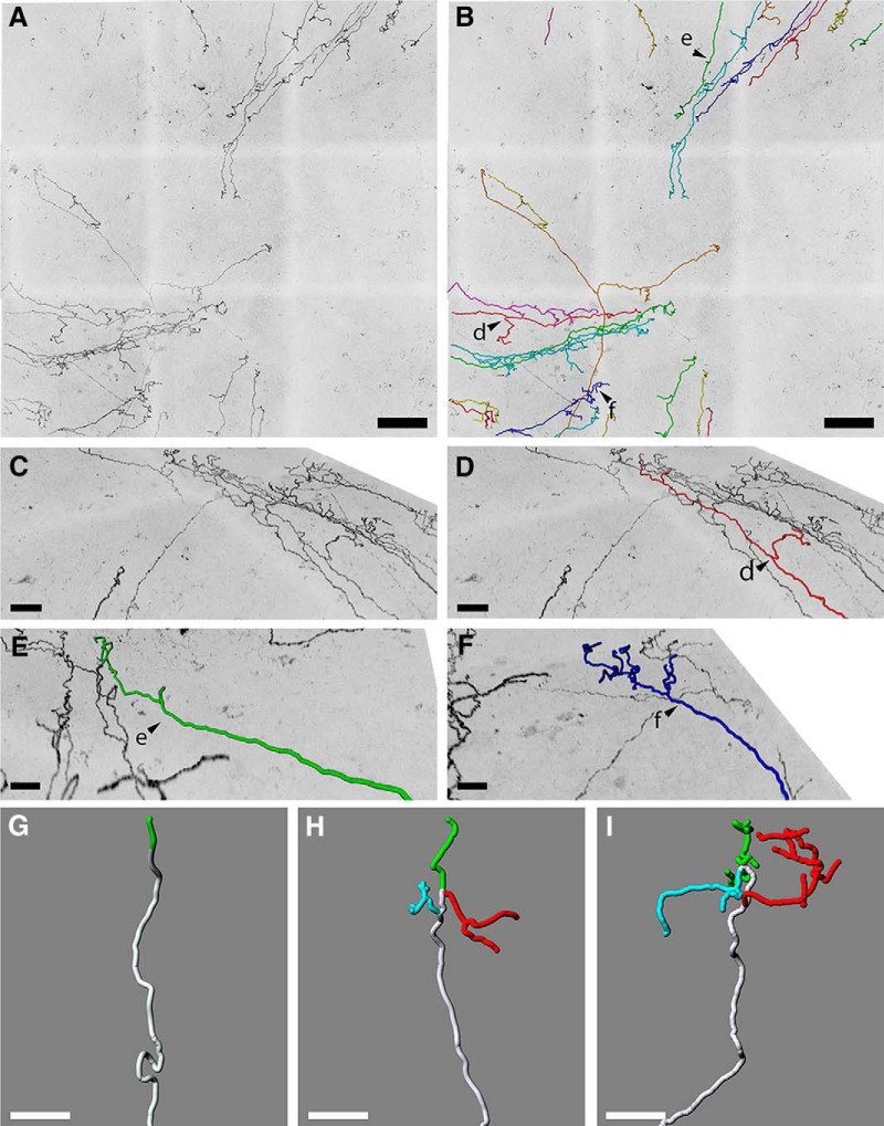
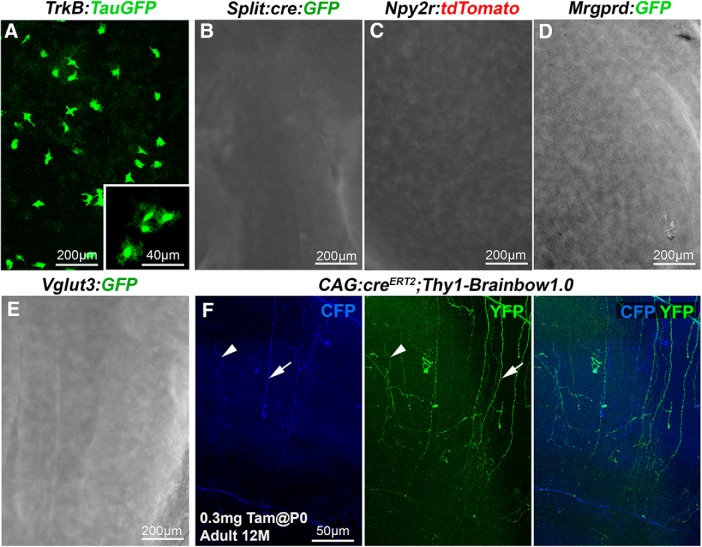
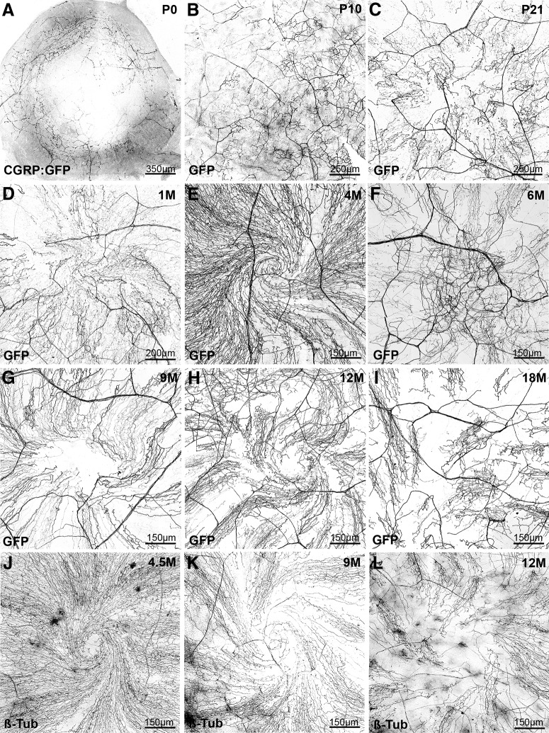
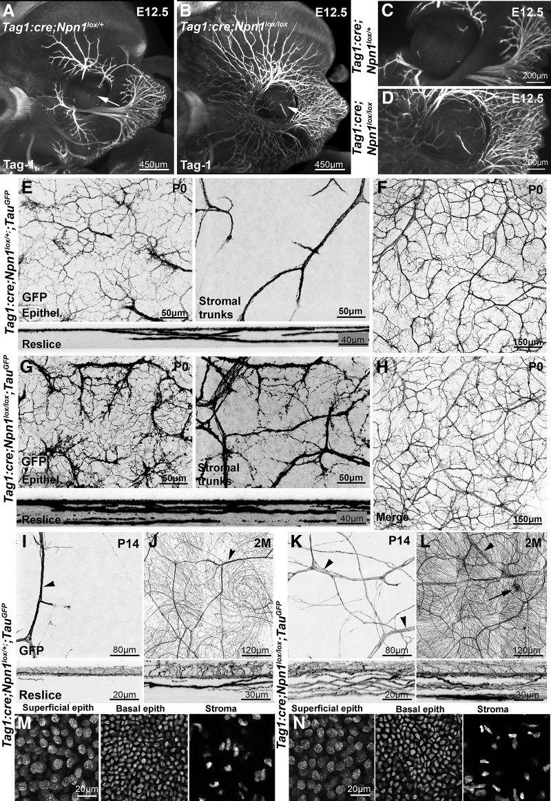
The cornea has the densest sensory innervation of the body, originating primarily from neurons in the trigeminal ganglion. The basic principles of cornea nerve patterning have been established many years ago using classic neuroanatomical methods, such as immunocytochemistry and electrophysiology. Our understanding of the morphology and distribution of the sensory nerves in the skin has considerably progressed over the past few years through the generation and analysis of a variety of genetically modified mouse lines. Surprisingly, these lines were not used to study corneal axons. Here, we have screened a collection of transgenic and knockin mice (of both sexes) to select lines allowing the visualization and genetic manipulation of corneal nerves. We identified multiple lines, including some in which different types of corneal axons can be simultaneously observed with fluorescent proteins expressed in a combinatorial manner. We also provide the first description of the morphology and arborization of single corneal axons and identify three main types of branching pattern. We applied this genetic strategy to the analysis of corneal nerve development and plasticity. We provide direct evidence for a progressive reduction of the density of corneal innervation during aging. We also show that the semaphorin receptor neuropilin-1 acts cell-autonomously to control the development of corneal axons and that early axon guidance defects have long-term consequences on corneal innervation.SIGNIFICANCE STATEMENT We have screened a collection of transgenic and knockin mice and identify lines allowing the visualization and genetic manipulation of corneal nerves. We provide the first description of the arborization pattern of single corneal axons. We also present applications of this genetic strategy to the analysis of corneal nerve development and remodeling during aging.

Keywords: aging; confocal microscopy; cornea; corneal innervation; mouse genetics; neuropilin.


Figures

Figure 1.
Figure 1.
Visualization of corneal peptidergic axons in CGRP:GFP mice. All panels (except D and F) are maximal intensity z-projection confocal stacks from whole-mount corneas. A, Schematic of the CGRP:GFP BAC transgenic construct. GFP was inserted downstream the promoter of the Calca gene, which encodes CGRP. B–G, Images from adult CGRP:GFP mice. B, Flat mount view of a whole-mount cornea, showing GFP expression in corneal nerves. C, Cornea immunolabeled with anti-GFP with DAPI counterstaining (blue). There is a perfect overlap (merge) between the endogenous GFP fluorescence (green) and the anti-GFP immunoreactivity (red). D, A reslice of the cornea (54-μm-thick optical section) showing the location of the GFP axons in the stroma, sub-basal plexus, and epithelium. E, Cornea immunolabeled with anti-CGRP (red). All CGRP axons are also GFP+. F, Cryostat section of the trigeminal ganglion stained with IB4 (blue) and immunolabeled for βIII-tubulin (red). GFP neurons only represent a subset of βIII-Tub+ trigeminal neurons. G, Cornea immunolabeled with anti-βIII-tubulin (red). Typical corneal axon leashes of almost parallel GFP+ axons (green) are seen. GFP is only expressed in a subset of corneal nerves. H, At P0, the endogenous GFP expression is weaker than after anti-GFP immunostaining (magenta). All corneal axons in this domain can be seen with anti-βIII-tubulin immunostaining (white, right). I, GFP+ axons in the P10 cornea immuno-labeled for βIII-tubulin.
Figure 2.
Figure 2.
Visualization of corneal axons in Wnt1:cre mice. All panels (except F and I) are maximal intensity z-projection confocal stacks from adult whole-mount corneas. A, Schematic description of the mouse lines. In Wnt1:cre knockin mice, Cre recombinase was placed downstream of the Wnt1 promoter. RosaTom: the tdTomato coding sequence was inserted in the Rosa locus downstream of a lox-STOP-lox cassette. In TauGFP mice, a lox-STOP-lox cassette preceding a myristoylated GFP sequence, followed by an Internal ribosome entry site (IRES) cDNA and the lacZ sequence with a nuclear localization signal (nls), was inserted by homologous recombination in the Tau locus. B, Islets of corneal cells express Tomato (red) in Wnt1:cre;RosaTom mice. C, D, The dense network of GFP+ corneal axons in Wnt1:cre;TauGFP mice. The apical vortex is shown in D. Inset, Terminal intraepithelial branches. E, Cornea immunolabeled with anti-βIII-tubulin antibodies (red). GFP and βIII-tubulin nicely overlap. F, Cryostat section of the trigeminal ganglion at the level of the ophthalmic V1 division stained DAPI (blue) and immunolabeled for β-galactosidase (red). GFP+ trigeminal neurons express β-gal in their nucleus. G, Description of the mouse lines. Wnt1:cre (see above). In TauSyn-GFP mice, a lox-STOP-lox cassette preceding a cDNA encoding Synaptophysin fused to GFP, followed by an Internal ribosome entry site (IRES) cDNA and the lacZ sequence with a nuclear localization signal (nls) was inserted by homologous recombination in the Tau locus. H, Beaded appearance of the GFP signal in Wnt1cre;TauSynGFP mice. I, A reslice of the cornea with DAPI counterstaining (blue).
Figure 3.
Figure 3.
Visualization of corneal axons in TAG-1:cre and En1:cre adult mice. B–E, I–K, Maximal intensity z-projection confocal stacks from adult whole-mount corneas. F, G, L, M, Confocal images of cryostat sections of trigeminal ganglia. A, Description of the mouse lines. RosaTom and TauGFP (see Fig. 2). In the TAG-1-cre BAC transgenic construct, Cre recombinase was inserted downstream the promoter of the Tag-1/Cntn2 gene in an artificial chromosome. B, Tomato is highly expressed by corneal cells in TAG-1:cre;RosaTom mice. C, D, The dense network of GFP+ corneal axons in TAG-1:cre;TauGFP mice. The apical vortex is shown in D. E, Cornea immunolabeled with anti-βIII-tubulin antibodies (red). GFP and βIII-tubulin perfectly overlap. F, G, In the trigeminal ganglion, GFP+ neurons express β-gal in their nucleus (F). All GFP+ neurons are also βIII-tubulin+, and some are also NF200+ (G). H, Description of the mouse lines. RosaTom and TauGFP (see Fig. 2). In En1:cre knockin mice, the first exon of the engrailed-1 gene was replaced by the Cre sequence using homologous recombination. I, Tomato is highly expressed by a large fraction of corneal cells in En1:cre;RosaTom mice. J, K, GFP+ corneal axons in En1:cre;TauGFP mice. The apical vortex is shown in K. L, M, In the trigeminal ganglion, all IB4+ and all βIII-immunoreactive neurons are GFP+ (L). GFP is also expressed in the CGRP+ and NF200+ populations (M).
Figure 4.
Figure 4.
Visualization of corneal axons in Islet1:cre adult mice. B, C, G, H, Maximal intensity z-projection confocal stacks from adult whole-mount corneas. A, Description of the mouse lines. RosaTom (see Fig. 2). In Islet1:cre knockin mice, the coding sequence of Cre was inserted in the isl1 gene by homologous recombination. B, C, In Islet1:cre;RosaTom mice, all corneal axons express Tomato (red). βIII-tubulin-immunoreactive axons (green) are also Tomato+ (see merge). D, E, Confocal images of cryostat sections of trigeminal ganglia. D, Colocalization of the GFP signal (green) and βIII-Tub immunoreactivity (red) in trigeminal neurons. E, All CGRP+ neurons (cyan) coexpress Tomato. F, Description of the mouse lines. Islet1:cre (see above). CGRP:GFP (see Fig. 1). RosaTom (see Fig. 2). G, GFP and Tomato expression in whole-mount cornea from an Islet1:cre;RosaTom;CGRP:GFP mouse. H, High magnification showing that Tomato (red) is expressed both by peptidergic (GFP+, green) and nonpeptidergic (GFP) axons. I, A reslice of the cornea (54-μm-thick optical section) showing the location of the fluorescent axons. Yellow represents GFP+/Tomato+ peptidergic nociceptor axons. Red represents nonpetidergic nociceptor axons.
Figure 5.
Figure 5.
Visualization of corneal axons in Ret:creER adult mice. B–I, Maximal intensity z-projection confocal stacks from adult whole-mount corneas. A, Description of the mouse lines RosaTom and TauGFP (see Fig. 2). In Ret:creER knockin mice, the coding sequence of creERT2 was inserted in the first exon of the Ret gene by homologous recombination. B, In the absence of tamoxifen, no GFP signal is detected in the cornea of Ret:creER;TauGFPmice. C–E, The number of GFP+ axons increases with the dose of tamoxifen injected (0.25 mg-1 mg). Corneas were collected 14 d (D14) or 60 d (D60) after injection. F, Immunostaining for anti-βIII-tubulin shows that GFP is only expressed in a fraction of βIII-Tub+ corneal axons. G–I, Corneas from Ret:creER;RosaTom mice injected with increasing doses of tamoxifen injected (0.25–3 mg). At the lowest dose (G), many Tomato+ corneal cells are seen and mask Tomato+ axons. H, I, At higher doses, highly fluorescent cells are seen in the limbal region, and more Tomato+ axons are observed.
Figure 6.
Figure 6.
Analysis of corneal nerves in Ret:creER compound mice. All images (except B and D) are maximal intensity z-projection confocal stacks from adult whole-mount corneas. A, Cornea from a Ret:creER;TauGFP;RosaTom mouse immunolabeled for βIII-tubulin. Some βIII-Tub+ axons (blue) also coexpress GFP and Tomato (and appear white). Other axons that only express GFP (right, green or cyan) or only Tomato (right, red or magenta). B, A reslice of the cornea (54-μm-thick optical section) illustrating the distribution of the fluorescent axons in the stroma and epithelium. C, Image of the apex of the cornea and axonal whorl from a Ret:creER;TauSyn-GFP;RosaTom mouse. The three types of axons are seen: GFP+, Tomato+, and a majority of GFP+/Tomato+ axons. D, A reslice of the cornea (54-μm-thick optical section). E, Description of the mouse lines. CGRP:GFP (see Fig. 1). RosaTom (see Fig. 2). Ret:creER (see Fig. 5). F, G, With a low dose of tamoxifen (0.25 mg), only a few Tomato+ axons and do not always overlap with GFP+ nociceptive axons. Middle, Arrowhead indicates the area seen on the high-magnification image of a single tomato+ terminal arbor (right). H, With a high dose of tamoxifen, most axons coexpress GFP and Tomato, but a few only express a single fluorescent protein.
Figure 7.
Figure 7.
Heterogeneous terminal arborization of corneal axons. A, Maximal intensity z-projection confocal stacks from adult Ret:creER;TauGFP whole-mount corneas injected with a low dose of tamoxifen (0.25 mg). B, Axons from A were analyzed with Imaris software using the Filament Tracer module. Arrowheads d, e and f indicate axons seen at higher magnification in panels D, E and F. C, High-magnification image showing single axons in a sagittal view. D, Single axon tracing showing ramifying nerve terminal. E, Single axon tracing showing simple nerve terminal. F, Single axon tracing showing complex nerve terminal. G–I, Reconstructions of superficial nerve terminals in the mouse corneal epithelium showing examples of simple (G), ramifying (H), and complex (I) nerve terminals based on 143 axons.
Figure 8.
Figure 8.
Other transgenic lines tested. Maximal intensity z-projection confocal stacks (A, F) or epifluorescence images (B–E) from adult whole-mount corneas. A–E, No fluorescent corneal axons were detected in TrkB:TauGFP, Split:cre:GFP, Npy2r:tdTomato, Mrgprd:GFP, and Vglut3:GFP mice. Note the presence of scattered GFP+ cells in TrkB:TauGFP line. F, Cornea from a 12-month-old CAG:creERT2;Thy1-Brainbow1.0 mouse injected with 0.3 mg of tamoxifen at P0. A few CFP+ axons (blue) and YFP+ (green) axons are seen. Left, Middle, Arrowhead indicates a CFP+/YFP+ double-labeled axon. Arrow indicates axons that are either YFP+ or CFP+.
Figure 9.
Figure 9.
Age-dependent evolution of the corneal innervation in CGRP:GFP mice. All images are maximal intensity z-projection confocal stacks from whole-mount corneas. A negative image was generated as fluorescent axons are more visible in black on a white background. A–D, Developmental time course of corneal innervation in CGRP:GFP mice during the first postnatal month. The progressive centripetal extension and polarization of the axonal leashes. E, At 4 months, the axonal vortex at the center of the cornea is well formed (compare with D). F–H, Abnormal pattern of innervation in the center of the cornea, frequently observed from 6 to 9 months. H, The lower density of GFP+ axons compared with E. I, cornea from an 18-month-old CGRP:GFP mouse. The axonal whorl is absent, axonal leashes are not seen in the center of the cornea, and polarity is perturbed. Larger areas do not contain GFP+ axons. J–L, WT corneas immunolabeled with anti-tubulin. The progressive thinning of corneal innervation is also seen from 9 months, as well as the disorganization of axonal leashes in a 1-year-old mouse.
Figure 10.
Figure 10.
Neuropilin-1 controls the development of corneal innervation. A–D, Light sheet microscopy 3D images of E12.5 TAG-1:cre;Npn1lox/+ (A, C) and TAG-1:cre;Npn1lox/lox (B, D) embryos, immunolabeled with anti-Tag-1 antibodies and cleared with 3DISCO. Tag-1+ sensory axons innervating the face are more numerous and highly defasciculated in TAG-1:cre;Npn1lox/lox embryo. Trigeminal axons have already invaded the cornea (arrows) in the mutant unlike in the heterozygous control. E, F, Confocal images of GFP+ axons in the cornea from a TAG-1:cre;Npn1lox/+;TauGFP newborn mouse at the level of the epithelium (E; Epithel.) or the stroma (E; stromal trunks) and the whole cornea (F). The GFP+ axons already form a dense network in the sub-basal plexus. A few large axonal trunks are found in the stroma. Bottom, A 54 μm reslice through the cornea stack. F, Maximal intensity z-projection confocal stack from a whole-mount TAG-1:cre;Npn1lox/+;TauGFP cornea. G, H, Confocal images of GFP+ axons in the cornea from a TAG-1:cre;Npn1lox/lox;TauGFP newborn mouse. The density of GFP+ axons and branches is strongly increased in the sub-basal plexus (G, left) compared with heterozygous controls. The stroma also contains a much higher number of large axonal trunks (right). Bottom, A 54 μm reslice through the cornea stack. H, Maximal intensity z-projection confocal stack from a whole-mount TAG-1:cre;Npn1lox/lox;TauGFP cornea. I–L, The density of GFP+ axonal branches and large nerve trunks (arrowheads) in the epithelium and stroma is still abnormally high in TAG-1:cre;Npn1lox/lox;TauGFP mice at P14 (K) and at 2 months (L) compared with aged-matched TAG-1:cre;Npn1lox/+;TauGFPmice (I,J). Occasional large accumulations of axons are also seen in the KO (L, arrow). Bottom, The 54 μm reslices of the confocal image stacks. DAPI counterstaining of adult corneas from TAG-1:cre;Npn1lox/+;TauGFP (M) and TAG-1:cre;Npn1lox/lox;TauGFP (N) mice. Density of superficial epithelial cells, basal epithelial cells, and keratocytes in the stroma is similar in mutant and control mice.

References

    1. Abraira VE, Ginty DD (2013) The sensory neurons of touch. Neuron 79:618–639. 10.1016/j.neuron.2013.07.051 - DOI - PMC - PubMed
    1. Alamri A, Bron R, Brock JA, Ivanusic JJ (2015) Transient receptor potential cation channel subfamily V member 1 expressing corneal sensory neurons can be subdivided into at least three subpopulations. Front Neuroanat 9:71. 10.3389/fnana.2015.00071 - DOI - PMC - PubMed
    1. Alamri AS, Wood RJ, Ivanusic JJ, Brock JA (2018) The neurochemistry and morphology of functionally identified corneal polymodal nociceptors and cold thermoreceptors. PLoS One 13:e0195108. 10.1371/journal.pone.0195108 - DOI - PMC - PubMed
    1. Alcalde I, Íñigo-Portugués A, González-González O, Almaraz L, Artime E, Morenilla-Palao C, Gallar J, Viana F, Merayo-Lloves J, Belmonte C (2018) Morphological and functional changes in TRPM8-expressing corneal cold thermoreceptor neurons during aging and their impact on tearing in mice. J Comp Neurol 520:633–655. 10.1002/cne.24454 - DOI - PubMed
    1. Bai L, Lehnert BP, Liu J, Neubarth NL, Dickendesher TL, Nwe PH, Cassidy C, Woodbury CJ, Ginty DD (2015) Genetic identification of an expansive mechanoreceptor sensitive to skin stroking. Cell 163:1783–1795. 10.1016/j.cell.2015.11.060 - DOI - PMC - PubMed

Publication types

LinkOut - more resources