. 2019 Feb 13;39(7):1320-1331.
doi: 10.1523/JNEUROSCI.1359-18.2018. Epub 2018 Dec 26.

Intermittent Hypoxia Disrupts Adult Neurogenesis and Synaptic Plasticity in the Dentate Gyrus

Affiliations

Intermittent Hypoxia Disrupts Adult Neurogenesis and Synaptic Plasticity in the Dentate Gyrus

Maggie A Khuu et al. J Neurosci. .

Abstract

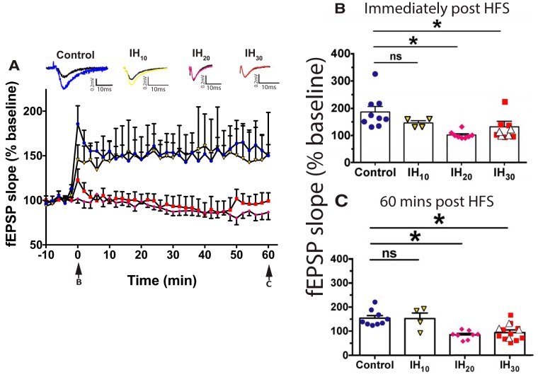
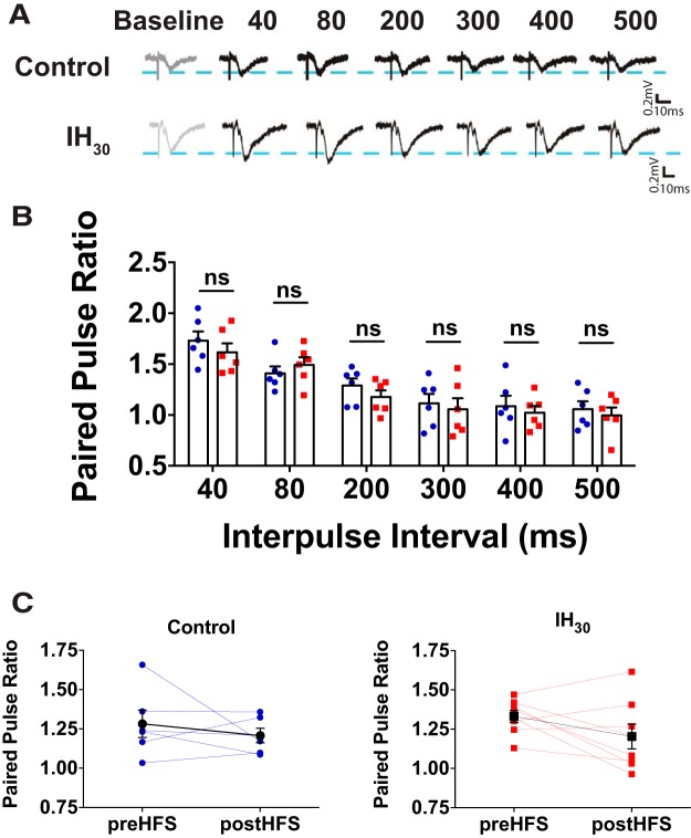
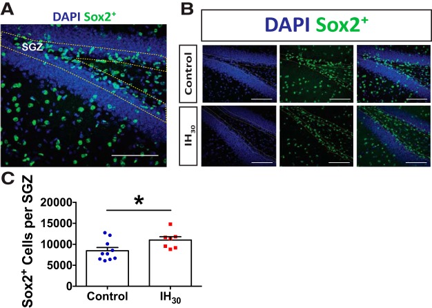
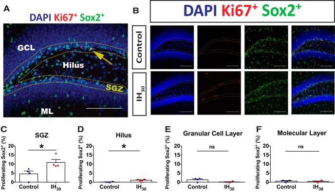
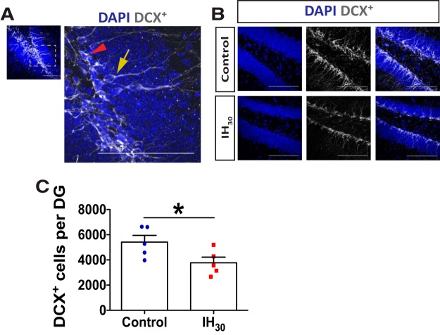
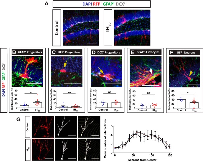
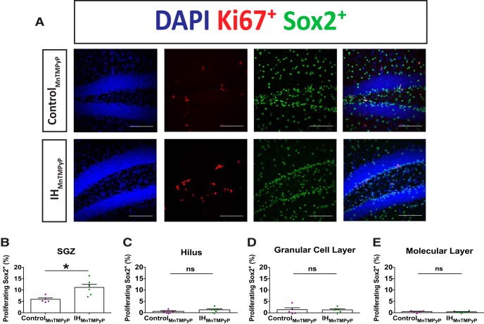
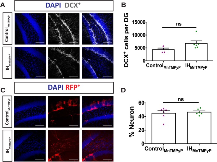
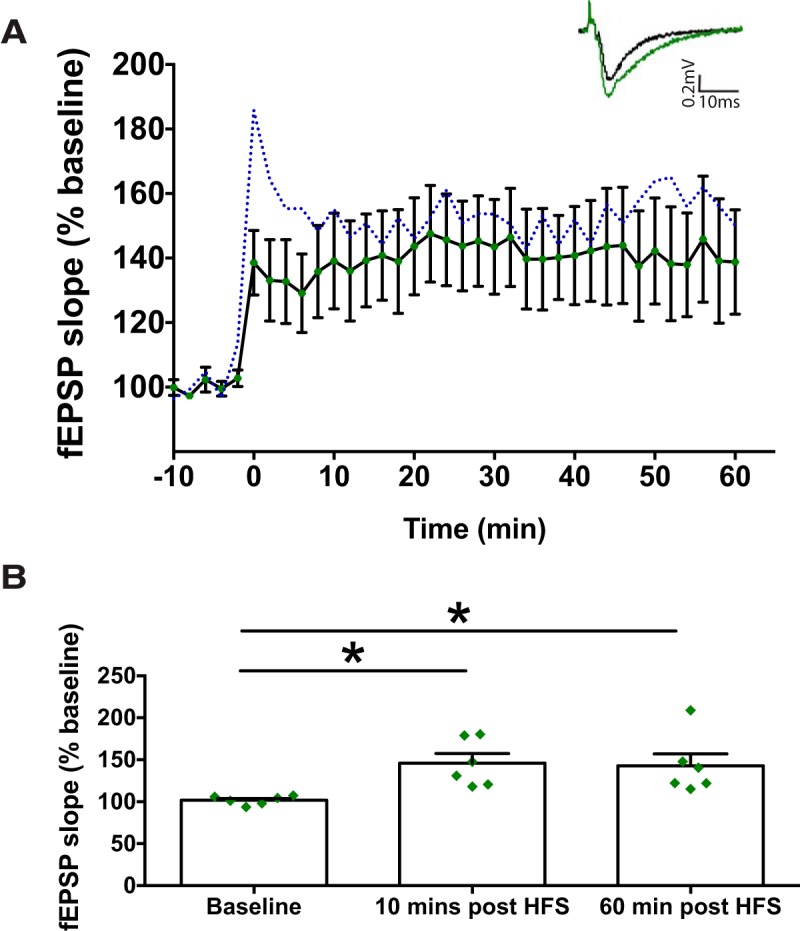
Individuals with sleep apnea often exhibit changes in cognitive behaviors consistent with alterations in the hippocampus. It is hypothesized that adult neurogenesis in the dentate gyrus is an ongoing process that maintains normal hippocampal function in many mammalian species, including humans. However, the impact of chronic intermittent hypoxia (IH), a principal consequence of sleep apnea, on hippocampal adult neurogenesis remains unclear. Using a murine model, we examined the impact of 30 d of IH (IH30) on adult neurogenesis and synaptic plasticity in the dentate gyrus. Although IH30 did not affect paired-pulse facilitation, IH30 suppressed long-term potentiation (LTP). Immunohistochemical experiments also indicate that IH perturbs multiple aspects of adult neurogenesis. IH30 increased the number of proliferating Sox2+ neural progenitor cells in the subgranular zone yet reduced the number of doublecortin-positive neurons. Consistent with these findings, cell lineage tracing revealed that IH30 increased the proportion of radial glial cells in the subgranular zone, yet decreased the proportion of adult-born neurons in the dentate gyrus. While administration of a superoxide anion scavenger during IH did not prevent neural progenitor cell proliferation, it mitigated the IH-dependent suppression of LTP and prevented adult-born neuron loss. These data demonstrate that IH causes both reactive oxygen species-dependent and reactive oxygen species-independent effects on adult neurogenesis and synaptic plasticity in the dentate gyrus. Our findings identify cellular and neurophysiological changes in the hippocampus that may contribute to cognitive and behavioral deficits occurring in sleep apnea.SIGNIFICANCE STATEMENT Individuals with sleep apnea experience periods of intermittent hypoxia (IH) that can negatively impact many aspects of brain function. Neurons are continually generated throughout adulthood to support hippocampal physiology and behavior. This study demonstrates that IH exposure attenuates hippocampal long-term potentiation and reduces adult neurogenesis. Antioxidant treatment mitigates these effects indicating that oxidative signaling caused by IH is a significant factor that impairs synaptic plasticity and reduces adult neurogenesis in the hippocampus.

Keywords: adult neurogenesis; hypoxia.


Figures

Figure 1.
Figure 1.
Prolonged IH exposure attenuates LTP within the dentate gyrus. A, LTP of the fEPSP following HFS in control (blue circles; n = 9 slices, 7 animals), IH10 (yellow triangles; n = 4 slices, 2 animals), IH20 (magenta diamonds; n = 8 slices, 3 animals), and IH30 (red squares; n = 10 slices, 7 animals) illustrate differences in potentiation following HFS. Representative traces of evoked fEPSPs are shown above the graph with baseline (black trace) and post-HFS induction indicated (color traces: control, blue; IH10, yellow; IH20, magenta; IH30, red). Arrows at the bottom indicate the time sampled for B and C. Calibration: 0.2 mV, 10 ms. B, Immediately following HFS, a difference among groups was observed (F(3,27) = 6.667, p = 0.0016). A post hoc Dunnett's test revealed no difference immediately following HFS between control and IH10 groups, yet did in both IH20 and IH30 groups in the fEPSP slope when compared with control. C, Sixty minutes post-HFS, a difference among groups was detected (F(3,27) = 9.529, p = 0.0002). Post hoc Dunnett's test revealed that, while no difference was present between the control and IH10 groups, the fEPSP in both the IH20 and IH30 groups was reduced compared with control group. In a subset of experiments (n = 4 slices, 3 animals), applying to a larger stimulation current during HFS did not to evoke LTP in the IH30 group (B and C, white triangles). *p < 0.05.
Figure 2.
Figure 2.
Prolonged IH exposure does not influence paired-pulse facilitation (PPF). A, Representative traces of evoked fEPSPs during PPF are shown. Calibration: 0.2 mV, 10 ms. B, PPF of the fEPSP was similar between control (blue circles, n = 6 slices; 4 animals) and IH30 (red squares, n = 6 slices, 5 animals) at all six interpulse intervals (IPIs) tested: 40 ms IPI (IPI 40), t(9.990) = 0.904, p = 0.386; IPI 80, t(9.890) = 0.813, p = 0.4352; IPI 200, t(9.963) = 1.169, p = 0.269; IPI 300, t(9.79) = 0.406, p = 0.693; IPI 400, t(8.512) = 0.515, p = 0.619; IPI 500, t(9.997) = 0.556, p = 0.591. C, PPF at IPI 50 was similar before and following high-frequency stimulation for control (blue, n = 6 slices, 3 animals; t(5) = 0.8110, p = 0.4542) and IH30 (red, n = 8 slices, 3 animals; t(7) = 1.777, p = 0.1188). Black-filled symbols represent mean for each group.
Figure 3.
Figure 3.
IH30 increases the number of neural progenitor cells. A, Representative section of dentate gyrus stained for Sox2 (green) and DAPI (blue). SGZ is shown outlined by yellow dotted lines. Scale bar, 100 μm. B, Representative images of Sox2+ labeling in control (top) and IH30 (bottom) animals. SGZ is outlined in yellow. Blue channel depicts DAPI-labeled nuclei on left. Green channel depicts Sox2+ labeling in middle, and a merge is on the right. Scale bars, 100 μm. C, The number of Sox2+ cells in the SGZ increases following IH30 (control, n = 10; IH30, n = 7; t(14.24) = 2.327, p = 0.035). *p < 0.05.
Figure 4.
Figure 4.
IH30 stimulates region-specific SOX2+ cell proliferation in the dentate gyrus. A, Representative section of dentate gyrus stained for Sox2 (green), Ki67 (red), and DAPI (blue). SGZ is outlined by yellow dotted lines. Counts were performed in the ML, GCL, SGZ, and hilus. The yellow arrow indicates a Ki67+/Sox2+ double-positive cell residing within the SGZ. Scale bar, 100 μm. B, Representative images of Ki67 and Sox2+ labeling in control (top) and IH30 (bottom) animals. SGZ is outlined in yellow. Blue channel depicts DAPI-labeled nuclei on left, red channel depicts Ki67+ labeling second from left, green channel depicts Sox2+ labeling second from right, and a merge is on the right. Scale bars, 100 μm. C–F, Quantified proportions of double-labeled Sox2+/Ki67+ cells (control, n = 4; IH30, n = 4) in the SGZ (t(5.993) = 2.747, p = 0.034), hilus (t(4.415) = 4.775, p = 0.0069), GCL (t(3.580) = 2.414, p = 0.0808), and ML (t(5.89) = 0.4592, p = 0.6625). *p < 0.05.
Figure 5.
Figure 5.
IH30 decreases the number of newly born neurons. A, Representative image of DCX+-labeled cells (gray) at low and high magnification. The yellow arrow shows a DCX+ immature neuron that was included in the analysis based on morphology. The red arrowhead points to a DCX+-labeled cell without a process extending into the GCL. Scale bars, 100 μm. B, Representative images of DCX+ labeling in control (top) and IH30 (bottom) animals. Blue channel depicts DAPI-labeled nuclei on left, gray channel depicts DCX+ labeling in middle, and a merge is on the right. Scale bars, 100 μm. C, IH30 reduced the number of DCX+ cells with neuronal morphology exhibiting clear dendritic projections from the dentate gyrus to the molecular layer (control, n = 5; IH30, n = 5; t(7.744) = 2.368, p = 0.046). *p < 0.05.
Figure 6.
Figure 6.
IH30 exposure alters neural progenitor cell fate within the dentate gyrus. A, Representative images of tissue stained for birth-labeled RFP+ cells (red), GFAP (green), DCX (gray), and DAPI (blue) from control (left) and IH30-exposed (right) mice. Scale bars, 100 μm. BF, The proportion of: RFP+/GFAP+-colabeled cells with radial glial morphology were significantly different between groups (control, n = 9; IH30, n = 11; t(15.16) = 2.635, p = 0.0186; B); RFP+ neural progenitor cells in the SGZ were unchanged between groups (control, n = 8; IH30, n = 11; t(12.60) = 0.4915, p = 0.6315; C); RFP+/DCX+-colabeled progenitor cells were unchanged between groups (control, n = 8; IH30, n = 11; t(17.13) = 0.7422, p = 0.4680; D); RFP+/GFAP+-colabeled cells with astrocytic morphology were not significantly different between the two groups (control, n = 8; IH30, n = 11; t(14.68) = 1.267, p = 0.2250; E); RFP+ cells that exhibit neuronal morphology were reduced in IH30 mice (control, n = 8; IH30, n = 11; t(13.97) = 2.730, p = 0.0163; F). Scale bars: B–F, 50 μm. G, Sholl analysis revealed that there were no significant changes in morphology as characterized by the number of intersections in dendritic arborization (control, n = 7; IH30, n = 10; F(30,320) = 0.750, p = 0.828). Representative images of neurons from control (top) and IH30 (bottom) used for analysis on left. Scale bars: 50 μm. *p < 0.05.
Figure 7.
Figure 7.
MnTMPyP administration reveals that neural progenitor cell proliferation is ROS independent. A, Representative images of Ki67 and Sox2+ labeling in controlMnTMPyP (top) and IHMnTMPyP (bottom) animals are shown. Blue channel depicts DAPI-labeled nuclei on left, red channel depicts Ki67+ labeling second from left, green channel depicts Sox2+ labeling second from right, and a merge is on the right. Scale bars, 100 μm. B–E, The proportion of Ki67+/Sox2+-colabeled cells (controlMnTMPyP, n = 5; IHMnTMPyP, n = 6) was increased following IHMnTMPyP in the SGZ (t(6.773) = 3.390, p = 0.0122; B), yet no differences were observed in the hilus (t(8.869) = 1.293, p = 0.2287; C), GCL (t(5.953) = 0.0863, p = 0.9340; D), and ML (t(8.532) = 0.6589, p = 0.5273; E). *p < 0.05.
Figure 8.
Figure 8.
MnTMPyP administration reveals that neuron development is ROS dependent. A, Representative images of DCX+ labeling in controlMnTMPyP (top) and IHMnTMPyP (bottom) animals are shown. Blue channel depicts DAPI-labeled nuclei on left, gray channel depicts DCX+ labeling in middle, and a merge is on the right. Scale bars, 100 μm. B, Immature granule neurons labeled with DCX+ showed no significant difference between IHMnTMPyP and controlMnTMPyP groups (controlMnTMPyP, n = 4; IHMnTMPyP, n = 6; t(7.708) = 2.144, p = 0.066). C, Representative images of RFP+ labeling in controlMnTMPyP (top) and IHMnTMPyP (bottom) animals. Blue channel depicts DAPI-labeled nuclei on left, red channel depicts RFP+ labeling in middle, and a merge is on the right. Scale bars, 100 μm. D, The percentages of RFP+ neurons were not different between IHMnTMPyP and controlMnTMPyP groups (controlMnTMPyP, n = 6; IHMnTMPyP, n = 6; t(7.361) = 0.402, p = 0.699).
Figure 9.
Figure 9.
MnTMPyP administration reveals that LTP is a ROS-dependent process. A, LTP of the fEPSP following HFS showed that synaptic plasticity was maintained in IHMnTMPyP-treated animals. The dashed blue line represents the mean from the control LTP experiments. Representative evoked EPSP traces illustrate pre-HFS (black trace) and post-HFS (green trace) induction. Calibration: 0.2 mV, 10 ms (inset). B, At 10 and 60 min post-HFS, there is a significant increase in EPSP slope when compared with pre-HFS baseline (F(2,10) = 7.627, p = 0.009). *p < 0.05.

Similar articles

Cited by

References

    1. Arendt A, Böttger G, Lehmann J (1983) Loss of neurons in the granular layer of the cerebellum in epilepsy (in German). Zentralblatt fur Allg Pathol u Pathol Anat 128:351–355. - PubMed
    1. Bartesaghi R, Guidi S, Ciani E (2011) Is it possible to improve neurodevelopmental abnormalities in Down syndrome? Rev Neurosci 22:419–455. 10.1515/RNS.2011.037 - DOI - PubMed
    1. Beebe DW, Gozal D (2002) Obstructive sleep apnea and the prefrontal cortex: towards a comprehensive model linking nocturnal upper airway obstruction to daytime cognitive and behavioral deficits. J Sleep Res 11:1–16. 10.1046/j.1365-2869.2002.00289.x - DOI - PubMed
    1. Brazel CY, Limke TL, Osborne JK, Miura T, Cai J, Pevny L, Rao MS (2005) Sox2 expression defines a heterogeneous population of neurosphere-forming cells in the adult murine brain. Aging Cell 4:197–207. 10.1111/j.1474-9726.2005.00158.x - DOI - PubMed
    1. Bruel-Jungerman E, Laroche S, Rampon C (2005) New neurons in the dentate gyrus are involved in the expression of enhanced long-term memory following environmental enrichment. Eur J Neurosci 21:513–521. 10.1111/j.1460-9568.2005.03875.x - DOI - PubMed

Publication types

MeSH terms

LinkOut - more resources