. 2019 Feb 22;294(8):2880-2891.
doi: 10.1074/jbc.RA118.003822. Epub 2018 Dec 26.

A cell-based high-throughput screening method based on a ubiquitin-reference technique for identifying modulators of E3 ligases

Affiliations

A cell-based high-throughput screening method based on a ubiquitin-reference technique for identifying modulators of E3 ligases

Maoyuan Tian et al. J Biol Chem. .

Abstract

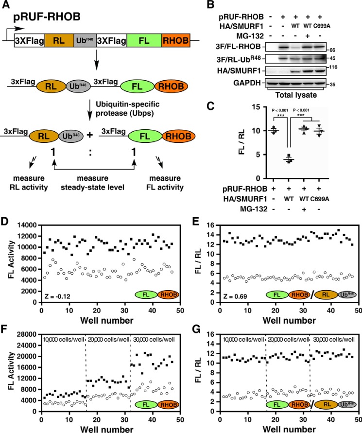
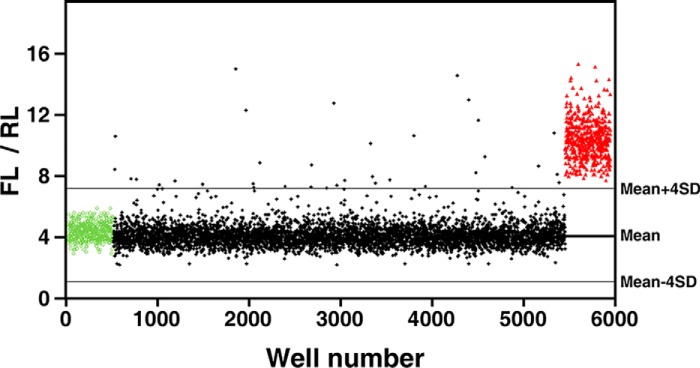
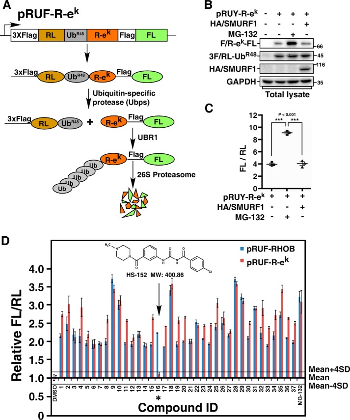
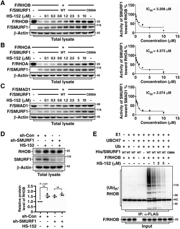
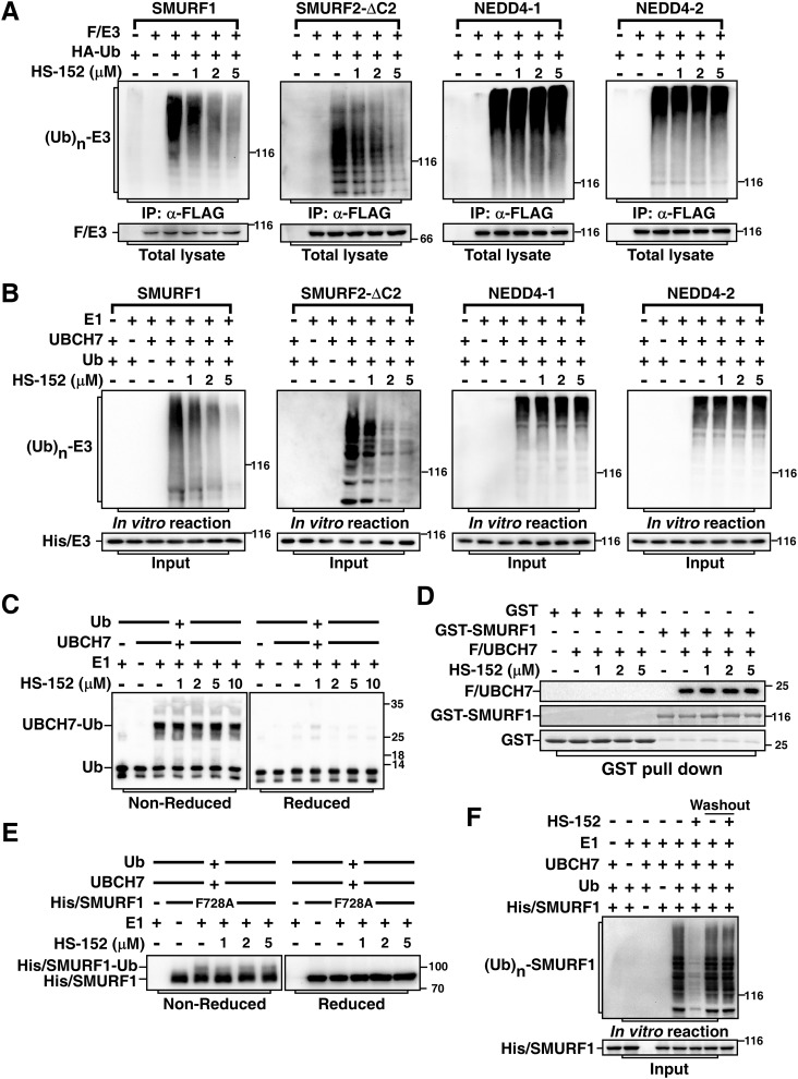
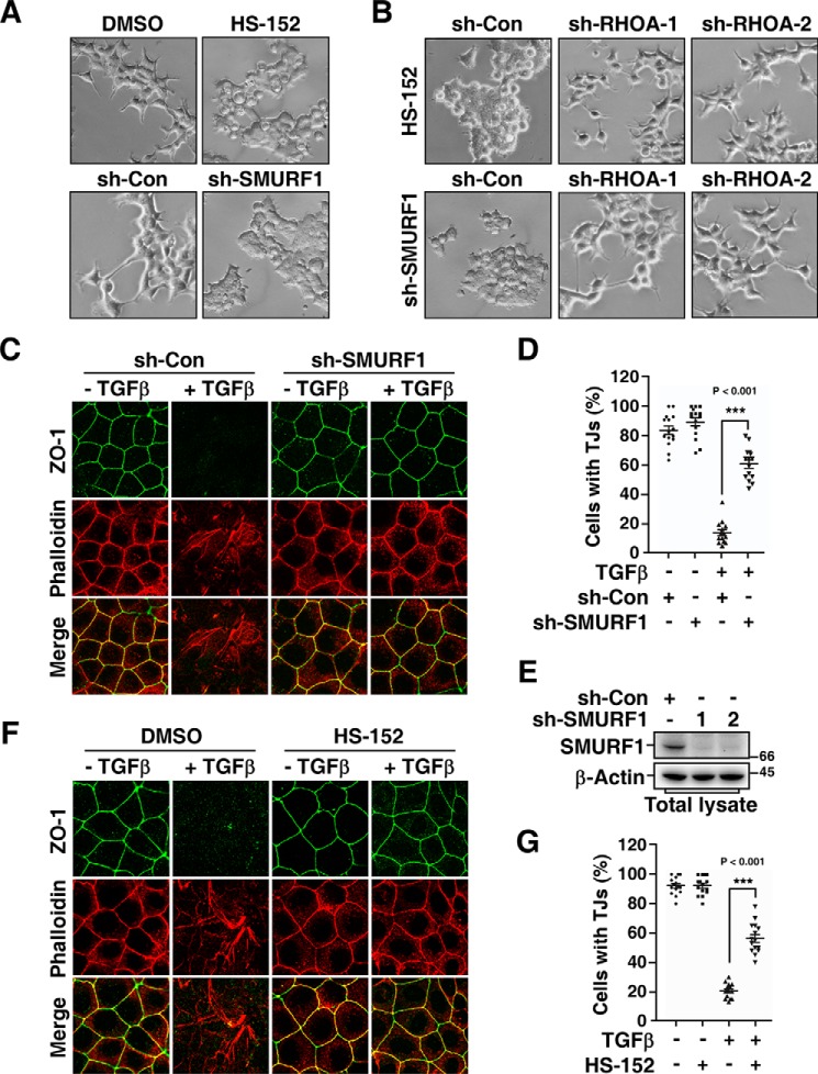
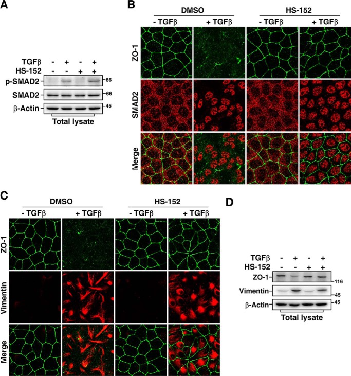
Accumulating evidence indicates that a wide range of E3 ubiquitin ligases are involved in the development of many human diseases. Searching for small-molecule modulators of these E3 ubiquitin ligases is emerging as a promising drug discovery strategy. Here, we report the development of a cell-based high-throughput screening method to identify modulators of E3 ubiquitin ligases by integrating the ubiquitin-reference technique (URT), based on a fusion protein of ubiquitin located between a protein of interest and a reference protein moiety, with a Dual-Luciferase system. Using this method, we screened for small-molecule modulators of SMAD ubiquitin regulatory factor 1 (SMURF1), which belongs to the NEDD4 family of E3 ubiquitin ligases and is an attractive therapeutic target because of its roles in tumorigenesis. Using RAS homolog family member B (RHOB) as a SMURF1 substrate in this screen, we identified a potent SMURF1 inhibitor and confirmed that it also blocks SMURF1-dependent degradation of SMAD family member 1 (SMAD1) and RHOA. An in vitro auto-ubiquitination assay indicated that this compound inhibits both SMURF1 and SMURF2 activities, indicating that it may be an antagonist of the catalytic activity of the HECT domain in SMURF1/2. Moreover, cell functional assays revealed that this compound effectively inhibits protrusive activity in HEK293T cells and blocks transforming growth factor β (TGFβ)-induced epithelial-mesenchymal transition (EMT) in MDCK cells, similar to the effects on these processes caused by SMURF1 loss. In summary, the screening approach presented here may have great practical potential for identifying modulators of E3 ubiquitin ligases.

Keywords: high-throughput screening (HTS); E3 ubiquitin ligase; ubiquitylation (ubiquitination); inhibitor; cellular regulation; protein degradation; epithelial-mesenchymal transition (EMT); auto-ubiquitination; chemical screen; HECT domain; SMAD ubiquitin regulatory factor 1 (SMURF1); ubiquitin-reference technique (URT).


Conflict of interest statement

The authors declare that they have no conflicts of interest with the contents of this article

Figures

Figure 1.
Figure 1.
Cell-based high-throughput URT-Dual-Luciferase Assay system for SMURF1. A, a schematic of the pRUF-RHOB construct. A fusion protein comprised of triple FLAG-tagged RL, UbR48 moiety, triple FLAG-tagged FL, and RHOB is cleaved in vivo by Ubps at the UbR48-RHOB junction to yield equimolars of triple FLAG-tagged RL-UbR48 and triple FLAG-tagged FL-RHOB. The triple FLAG-tagged FL-RHOB is a substrate of SMURF1 and will be degraded in the presence of SMURF1. B, analysis of the steady-state levels of FL-RHOB by immunoblotting. HEK293T cells were transfected with pRUF-RHOB and HA-tagged SMURF1 (HA/SMURF1), WT, or catalytically inactive mutant C699A, as indicated. After overnight treatment with or without 5 μm MG-132, total cell lysates were subjected to immunoblotting using indicated antibodies to determine the steady-state protein levels. C, luciferase assay of FL-RHOB. HEK293T cells were transfected with pRUF-RHOB and HA/SMURF1 WT or C699A, and treated with or without 5 μm MG-132 as in (B) and then applied to Dual-Glo Luciferase Assay to measure the activities of FL and RL. Results were plotted as the ratio of FL activity to RL activity (FL/RL). D and E, evaluation of the screening system. HEK293T cells co-transfected with pRUF-RHOB and WT HA/SMURF1 were treated overnight with DMSO (open circles) and 5 μm MG-132 (filled squares) as negative or positive controls, respectively. Luciferase activities were measured and plotted using either FL activity alone (D) or the FL/RL ratio (E). F and G, the URT system effectively corrects variation of cell numbers. HEK293T cells co-transfected with pRUF-RHOB and WT HA/SMURF1 were seeded with varying number of cells as indicated and treated with DMSO or MG-132 as in (D). Luciferase activities were measured and plotted as FL alone (F) or FL/RL (G).
Figure 2.
Figure 2.
High-throughput screening for SMURF1 inhibitors. HEK293T cells co-transfected with pRUF-RHOB and WT HA/SMURF1 were treated overnight with 10 μm of each compound from the compound library. DMSO (green circles) and 5 μm MG-132 (red triangles) were used as negative or positive controls, respectively. Activities of FL and RL were measured and results plotted as FL/RL. Solid lines represent the mean and the mean ± 4 × S.D. of all assay points excluding MG-132–treated wells.
Figure 3.
Figure 3.
Counterscreen using the N-end rule pathway. A, a schematic of the pRUF–R-ek construct. The pRUF–R-ek is designed similarly to pRUF-RHOB (Fig. 1A) except that triple FLAG-tagged FL-RHOB is replaced by R-ek–FLAG-FL. B, immunoblotting assay of the steady-state levels of R-ek–FL. HEK293T cells were transfected with pRUF–R-ek with or without HA/SMURF1 as indicated. After 3 h treatment with or without 10 μm MG-132, steady-state protein levels were determined by immunoblotting total cell lysates using indicated antibody. C, luciferase assay of R-ek–FL. HEK293T cells were transfected with or without HA/SMURF1 and pRUF–R-ek, and treated with or without MG-132 as in (B). The activities of FL and RL were then measured and plotted as FL/RL. D, counterscreen of selected compounds from the primary screen. HEK293T cells transfected with pRUF–R-ek were treated 6 h with 10 μm of each of the 37 compounds identified in the primary screen. DMSO and MG-132 were used as negative and positive controls, respectively. The effect of each compound on the FL-RHOB/RL or R-ek–FL/RL ratio was plotted relative to the mean of the eight DMSO-treated control wells, respectively. Solid lines represent the mean ± 4 × S.D. of the DMSO controls, as indicated. Arrow and asterisk show the compound that had minimal effect on R-ek–FL/RL ratio.
Figure 4.
Figure 4.
HS-152 inhibits SMURF1-mediated ubiquitination and degradation. A, HS-152 inhibits SMURF1-mediated RHOB degradation in a dose-dependent manner. HEK293T cells were transfected with FLAG-tagged RHOB (F/RHOB) and WT or C699A FLAG-tagged SMURF1 (F/SMURF1) as indicated. After overnight treatment with or without different doses of HS-152, total cell lysates were subjected to immunoblotting using indicated antibodies. The steady-state protein levels were quantified using Image Lab software (Bio-Rad) with β-actin as a loading control. Results were plotted in right panel as the levels of F/RHOB in cells co-transfected with WT F/SMURF1 and F/RHOB at each dose of HS-152 treatment relative to the level of F/RHOB in cells transfected with F/RHOB alone and without HS-152 treatment. B, HS-152 inhibits SMURF1-mediated RHOA degradation in a dose-dependent manner. HEK293T cells transfected with indicated FLAG-tagged RHOA (F/RHOA) and F/SMURF1 (WT or C699A) were treated with different doses of HS-152 and subjected to immunoblotting and then quantified and plotted as in (A). C, HS-152 inhibits SMURF1-mediated SMAD1 degradation in a dose-dependent manner. HEK293T cells transfected with indicated FLAG-tagged SMAD1 (F/SMAD1) and F/SMURF1 (WT or C699A) were treated with a different dose of HS-152 and subjected to immunoblotting and then quantified and plotted as in (A). D, HS-152 up-regulates endogenous RHOB levels through SMURF1. HEK293T cells transfected with control shRNA (sh-Con) or shRNA against SMURF1 (sh-SMURF1) and treated 4 h with or without 2 μm HS-152 and then subjected to immunoblotting assay. The lower panel presents quantitative analysis of Western blotting results (mean ± S.D. of three independent experiments). E, HS-152 inhibits SMURF1-mediated RHOB ubiquitination in vitro. FLAG-tagged RHOB (F/RHOB) and His-tagged SMURF1 (His/SMURF1) expressed and purified from bacteria were subjected to an in vitro ubiquitination assay in the absence or presence of different doses of HS-152 as indicated. The reaction products were then subjected to anti-FLAG immunoprecipitation (IP) followed by immunoblotting assay to detect ubiquitin-conjugated RHOB ((Ub)n-RHOB) using an anti-ubiquitin antibody.
Figure 5.
Figure 5.
HS-152 inhibits catalytic activities of SMURFs. A, HS-152 inhibits auto-ubiquitination of SMURF1 and SMURF2-ΔC2 in cells. HEK293T cells were transfected with HA-tagged ubiquitin (HA/Ub) and FLAG-tagged SMURF1, SMURF2-ΔC2, NEDD4–1, or NEDD4–2 as indicated. The cells were treated 3 h with 40 μm MG-132 and then subjected to anti-FLAG immunoprecipitation (IP) followed by immunoblotting assay to detect ubiquitin-conjugated E3s ((Ub)n-E3) using anti-HA antibody. B, HS-152 inhibits auto-ubiquitination of SMURF1 and SMURF2-ΔC2 in vitro. Purified SMURF1, SMURF2-ΔC2, NEDD4–1, and NEDD4–2 were subjected to an in vitro auto-ubiquitination assay in the absence or presence of HS-152 at the indicated concentrations and ubiquitin-conjugated E3s ((Ub)n-E3) were detected by immunoblotting with anti-ubiquitin antibody. C, HS-152 does not inhibit E2 UBCH7 ubiquitin conjugation. E2 UBCH7 conjugation by E1 enzyme was conducted with varying concentrations of HS-152 as indicated. Reactions were stopped in nonreducing SDS sample buffer and then were applied to immunoblotting under nonreducing condition (left panel), or were reduced using DTT prior to SDS-PAGE (right panel). Ubiquitin and ubiquitin conjugated to UBCH7 via the reductant-sensitive thioester bond are indicated. D, HS-152 does not affect binding of E2 to Smurf1. Purified Smurf1 and UBCH7 were subjected to GST pulldown assay in the absence or presence of indicated amount of HS-152. E, HS-152 does not affect formation of ubiquitin-thioester intermediate in vitro. Purified Smurf1-F728A mutant was subjected to an in vitro auto-ubiquitination assay in the absence or presence of indicated amount HS-152. Reactions were stopped as in (C) to examine the thioester bond formation. F, HS-152 is a reversible inhibitor of SMURF1. Purified SMURF1 was incubated with 5 μm HS-152 for 20 min and then carried out with or without washing three times, as indicated, before subjected to an in vitro auto-ubiquitination assay.
Figure 6.
Figure 6.
HS-152 inhibits cell protrusive activity and TGFβ-induced EMT. A, HS-152 has a similar effect with SMURF1 shRNA on inhibiting protrusive activity of HEK293T cells. Twenty-four h after being transfected with control shRNA (sh-Con) or shRNA against SMURF1 (sh-SMURF1) (lower panel), or overnight after being treated with or without 1 μm HS-152 (upper panel) as indicated, HEK293T cell morphology was imaged by phase contrast microscopy. B, HS-152–caused loss of protrusion depends on RHOA. HEK293T cell morphology was imaged by phase contrast microscopy 24 h after co-transfection with the indicated combination of sh-SMURF1 and sh-Con or shRNA against RHOA (sh-RHOA) (lower panel), or treated another 4 h with DMSO or 1 μm HS-152 20 h after transfection with sh-Con or sh-RHOA (upper panel) as indicated. C and D, knockdown of SMURF1 blocks TGFβ-induced EMT. MDCK cells transduced with lentivirus encoding sh-Con or sh-SMURF1 were treated 20 h with or without 100 pm TGFβ, and then subjected to immunofluorescence assay. ZO-1 staining was detected with an anti-ZO-1 antibody (green), and F-actin was visualized with Texas red-conjugated phalloidin (red) (C). The percentages of cells with tight junctions (TJs) were plotted in (D). Five random areas were counted for each experiment and data of three independent experiments were assessed and represented as mean ± S.D. (D). E, knockdown efficiency of SMURF1. MDCK cells transduced with lentivirus encoding sh-Con or sh-SMURF1 were subjected to immunoblotting assay to examine the knockdown efficiency of SMURF1. F and G, HS-152 inhibits TGFβ-induced EMT. MDCK cells pretreated 12 h with DMSO or 0.05 μm HS-152 were treated another 20 h with or without 100 pm TGFβ and then subjected to immunofluorescence assay (F). Quantification of cells with tight junctions was as in (D).
Figure 7.
Figure 7.
HS-152 does not interfere with TGFβ/SMAD pathway. A, HS-152 does not inhibit TGFβ-induced phosphorylation of SMAD2. MDCK cells pretreated 12 h with DMSO or 0.05 μm HS-152 were treated 20 h with or without 100 pm TGFβ and then subjected to immunoblotting of total cell lysates with indicated antibodies. B, HS-152 does not hinder the nuclear accumulation of SMAD2 in response to TGFβ treatment. MDCK cells pretreated 12 h with or without 0.05 μm HS-152 were treated another 20 h with or without 100 pm TGFβ, and then subjected to immunofluorescence assay. ZO-1 staining was detected with an anti-ZO-1 antibody (green), and SMAD2 was detected with an anti-SMAD2 antibody (red). C, HS-152 does not block TGFβ-induced vimentin expression. MDCK cells treated as in (B) were subjected to immunofluorescence assay to detect ZO-1 (green) and vimentin (red) staining using anti–ZO-1 and anti-vimentin antibodies, respectively. D, HS-152 blocks TGFβ-induced down-regulation of ZO-1 but not up-regulation of vimentin. MDCK cells treated as in (B) were subjected to immunoblotting assay with indicated antibodies.

Similar articles

Cited by

References

    1. Hershko A., and Ciechanover A. (1998) The ubiquitin system. Annu. Rev. Biochem. 67, 425–479 10.1146/annurev.biochem.67.1.425 - DOI - PubMed
    1. Pickart C. M. (2001) Mechanisms underlying ubiquitination. Annu. Rev. Biochem. 70, 503–533 10.1146/annurev.biochem.70.1.503 - DOI - PubMed
    1. Varshavsky A. (2017) The ubiquitin system, autophagy, and regulated protein degradation. Annu. Rev. Biochem. 86, 123–128 10.1146/annurev-biochem-061516-044859 - DOI - PubMed
    1. Zou X., Levy-Cohen G., and Blank M. (2015) Molecular functions of NEDD4 E3 ubiquitin ligases in cancer. Biochim. Biophys. Acta 1856, 91–106 10.1016/j.bbcan.2015.06.005 - DOI - PubMed
    1. Lin X., Liang M., and Feng X. H. (2000) Smurf2 is a ubiquitin E3 ligase mediating proteasome-dependent degradation of Smad2 in transforming growth factor-β signaling. J. Biol. Chem. 275, 36818–36822 10.1074/jbc.C000580200 - DOI - PubMed

Publication types

LinkOut - more resources