. 2019 Mar 1;294(9):3117-3124.
doi: 10.1074/jbc.RA118.005923. Epub 2018 Dec 26.

O-GlcNAcylation regulates integrin-mediated cell adhesion and migration via formation of focal adhesion complexes

Affiliations

O-GlcNAcylation regulates integrin-mediated cell adhesion and migration via formation of focal adhesion complexes

Zhiwei Xu et al. J Biol Chem. .

Abstract

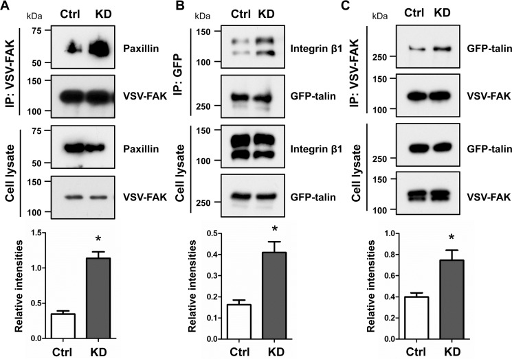
O-GlcNAcylation is a post-translational modification of a protein serine or threonine residue catalyzed by O-GlcNAc transferase (OGT) in the nucleus and cytoplasm. O-GlcNAcylation plays important roles in the cellular signaling that affect the different biological functions of cells, depending upon cell type. However, whether or not O-GlcNAcylation regulates cell adhesion and migration remains unclear. Here, we used the doxycycline-inducible short hairpin RNA (shRNA) system to establish an OGT knockdown (KD) HeLa cell line and found that O-GlcNAcylation is a key regulator for cell adhesion, migration, and focal adhesion (FA) complex formation. The expression levels of OGT and O-GlcNAcylation were remarkably suppressed 24 h after induction of doxycycline. Knockdown of OGT significantly promoted cell adhesion, but it suppressed the cell migration on fibronectin. The immunostaining with paxillin, a marker for FA plaque, clearly showed that the number of FAs was increased in the KD cells compared with that in the control cells. The O-GlcNAcylation levels of paxillin, talin, and focal adhesion kinase were down-regulated in KD cells. Interestingly, the complex formation between integrin β1, focal adhesion kinase, paxillin, and talin was greatly increased in KD cells. Consistently, levels of active integrin β1 were significantly enhanced in KD cells, whereas they were decreased in cells overexpressing OGT. The data suggest a novel regulatory mechanism for O-GlcNAcylation during FA complex formation, which thereby affects integrin activation and integrin-mediated functions such as cell adhesion and migration.

Keywords: O-GlcNAcylation; O-linked N-acetylglucosamine (O-GlcNAc) transferase (OGT); cell adhesion; cell migration; focal adhesions; glycosylation; integrin.


Conflict of interest statement

The authors declare that they have no conflicts of interest with the contents of this article

Figures

Figure 1.
Figure 1.
Knockdown of OGT suppressed O-GlcNAcylation and enhanced cell spreading in HeLa cells. A and B, the expression levels of OGT and O-GlcNAcylation from cell lysates of DOX-controlled OGT KD HeLa cells were verified with concentrations of DOX at 0, 0. 1, 0.5, 1.0, and 5.0 μg/ml for 72 h (A) or at the indicated time with 0.1 μg/ml DOX (B). The control (Ctrl) indicates the cells treated without DOX. Cell lysates from the indicated cells were subjected to Western blotting with the O-GlcNAc (CTD110.6), OGT, and α-tubulin antibodies. C, representative images of cell spreading are shown after incubation for 48 h. Cells were incubated with (KD) or without (Ctrl) 0.1 μg/ml DOX for 24 or 48 h on a normal culture dish, after which the cell areas were measured. Values represent the mean ± S.E. (error bars) (n = 50). **, p < 0.01 (Welch's correction t test). Scale bars, 15 μm. Experiments were independently repeated at least two times.
Figure 2.
Figure 2.
Reduced O-GlcNAcylation promoted cell adhesion and FA formation but decreased cell migration. HeLa cells were cultured in the presence (KD) or absence (Ctrl) of DOX for 24 h. A, 20 min after replating cells onto FN-coated 96-well plates, the attached cells were fixed, and then the nuclei were stained and counted. Representative fields were photographed via fluorescent microscopy. Scale bars, 30 μm. Values represent the mean ± S.E. (error bars) (n = 11). **, p < 0.01 (Welch's correction t test). B, cells were allowed to spread on FN-coated coverslips for 1 h. Cells were then stained with anti-paxillin antibody (green) and TO-PRO-3 (blue). The numbers of focal adhesions were quantified by ImageJ software. Scale bars, 5 μm. Values represent the mean ± S.E. (n = 11). **, p < 0.01 (Welch's correction t test). C, cell motility was observed by time-lapse video microscopy. Values represent the mean ± S.E. (n = 30). **, p < 0.01 (Welch's correction t test). Experiments were independently repeated at least two times.
Figure 3.
Figure 3.
Decreased O-GlcNAcylation levels of paxillin, talin, and FAK in OGT-KD cells. HeLa cells (A) or cells transfected with talin (B) or FAK (C) were incubated without (Ctrl) or with (KD) DOX. The cell extracts were immunoprecipitated (IP) with the indicated antibodies and Western blotted with anti-O-GlcNAc or the indicated antibodies, respectively. Experiments were independently repeated at least three times.
Figure 4.
Figure 4.
Confirmation of O-GlcNAcylation on talin, FAK, and paxillin. Cell lysates of 293T cells transfected with talin (A), FAK (B), or WT HeLa cells (C) were immunoprecipitated (IP) with anti-GFP, anti-VSV, or anti-paxillin antibodies, respectively, followed by click chemistry labeling of O-GlcNAc residues with (+) or without (−) GalT and UDP-GalNAz, and were detected using an ABC kit, as described under “Experimental procedures.” Experiments were independently repeated at least two times.
Figure 5.
Figure 5.
Increased focal adhesion complex formation in OGT KD cells. Cell lysates from the control (Ctrl) and KD HeLa cells that were transfected with expression plasmids of FAK (A), talin (B), or both FAK and talin (C) were immunoprecipitated (IP) by the indicated antibodies and then subjected to Western blotting as described under “Experimental procedures.” The relative ratios are shown at the bottom (n = 3 individual experiments). Values represent the mean ± S.E. (error bars). *, p < 0.05 (Welch's correction t test). Cell lysates were used as input. Experiments were independently repeated at least three times.
Figure 6.
Figure 6.
Comparison of the expression levels of active integrin β1 among the control (Ctrl), KD, and OGT-overexpressing cells. A, a representative immunostaining pattern with anti-active β1 or anti-β1 antibodies in the control, KD, and OGT-overexpressing (OGT) HeLa cells. Cells were cultured on FN-coated coverslips for 1 h and then subjected to immunostaining analyses. The relative fluorescence intensities of KD and OGT-overexpressing cells were compared with the control, and relative fluorescence intensity was 1.0 for the control cells. Scale bar, 5 μm. B, the expression levels of active and total integrin β1 were verified by immunoblotting with the indicated antibodies in control and KD HeLa cells or parent (WT) or transiently OGT-overexpressing HeLa cells. The relative ratios are shown at the bottom (n = 10 random fields of view). Values represent the mean ± S.E. (error bars). *, p < 0.05 (Welch's correction t test); n.s, not significant (p > 0.05). Experiments were independently repeated at least two times.
Figure 7.
Figure 7.
Proposed molecular mechanism for the regulation of cell adhesion and migration by O-GlcNAcylation. During cell adhesion, integrin may form a complex with focal adhesion proteins, such as FAK, talin, and paxillin, to connect with an actin cytoskeleton, which mediates appropriate cell adhesion to promote cell migration. It is well-known that focal adhesion assembly and disassembly processes are fundamental for efficient cell migration. Most focal adhesion proteins can be modified by O-GlcNAc and O-phosphate on serine or threonine residues. In the present study, suppression of the O-GlcNAcylation of paxillin, talin, and FAK aberrantly enhanced integrin activation, integrin-mediated cell adhesion, and complex formation, which in turn led to an inhibition of cell migration, which strongly suggests that OGT functions as a key regulator for cell adhesion.

Similar articles

Cited by

References

    1. Zachara N. E., and Hart G. W. (2006) Cell signaling, the essential role of O-GlcNAc! Biochim. Biophys. Acta 1761, 599–617 10.1016/j.bbalip.2006.04.007 - DOI - PubMed
    1. Vosseller K., Sakabe K., Wells L., and Hart G. W. (2002) Diverse regulation of protein function by O-GlcNAc: a nuclear and cytoplasmic carbohydrate post-translational modification. Curr. Opin. Chem. Biol. 6, 851–857 10.1016/S1367-5931(02)00384-8 - DOI - PubMed
    1. Tan E. P., Duncan F. E., and Slawson C. (2017) The sweet side of the cell cycle. Biochem. Soc. Trans. 45, 313–322 10.1042/BST20160145 - DOI - PMC - PubMed
    1. Singh J. P., Zhang K., Wu J., and Yang X. (2015) O-GlcNAc signaling in cancer metabolism and epigenetics. Cancer Lett. 356, 244–250 10.1016/j.canlet.2014.04.014 - DOI - PMC - PubMed
    1. Itkonen H. M., Minner S., Guldvik I. J., Sandmann M. J., Tsourlakis M. C., Berge V., Svindland A., Schlomm T., and Mills I. G. (2013) O-GlcNAc transferase integrates metabolic pathways to regulate the stability of c-MYC in human prostate cancer cells. Cancer Res. 73, 5277–5287 10.1158/0008-5472.CAN-13-0549 - DOI - PubMed

Publication types

MeSH terms