. 2019 Feb 22;294(8):2651-2664.
doi: 10.1074/jbc.RA118.005861. Epub 2018 Dec 26.

Coupled monoubiquitylation of the co-E3 ligase DCNL1 by Ariadne-RBR E3 ubiquitin ligases promotes cullin-RING ligase complex remodeling

Affiliations

Coupled monoubiquitylation of the co-E3 ligase DCNL1 by Ariadne-RBR E3 ubiquitin ligases promotes cullin-RING ligase complex remodeling

Ian R Kelsall et al. J Biol Chem. .

Abstract

Cullin-RING E3 ubiquitin ligases (CRLs) are large and diverse multisubunit protein complexes that contribute to about one-fifth of ubiquitin-dependent protein turnover in cells. CRLs are activated by the attachment of the ubiquitin-like protein neural precursor cell expressed, developmentally down-regulated 8 (NEDD8) to the cullin subunits. This cullin neddylation is essential for a plethora of CRL-regulated cellular processes and is vital for life. In mammals, neddylation is promoted by the five co-E3 ligases, defective in cullin neddylation 1 domain-containing 1-5 (DCNL1-5); however, their functional regulation within the CRL complex remains elusive. We found here that the ubiquitin-associated (UBA) domain-containing DCNL1 is monoubiquitylated when bound to CRLs and that this monoubiquitylation depends on the CRL-associated Ariadne RBR ligases TRIAD1 (ARIH2) and HHARI (ARIH1) and strictly requires the DCNL1's UBA domain. Reconstitution of DCNL1 monoubiquitylation in vitro revealed that autoubiquitylated TRIAD1 mediates binding to the UBA domain and subsequently promotes a single ubiquitin attachment to DCNL1 in a mechanism previously dubbed coupled monoubiquitylation. Moreover, we provide evidence that DCNL1 monoubiquitylation is required for efficient CRL activity, most likely by remodeling CRLs and their substrate receptors. Collectively, this work identifies DCNL1 as a critical target of Ariadne RBR ligases and coupled monoubiquitylation of DCNL1 as an integrated mechanism that affects CRL activity and client-substrate ubiquitylation at multiple levels.

Keywords: post-translational modification; ubiquitin; ubiquitin ligase; ubiquitylation (ubiquitination); cell signaling; ARIH1; ARIH2; coupled monoubiquitylation; DCNL1; NEDD8 E3 ligase.


Conflict of interest statement

This work was supported by RCUK, Medical Research Council, and Max-Planck-Gesellschaft. The authors declare that they have no conflicts of interest with the contents of this article

Figures

Figure 1.
Figure 1.
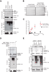
TRIAD1 and HHARI are required for cellular DCNL1 monoubiquitylation. A and B, IP of CUL5 complexes from cells expressing GFP–control, GFP–TRIAD1, or catalytically inactive GFP–TRIAD (C310S) and detection of co-precipitated proteins by immunoblot analysis as indicated. Nonspecific IgG was used as negative control. *, nonspecific band. C, subfractionation of HEK293 cell extracts (Total) into cytosolic high-salt nuclear extract (NEX), and soluble chromatin (CHEX) fractions. Detection of endogenous DCNL1 and CRLs was done by immunoblot as indicated. Histone H2A served as a chromatin marker. D, HA-IP of HEK293 cells expressing HA–DCNL1 or HA-CUL5 were treated with either ubiquitin-deconjugating enzyme USP2 or the deneddylating enzyme NEDP1 followed by anti-HA immunoblot analysis. E, immunoblot analysis of endogenous DCNL1-Ub in cytosolic fractions of cells expressing WT TRIAD1 (WT), catalytically inactive variants TRIAD1 (C310S), TRIAD1 (C310S, R371A, E382A, and E455A), and constitutively ligase-active variant TRIAD1 (R371A, E382A, and E455A). F, immunoblot analysis of endogenous DCNL1-Ub in cytosolic fractions of cells expressing WT HHARI (WT), catalytically inactive HHARI (C357S) or HHARI (C357S, F430A, E431A, and E503A), and constitutive ligase-active HHARI (F430A, E431A, and E503A).
Figure 2.
Figure 2.
TRIAD1 and HHARI monoubiquitylate DCNL1 in vitro. A, reconstitution of DCNL1 monoubiquitylation with purified recombinant TRIAD1, neddylated CUL5–RBX2 (N8–CUL5–RBX2), and UBCH7. Products of complete and drop-out (−) reactions were separated on SDS-PAGE and detected by Coomassie stain as well as immunoblot analysis as indicated. B, quantitative ubiquitylation assay with fluorescein-labeled ubiquitin (Ub5′-IAF) in the absence or presence of N8–CUL5–RBX2. Samples were taken at indicated time points and resolved on SDS-PAGE and scanned at 520 nm to visualize reaction products. C, quantitation of DCNL1-Ub signal from B using ImageJ software. Standard error of the mean is given from two independent replicates. D, DCNL1 monoubiquitylation with purified recombinant WT MBP–TRIAD1 or catalytically dead mutant MBP–TRIAD1 (C310A) with drop-out (−) UBCH7 and DCNL1 controls. Reaction products were separated on SDS-PAGE and detected by Coomassie stain as well as immunoblot analysis as indicated. E, DCNL1 monoubiquitylation reactions with HHARI that lacks the autoinhibitory Ariadne domain HHARI (ΔARI) using UBCH7 or UBCH5c as E2 enzymes including drop-out (−) E2, E3, and DCNL1 controls. Reaction products were separated on SDS-PAGE and detected by Coomassie stain and immunoblot analysis as indicated.
Figure 3.
Figure 3.
Autoubiquitylated TRIAD1 associates with the UBA domain of DCNL1. A, in vitro binding assay to assess binding between DCNL1, UBA-deleted DCNL1 (PONY), and isolated UBA domain and either ubiquitin (Ub)- or NEDD8-agarose beads. Samples of input and beads-bound (Pellet) fraction were analyzed by individual anti-DCNL1 immunoblots. TRIAD1 with NEDD8-binding preference was used as comparison. B, Halo-tagged DCNL1 and mutated DCNL1 comprising a Ub-binding–deficient UBA domain (input visualized by Coomassie-stained SDS-PAGE), were incubated with ubiquitin tetramers (Ub4) of seven different linkage types. Bound fractions of ubiquitin tetramers were detected and shown by silver-stained SDS-PAGE. C and D, cell lysates of mock- or MG132-treated HEK293 cells expressing the indicated GFP-tagged variants of TRIAD1 or HHARI were analyzed by anti-GFP immunoblot analysis. β-Actin served as loading control. E and F, GFP precipitates of HEK293 cells expressing GFP–TRIAD1, GFP-HHARI, or their catalytic Cys to Ser mutant variants were mock-treated and incubated with USP2 or heat-inactivated USP2, followed by anti-TRIAD1 (E) and anti-HHARI (F) immunoblot analysis. G, in vitro MBP-pulldown experiment with mock or autoubiquitylated MBP–TRIAD1 and DCNL1, DCNL1 (PONY), and DAD patch-mutated (DADmut) DCNL1. DCNL1 input was monitored by Coomassie-stained SDS-PAGE, and MBP-pellet samples were analyzed by immunoblot.
Figure 4.
Figure 4.
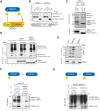
DCNL1 monoubiquitylation depends on UBA domain and is stimulated by autoubiquitylated TRIAD1. A, working model proposing that DCNL1 monoubiquitylation is promoted by an interaction between DCNL1's UBA and autoubiquitylated TRIAD1. B, HA immunoprecipitates of HEK293 cells stably expressing HA-tagged DCNL1 (WT), DCNL1 (DADMUT), or DCNL1 (PONY) were analyzed by immunoblot analysis as indicated. C, in vitro ubiquitylation reaction with purified recombinant MBP–TRIAD1, N8–CUL5–RBX2, His6-ubiquitin, and UBCH7 comparing DCNL1 and DCNL1(PONY) as substrates, including non-UBCH7 control. Reaction products were separated on SDS-PAGE and detected by Coomassie stain as well as immunoblot analysis as indicated. D, purified recombinant TRIAD1, N8–CUL5–RBX2, UBCH7, with either mock, HA-ubiquitin (WT), or mutant HA-ubiquitin (I44A) and with either DCNL1 or DCNL1 (PONY) as substrates were subjected to an ubiquitylation reaction. Reaction products were separated on reducing SDS-PAGE followed by anti-HA immunoblot analysis. Equal input of DCNL1 and DCNL1 (PONY) was verified by Coomassie-stained SDS-PAGE. Monoubiquitylated DCNL1 and DCNL1 (PONY) are indicated by * and #, respectively. E, E3-free DCNL1 ubiquitylation reaction with either UBCH5c or UBCH7, including E2 drop-out control. Reaction products were separated on SDS-PAGE followed by anti-HA immunoblot analysis or Coomassie staining to verify the presence of DCNL1. F and G, composition of ubiquitylation reactions as in D, but the reaction was split into two steps. F, TRIAD1 was either mock or autoubiquitylated with HA-ubiquitin (HA-UbWT) before addition of DCNL1. G, TRIAD1 was autoubiquitylated with either HA-ubiquitin (HA-UbWT) or mutant HA-ubiquitin (HA-UbI44A) before addition of DCNL1.
Figure 5.
Figure 5.
DCNL1 activity is independent of its UBA domain and not altered by monoubiquitylation. Reconstitution of CUL5–RBX2 neddylation with recombinant purified NEDD8-E1, N-terminally acetylated UBE2F, and WT DCNL1 or DCNL1 variants. Side-by-side comparisons of CUL5 neddylation in the absence of DCNL1 (mock) and between DCNL1 and DAD-patch mutant of DCNL1 (A), and DCNL1 (PONY) (B), monoubiquitylated DCNL1-Ub (C), and ubiquitin C-terminally-fused DCNL1-UbCT (D) are shown. CUL5 neddylation was monitored by silver-stained SDS-PAGE and immunoblot analysis with anti-CUL5 antibody. Ratio of N8-CUL5 (in %) was determined from silver-stained SDS-PAGE using ImageJ software. Standard error of the mean is given from at least two independent replications.
Figure 6.
Figure 6.
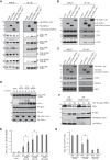
Monoubiquitylated DCNL1 modulates CRL complex composition. A–C, immunoblot analysis of anti-HA-IP and cell lysates (INPUT) from HEK293 cells expressing mock, HA–DCNL1, HA–DCNL1 (PONY), or HA–DCNL1-UbCT was used to monitor co-precipitated cullins (A), CSN subunits (B), as well as CAND1 and CRL substrate receptors (C). D, HEK293 cells expressing HA–DCNL1 or HA–DCNL1-UbCT were either mock-treated or pretreated with MG132 or MLN4924 as indicated, and cell lysates were assessed for Hif1α expression by immunoblot analysis. E, quantitation of Hif1α shown in D using ImageJ software. Standard error of the mean is given from three independent experiments. t tests have been performed for indicated data sets. *, p ≤ 0.05 statistically significant. F, HEK293 cells expressing HA–DCNL1 or HA–DCNL1-UbCT were stimulated with TNFα for indicated times, and cell lysates were prepared and analyzed for the expression of IκBα and phospho-Ser-536–p65 (P-p65) by immunoblotting. G, quantitation of IκBα shown in F using ImageJ software. Standard error of the mean is given from three independent experiments. t tests have been performed for indicated data sets. *, p ≤ 0.05 statistically significant.

Similar articles

Cited by

References

    1. Lydeard J. R., Schulman B. A., and Harper J. W. (2013) Building and remodelling Cullin-RING E3 ubiquitin ligases. EMBO Rep. 14, 1050–1061 10.1038/embor.2013.173 - DOI - PMC - PubMed
    1. Soucy T. A., Smith P. G., Milhollen M. A., Berger A. J., Gavin J. M., Adhikari S., Brownell J. E., Burke K. E., Cardin D. P., Critchley S., Cullis C. A., Doucette A., Garnsey J. J., Gaulin J. L., Gershman R. E., et al. (2009) An inhibitor of NEDD8-activating enzyme as a new approach to treat cancer. Nature 458, 732–736 10.1038/nature07884 - DOI - PubMed
    1. Petroski M. D., and Deshaies R. J. (2005) Function and regulation of cullin-RING ubiquitin ligases. Nat. Rev. Mol. Cell Biol. 6, 9–20 10.1038/nrm1547 - DOI - PubMed
    1. Deshaies R. J., Emberley E. D., and Saha A. (2010) Control of cullin-ring ubiquitin ligase activity by nedd8. Subcell. Biochem. 54, 41–56 10.1007/978-1-4419-6676-6_4 - DOI - PubMed
    1. Scott D. C., Monda J. K., Grace C. R., Duda D. M., Kriwacki R. W., Kurz T., and Schulman B. A. (2010) A dual E3 mechanism for Rub1 ligation to Cdc53. Mol. Cell 39, 784–796 10.1016/j.molcel.2010.08.030 - DOI - PMC - PubMed

Publication types