

Fusion Imaging of Contrast-enhanced Ultrasound With CT or MRI for Kidney Lesions
- PMID: 30587624
- PMCID: PMC6364056
- DOI: 10.21873/invivo.11460
Fusion Imaging of Contrast-enhanced Ultrasound With CT or MRI for Kidney Lesions
Abstract
Aim: To evaluate the feasibility of ultrasound (US) computed tomography (CT) or magnetic resonance imaging (MRI) fusion imaging (FI) for localization and assessment of kidney lesions.
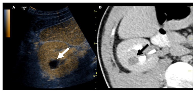
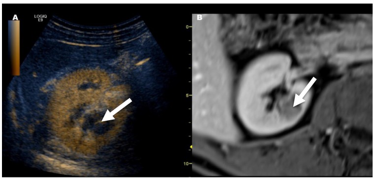
Materials and methods: Twenty-eight patients with kidney lesions previously detected on CT or MRI were included in this retrospective study. All 28 patients with kidney lesions, which were indefinable (42.9%) or hard to localize (57.1%) on gray-scale US alone, underwent FI of US with CT/MRI datasets. In 23 (82%) patients with indeterminate kidney lesions, FI including contrast-enhanced US was conducted.
Results: FI was successfully performed in 25 out of 28 (89.3%) patients. FI with contrast-enhanced US was able to clarify the previously detected kidney lesions in 21 out of 23 patients (91.3%).
Conclusion: FI is a feasible technique for localizing kidney lesions that are hard to define by grayscale US alone and the additional application of contrast-enhanced US is useful in clarifying indeterminate CT or MRI findings.
Keywords: Fusion imaging; computed tomography; contrast-enhanced ultrasound; kidney; magnetic resonance imaging; ultrasound.
Copyright© 2019, International Institute of Anticancer Research (Dr. George J. Delinasios), All rights reserved.
Figures

Similar articles
-
Do Incidental Hyperechoic Renal Lesions Measuring Up to 1 cm Warrant Further Imaging? Outcomes of 161 Lesions.AJR Am J Roentgenol. 2017 Aug;209(2):346-350. doi: 10.2214/AJR.16.17490. Epub 2017 Jun 13. AJR Am J Roentgenol. 2017. PMID: 28609114
-
Multimodality imaging using ultrasound image fusion in renal lesions.Clin Hemorheol Microcirc. 2012;50(1-2):79-89. doi: 10.3233/CH-2011-1445. Clin Hemorheol Microcirc. 2012. PMID: 22538537
-
MRI and contrast enhanced ultrasound (CEUS) image fusion of renal lesions.Clin Hemorheol Microcirc. 2016;64(3):457-466. doi: 10.3233/CH-168116. Clin Hemorheol Microcirc. 2016. PMID: 27886003
-
Imaging in Suspected Renal-Cell Carcinoma: Systematic Review.Clin Genitourin Cancer. 2019 Apr;17(2):e345-e355. doi: 10.1016/j.clgc.2018.07.024. Epub 2018 Aug 11. Clin Genitourin Cancer. 2019. PMID: 30528378
-
Contrast-Enhanced Ultrasound in Renal Imaging and Intervention.Curr Urol Rep. 2019 Oct 17;20(11):73. doi: 10.1007/s11934-019-0936-y. Curr Urol Rep. 2019. PMID: 31624973 Review.
Cited by
-
Local recurrence of renal cell carcinoma successfully treated with fusion imaging-guided percutaneous thermal ablation.Ecancermedicalscience. 2020 Jul 13;14:1070. doi: 10.3332/ecancer.2020.1070. eCollection 2020. Ecancermedicalscience. 2020. PMID: 32728386 Free PMC article.
-
Dynamic Contrast Enhanced-MR CEST Urography: An Emerging Tool in the Diagnosis and Management of Upper Urinary Tract Obstruction.Tomography. 2021 Mar 2;7(1):80-94. doi: 10.3390/tomography7010008. Tomography. 2021. PMID: 33801533 Free PMC article. Review.
-
An overview of non-invasive imaging modalities for diagnosis of solid and cystic renal lesions.Med Biol Eng Comput. 2020 Jan;58(1):1-24. doi: 10.1007/s11517-019-02049-z. Epub 2019 Nov 21. Med Biol Eng Comput. 2020. PMID: 31748942 Review.
-
Artificial Intelligence Algorithm-Based Computed Tomography Image of Both Kidneys in Diagnosis of Renal Dysplasia.Comput Math Methods Med. 2022 Jan 27;2022:5823720. doi: 10.1155/2022/5823720. eCollection 2022. Comput Math Methods Med. 2022. Retraction in: Comput Math Methods Med. 2023 Dec 6;2023:9834943. doi: 10.1155/2023/9834943. PMID: 35126629 Free PMC article. Retracted. Clinical Trial.
-
Artificial Intelligence (AI) Assisted CT/MRI Image Fusion Technique in Preoperative Evaluation of a Pelvic Bone Osteosarcoma.Front Oncol. 2020 Aug 4;10:1209. doi: 10.3389/fonc.2020.01209. eCollection 2020. Front Oncol. 2020. PMID: 32850355 Free PMC article.
References
-
- Helenon O, Correas JM, Balleyguier C, Ghouadni M, Cornud F. Ultrasound of renal tumors. Eur Radiol. 2001;11(10):1890–1901. - PubMed
-
- Helck A, D’Anastasi M, Notohamiprodjo M, Thieme S, Sommer W, Reiser M, Clevert DA. Multimodality imaging using ultrasound image fusion in renal lesions. Clin Hemorheol Microcirc. 2012;50(1-2):79–89. - PubMed
-
- Lee MW, Rhim H, Cha DI, Kim YJ, Choi D, Kim YS, Lim HK. Percutaneous radiofrequency ablation of hepatocellular carcinoma: Fusion imaging guidance for management of lesions with poor conspicuity at conventional sonography. Am J Roentgenol. 2012;198(6):1438–1444. - PubMed
-
- Nakano S, Kousaka J, Fujii K, Yorozuya K, Yoshida M, Mouri Y, Akizuki M, Tetsuka R, Ando T, Fukutomi T, Oshima Y, Kimura J, Ishiguchi T, Arai O. Impact of real-time virtual sonography, a coordinated sonography and MRI system that uses an image fusion technique, on the sonographic evaluation of MRI-detected lesions of the breast in second-look sonography. Breast Cancer Res Treat. 2012;134(3):1179–1188. - PubMed
MeSH terms
Substances
LinkOut - more resources
Full Text Sources
Medical