. 2018 Dec 26;8(1):1.
doi: 10.3390/pathogens8010001.

Identification of Residues in Lassa Virus Glycoprotein Subunit 2 That Are Critical for Protein Function

Affiliations

Identification of Residues in Lassa Virus Glycoprotein Subunit 2 That Are Critical for Protein Function

Katherine A Willard et al. Pathogens. .

Abstract

Lassa virus (LASV) is an Old World arenavirus, endemic to West Africa, capable of causing hemorrhagic fever. Currently, there are no approved vaccines or effective antivirals for LASV. However, thorough understanding of the LASV glycoprotein and entry into host cells could accelerate therapeutic design. LASV entry is a two-step process involving the viral glycoprotein (GP). First, the GP subunit 1 (GP1) binds to the cell surface receptor and the viral particle is engulfed into an endosome. Next, the drop in pH triggers GP rearrangements, which ultimately leads to the GP subunit 2 (GP2) forming a six-helix-bundle (6HB). The process of GP2 forming 6HB fuses the lysosomal membrane with the LASV envelope, allowing the LASV genome to enter the host cell. The aim of this study was to identify residues in GP2 that are crucial for LASV entry. To achieve this, we performed alanine scanning mutagenesis on GP2 residues. We tested these mutant GPs for efficient GP1-GP2 cleavage, cell-to-cell membrane fusion, and transduction into cells expressing α-dystroglycan and secondary LASV receptors. In total, we identified seven GP2 mutants that were cleaved efficiently but were unable to effectively transduce cells: GP-L280A, GP-L285A/I286A, GP-I323A, GP-L394A, GP-I403A, GP-L415A, and GP-R422A. Therefore, the data suggest these residues are critical for GP2 function in LASV entry.

Keywords: Lassa virus; arenavirus; fusion protein; viral entry; viral fusion; viral glycoprotein.


Conflict of interest statement

The authors declare no conflict of interest.

Figures
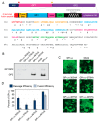
Figure 1
Figure 1
Functional analysis of LASV GP2 HA mutants. (a) Schematic of LASV GPC and amino acid sequence of GP2. The signal peptidase cleaves the SSP (red arrow) whereas SKI/S1P cleaves GP1-GP2 (yellow arrow). The known GP2 domains have been color coded, N-terminal fusion peptide (red); internal fusion loop (orange); heptad repeat 1 (blue); the T-loop (magenta); heptad repeat 2 (green); and the transmembrane domain is in italics (grey). The HA tags were inserted before the amino acids labeled with a ^, * denote charged, and # hydrophobic amino acids examined with alanine scanning. (b) Representative image of surface biotinylation to assess LASV glycoprotein processing to form GP2. GP1GP2 is the uncleaved glycoprotein precursor. LASV GPFLAG was detected with an anti-FLAG antibody, M2, against the C-terminal GP2 3x FLAG tag. (c) Representative images of HA mutants in the cell-to-cell fusion assay. GPFLAG is the parental LASV glycoprotein and mock represents cells transfected with only GFP (no glycoprotein). (d) LASV GP2 HA mutant cleavage and fusion efficiencies compared to parental GPFLAG. Error bars represent the standard error of the mean (SEM) from at least three independent trials.
Figure 2
Figure 2
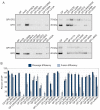
Functional analysis of charged residues in GP2. (a) Cell surface proteins were cross-linked to biotin and purified with streptavidin beads. Purified proteins were separated on SDS-PAGE and probed with an anti-FLAG antibody to detect GP, representative immunoblots are shown. (b) LASV GP2 mutant cleavage and fusion efficiencies compared to parental GPFLAG. Error bars represent the SEM from at least three independent trials.
Figure 3
Figure 3
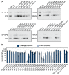
Functional analysis of conserved hydrophobic GP2 mutations. (a) Cell surface proteins were cross-linked to biotin and purified with streptavidin beads. Purified proteins were separated on SDS-PAGE and probed with anti-GP2 antibody (22.5D) representative immunoblots are shown. (b) LASV GP2 mutant cleavage and fusion efficiencies compared to parental GP. Error bars represent the SEM from at least three independent trials.
Figure 4
Figure 4
Transduction efficiencies of parental-like charged (a), parental-like hydrophobic (b), and fusion-defective (c) GP2 mutants. HAP1 and HAP1-ΔDAG1 cells were transduced with VSVΔG-LASV GP constructs encoding GFP. Transduction was quantified using flow cytometry by gating for GFP-positive cells. Transduction efficiency for each construct was normalized to parental LASV GP particle transduction for each cell line. All data are based on the average and standard error of the mean of at least three replicate experiments.
Figure 5
Figure 5
Mapping the fusion-defective charged and hydrophobic mutations on pre-fusion LASV GP2 and post-fusion LCMV homology model. (a) The LASV GP1-GP2 monomeric pre-fusion crystal structure (PDB 5vk2) [29] The GP1 subunit is shown in purple and the GP2 subunit is shown in green. The residues targeted in this study are highlighted, charged residues (red) and hydrophobic (blue). Residues found to be critical for GP2 fusion activity are shown in spheres (L280, L285/I286, I323, L394, I403, and L415) (b) The LCMV (an Old World arenavirus closely-related to LASV) GP2 post-fusion crystal structure (PDB 3mko) [19]. Homologous residues are highlighted as in part (a). (c) Amino acid alignment of LCMV GP2 region crystallized with corresponding region of LASV GP2. Identical residues are indicated with a (*), whereas conservative replacements are indicated by (:). Residues labeled in the structure are in bold-italics and contain a dash above the residues. All structures were rendered with PyMol.

Similar articles

Cited by

References

    1. Maes P., Alkhovsky S.V., Bao Y., Beer M., Birkhead M., Briese T., Buchmeier M.J., Calisher C.H., Charrel R.N., Choi I.R., et al. Taxonomy of the family Arenaviridae and the order Bunyavirales: Update 2018. Arch. Virol. 2018;163:2295–2310. doi: 10.1007/s00705-018-3843-5. - DOI - PubMed
    1. Ogbu O., Ajuluchukwu E., Uneke C.J. Lassa fever in West African sub-region: An overview. J. Vector Borne Dis. 2007;44:1–11. - PubMed
    1. Fichet-Calvet E., Rogers D.J. Risk maps of Lassa fever in West Africa. PLoS Negl. Trop. Dis. 2009;3:e388. doi: 10.1371/journal.pntd.0000388. - DOI - PMC - PubMed
    1. Lassa Fever—Nigeria. [(accessed on 2 July 2018)]; Available online: http://www.who.int/csr/don/20-april-2018-lassa-fever-nigeria/en/
    1. Olayemi A., Cadar D., Magassouba N., Obadare A., Kourouma F., Oyeyiola A., Fasogbon S., Igbokwe J., Rieger T., Bockholt S., et al. New Hosts of The Lassa Virus. Sci. Rep. 2016;6:25280. doi: 10.1038/srep25280. - DOI - PMC - PubMed

LinkOut - more resources