. 2018 Dec 26;20(1):84.
doi: 10.3390/ijms20010084.

The Unique Mechanisms of Cellular Proliferation, Migration and Apoptosis are Regulated through Oocyte Maturational Development-A Complete Transcriptomic and Histochemical Study

Affiliations

The Unique Mechanisms of Cellular Proliferation, Migration and Apoptosis are Regulated through Oocyte Maturational Development-A Complete Transcriptomic and Histochemical Study

Błażej Chermuła et al. Int J Mol Sci. .

Abstract

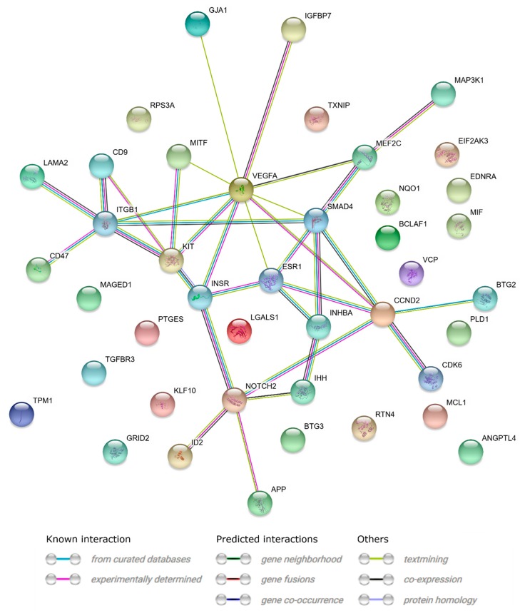
The growth and development of oocyte affect the functional activities of the surrounding somatic cells. These cells are regulated by various types of hormones, proteins, metabolites, and regulatory molecules through gap communication, ultimately leading to the development and maturation of oocytes. The close association between somatic cells and oocytes, which together form the cumulus-oocyte complexes (COCs), and their bi-directional communication are crucial for the acquisition of developmental competences by the oocyte. In this study, oocytes were extracted from the ovaries obtained from crossbred landrace gilts and subjected to in vitro maturation. RNA isolated from those oocytes was used for the subsequent microarray analysis. The data obtained shows, for the first time, variable levels of gene expression (fold changes higher than |2| and adjusted p-value < 0.05) belonging to four ontological groups: regulation of cell proliferation (GO:0042127), regulation of cell migration (GO:0030334), and regulation of programmed cell death (GO:0043067) that can be used together as proliferation, migration or apoptosis markers. We have identified several genes of porcine oocytes (ID2, VEGFA, BTG2, ESR1, CCND2, EDNRA, ANGPTL4, TGFBR3, GJA1, LAMA2, KIT, TPM1, VCP, GRID2, MEF2C, RPS3A, PLD1, BTG3, CD47, MITF), whose expression after in vitro maturation (IVM) is downregulated with different degrees. Our results may be helpful in further elucidating the molecular basis and functional significance of a number of gene markers associated with the processes of migration, proliferation and angiogenesis occurring in COCs.

Keywords: cellular competence; microarray; oocytes; pig.


Conflict of interest statement

The authors declare no conflict of interest.

Figures

Figure 1
Figure 1
Heat map representations of differentially expressed genes belonging to the “cell proliferation”, “regulation of programmed cell death”, and “regulation of cell migration” GO BP (gene ontology Biological Process) terms. Arbitrary signal intensity acquired from microarray analysis is represented by colors (green, higher; red, lower expression). Log2 signal intensity values for any single gene were resized to Row Z-Score scale (from −2, the lowest expression to +2, the highest expression for the single gene, each). Each copy of the analyzed groups represents a different biological sample.
Figure 2
Figure 2
The circle plot showing the differently expressed genes and z-score of “cell proliferation”, “regulation of programmed cell death”, and “regulation of cell migration” GOs. The outer circle shows a scatter plot for each term of the fold change of the assigned genes. Red dots display down-regulation. The inner circle shows the z-score of each GO BP term. The width of each bar corresponds to the number of genes within GO BP term and the color corresponds to the z-score.
Figure 3
Figure 3
The representation of the mutual relationship between differentially expressed genes that belongs to the “cell proliferation”, “regulation of programmed cell death”, and “regulation of cell migration”. The ribbons indicate which gene belongs to which categories. The middle circle represents logarithm from fold change (LogFC) between D7/D1, D15/D1 and D30/D1 respectively. The genes were sorted by LogFC from the most to the least changed gene.
Figure 4
Figure 4
Heatmap showing the gene occurrence between differentially expressed genes that belong to the “cell proliferation”, “regulation of programmed cell death”, and “regulation of cell migration”. The red color is associated with gene occurrence in the GO Term. The intensity of the color is corresponding to the amount of GO BP terms that each gene belongs to.
Figure 5
Figure 5
STRING-generated interaction network between genes that belongs to the “cell proliferation”, “regulation of programmed cell death”, and “regulation of cell migration”. The intensity of the edges reflects the strength of the interaction score.
Figure 6
Figure 6
Functional interaction (FI) between the analyzed downregulated genes that belong to the “cell proliferation”, “regulation of programmed cell death”, and “regulation of cell migration”. In the following figure “->“ stands for activating/catalyzing, “-|” for inhibition, “-” for FIs extracted from complexes or inputs, and “---” for predicted FIs.
Figure 7
Figure 7
Representative sections of pubertal crossbred Landrace gilt ovaries stained with (H) and (E). (AC). primordial and primary follicles, (DF). secondary follicle, (GI). Graafian follicle. Arrows: 1—primary follicle, 2—primordial follicle, 3,7,11—primary oocyte, 4,12,20—zona pellucida, 5,14,24—granulosa cells, 6,15—theca cells, 8—follicular cells, 9—cortical stroma, 10—secondary follicle, 13,23—antrum, 16,25—theca externa, 17—tunica albuginea, 18—Graafian follicle, 19—secondary oocyte, 21—corona radiata, 22—cumulus oophorus.
Figure 8
Figure 8
The results of the RT-qPCR validation of the analyzed genes, presented in a form of bar graph.
Figure 9
Figure 9
Experimental design, presenting procedures and numbers of oocytes in each stage of selection, applied to every biological replicate.

Similar articles

Cited by

References

    1. Hunt P.A., Hassold T.J. Human female meiosis: What makes a good egg go bad? Trends Genet. 2008;24:86–93. doi: 10.1016/j.tig.2007.11.010. - DOI - PubMed
    1. Viswanathan S.R., Mermel C.H., Lu J., Lu C.-W., Golub T.R., Daley G.Q. microRNA Expression during Trophectoderm Specification. PLoS ONE. 2009;4:e6143. doi: 10.1371/journal.pone.0006143. - DOI - PMC - PubMed
    1. Sinha P.B., Tesfaye D., Rings F., Hossien M., Hoelker M., Held E., Neuhoff C., Tholen E., Schellander K., Salilew-Wondim D. MicroRNA-130b is involved in bovine granulosa and cumulus cells function, oocyte maturation and blastocyst formation. J. Ovarian Res. 2017;10:37. doi: 10.1186/s13048-017-0336-1. - DOI - PMC - PubMed
    1. Kempisty B., Ziółkowska A., Ciesiółka S., Piotrowska H., Antosik P., Bukowska D., Nowicki M., Brüssow K.P., Zabel M. Study on connexin gene and protein expression and cellular distribution in relation to real-time proliferation of porcine granulosa cells. J. Biol. Regul. Homeost. Agents. 2014;28:625–635. - PubMed
    1. Diaz F.J., Wigglesworth K., Eppig J.J. Oocytes are required for the preantral granulosa cell to cumulus cell transition in mice. Dev. Biol. 2007;305:300–311. doi: 10.1016/j.ydbio.2007.02.019. - DOI - PMC - PubMed

MeSH terms