. 2018 Dec 26;20(1):85.
doi: 10.3390/ijms20010085.

LT-IIc, A Bacterial Type II Heat-Labile Enterotoxin, Induces Specific Lethality in Triple Negative Breast Cancer Cells by Modulation of Autophagy and Induction of Apoptosis and Necroptosis

Affiliations

LT-IIc, A Bacterial Type II Heat-Labile Enterotoxin, Induces Specific Lethality in Triple Negative Breast Cancer Cells by Modulation of Autophagy and Induction of Apoptosis and Necroptosis

Patricia Masso-Welch et al. Int J Mol Sci. .

Abstract

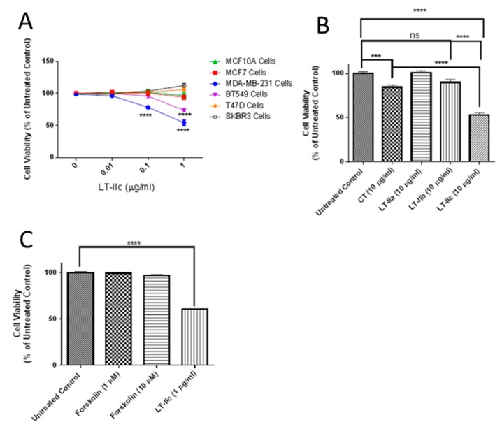
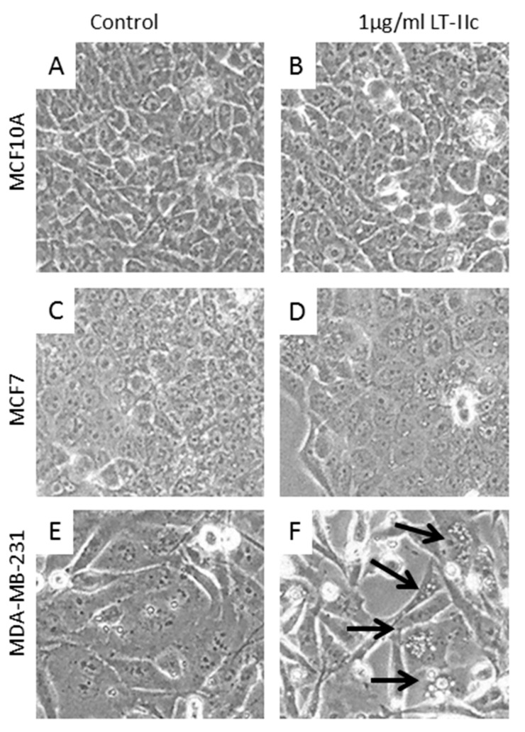
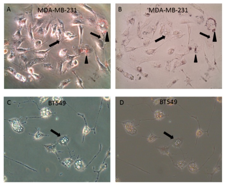
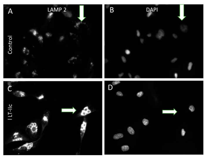
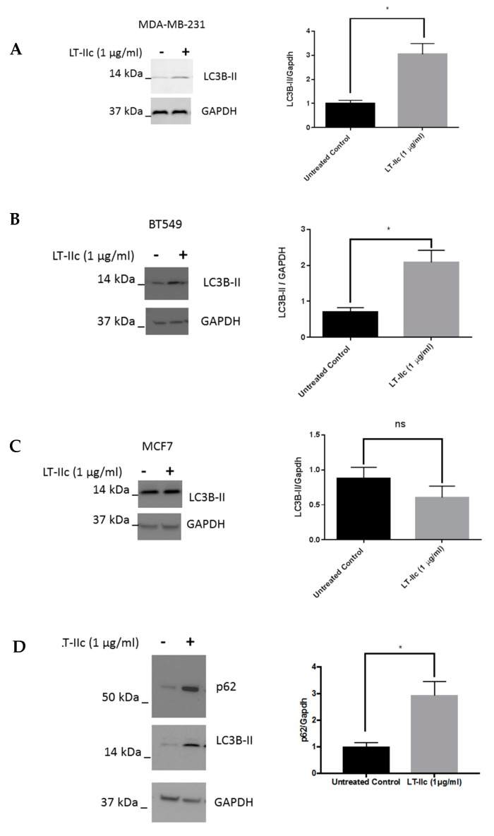
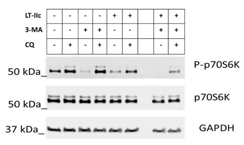
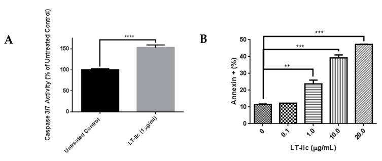
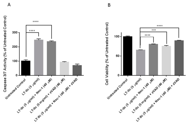
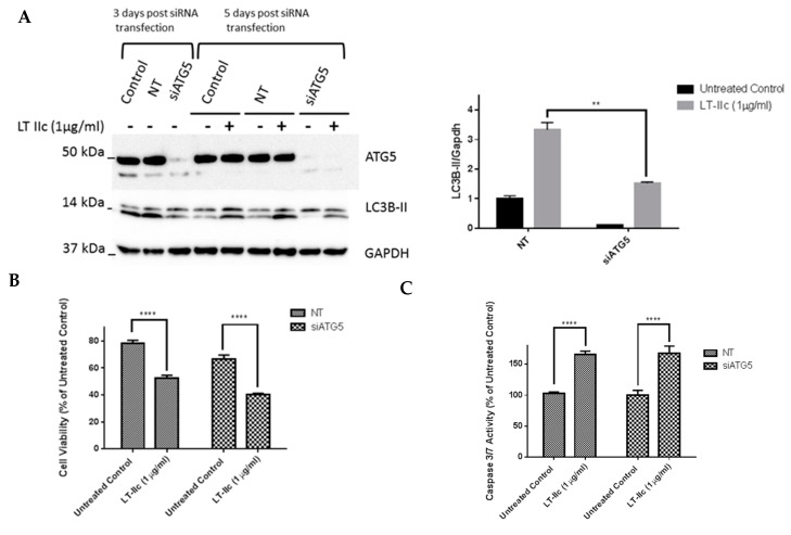
Triple negative breast cancer (TNBC) remains a serious health problem with poor prognosis and limited therapeutic options. To discover novel approaches to treat TNBC, we screened cholera toxin (CT) and the members of the bacterial type II heat-labile enterotoxin family (LT-IIa, LT-IIb, and LT-IIc) for cytotoxicity in TNBC cells. Only LT-IIc significantly reduced viability of the TNBC cell lines BT549 and MDA-MB-231 (IC50 = 82.32 nM). LT-IIc had no significant cytotoxic effect on MCF10A (IC50 = 2600 nM), a non-tumorigenic breast epithelial cell line, and minimal effects on MCF7 and T47D, ER⁺ cells, or SKBR-3 cells, HER2⁺ cells. LT-IIc stimulated autophagy through inhibition of the mTOR pathway, while simultaneously inhibiting autophagic progression, as seen by accumulation of LC3B-II and p62. Morphologically, LT-IIc induced the formation of enlarged LAMP2+ autolysosomes, which was blocked by co-treatment with bafilomycin A1. LT-IIc induced apoptosis as demonstrated by the increase in caspase 3/7 activity and Annexin V staining. Co-treatment with necrostatin-1, however, demonstrated that the lethal response of LT-IIc is elicited, in part, by concomitant induction of necroptosis. Knockdown of ATG-5 failed to rescue LT-IIc-induced cytotoxicity, suggesting LT-IIc can exert its cytotoxic effects downstream or independently of autophagophore initiation. Collectively, these experiments demonstrate that LT-IIc acts bifunctionally, inducing autophagy, while simultaneously blocking autolysosomal progression in TNBC cells, inducing a specific cytotoxicity in this breast cancer subtype.

Keywords: LT-IIc; apoptosis; autophagy; bacterial enterotoxins; breast cancer; heat-labile enterotoxins; necroptosis; triple-negative breast cancer.


Conflict of interest statement

The authors declare no conflict of interest.

Figures

Figure 1
Figure 1
Effects of LT-IIc on breast cancer cell viability and morphology. (A) LT-IIc was specifically toxic to BT549 and MDA-MB-231 TNBC cell lines, but not MCF10A, MCF7, or SK-BR3 cells. All cell lines were treated with 0, 0.01, 0.1, 1, or 10 µg/mL LT-IIc for 48 h, followed by MTT assay. Data points represents the means ± SEM of three independent experiments. (B) LT-IIc, CT, LT-IIa, and LT-IIb (10 µg/mL) were tested for durable cytotoxic effects by pulsing MDA-MB-231 breast cancer cells for 24 h, followed by a wash-out period of an additional 24 h. Cytotoxicity, assessed using MTT, showed the greatest effect in cells treated with LT-IIc. Bars represent mean ± SEM from two independent experiments with eight replicates each. (C) The cytotoxic effects of LT-IIc enterotoxin were not mimicked by forskolin, an activator of adenylate cyclase. MDA-MB-231 cells were treated with LT-IIc (1 µg/mL) or forskolin (1 or 10 µM) for 48 h, followed by assessment of cell viability using MTT assay. The data represent the mean ± SEM of three replicates. Key: *** p < 0.001; **** p < 0.0001. ns (non-significant).
Figure 2
Figure 2
LT-IIc did not induce morphologic change in the immortalized human breast epithelial MCF10A (A,B), or ER positive MCF7 human breast cancer cell lines (C,D). LT-IIc induced extensive intracellular vacuolation (arrows) in MDA-MB-231 (E,F). All cells were treated with 1 µg/mL LT-IIc for 24 h. Cell images were obtained using a 40× objective under phase contrast illumination.
Figure 3
Figure 3
LT-IIc-induced vacuolation is not due to intracellular lipid accumulation. (A) Oil Red O staining revealed perinuclear pool of small lipid droplets (arrowheads) in all MDA-MB-231 cells treated with 1 µg/mL LT-IIc for 24 h, viewed under phase contrast (A) or bright field, after staining with Oil Red O (B). This small perinuclear lipid pool was independent of the large vacuoles (arrows). BT549 treated with 1 µg/mL LT-IIc lacked perinuclear lipid droplets seen in MDA-MB-231 cells. Large intracellular vacuoles were Oil Red O negative (arrows). (C,D) BT549 cells lacked perinuclear lipid droplets seen in MDA-MB-231 cells. Large intracellular vacuoles were oil red O negative. All images were obtained using 40× objective magnification.
Figure 4
Figure 4
Enlarged intracellular vacuoles induced by LT-IIc are positive for LAMP-2. (A,B): Untreated MDA-MB-231 cells display punctate cytoplasmic staining for small lysosomes expressing LAMP-2 (A, arrow); DAPI staining for nuclei in same field (B). (C,D): LT-IIc-treated cells at 6 h possess multiple enlarged LAMP-2-stained bodies (C, arrow); DAPI staining of the same field (D). All images were taken under a 40× objective, using identical manual exposure times for control versus treated cells.
Figure 5
Figure 5
The effect of LT-IIc on autophagy in breast cancer cells. MDA-MB-231, BT549, and MCF7 cells were treated with 1 µg/mL LT-IIc for 24 h, prior to generating lysates for Western blotting. (A) Representative Western blot for LC3B-II with GAPDH as a loading control is shown (N = 3 independent experiments) for MDA-MB-231 (A), BT549 (B), and MCF7 cells (C). (D) Lysates from MDA-MB-231 cells treated ±1 µg/mL LT-IIc were analyzed for the expression of p62 protein levels by Western blotting, using GAPDH as a loading control. Representative blot of three independent experiments. Quantitation of blots was performed using Image J. Error bars represent SEM. Key: * p < 0.05.
Figure 6
Figure 6
Effects of bafilomycin A1 on LT-IIc efficacy in MDA-MB-231 cells. (A) MDA-MB-231 cells were treated with LT-IIc (1 µg/mL) in the presence or absence of bafilomycin A1 (10 nM) for 48 h. (B) The level of expression of LC3B-II was analyzed using Western blotting and the blots were quantified using ImageJ software. (C) MDA-MB-231 cells were treated with LT-IIc (1 µg/mL) in the presence or absence of bafilomycin A1 (10 nM) for 24 h and cell morphology was evaluated using microscopic analysis (10× magnification). Arrows indicate vacuoles observed in LT-IIc treated cells not exposed to bafilomycin A1. (D) Bafilomycin A1 and LT-IIc showed similar effects on MDA-MB-231 cytotoxicity (measured by MTT assay). Co-treatment significantly enhanced cytotoxicity (compared to LT-IIc plus DMSO control). All panels represent at least three independent replicates from three independent experiments. Bars represent means ± standard error of the mean. ** p < 0.01; *** p < 0.0001; **** p < 0.00001.
Figure 7
Figure 7
Effect of LT-IIc on p70S6K phosphorylation. MDA-MB-231 cells were treated with LT-IIc (5 µg/mL) ± 25 µM chloroquine (CQ) and/or 3-methyladinine (3-MA) (10 mM) as indicated. After 24 h treatment, cell lysates were harvested and analyzed by immunoblotting. Representative blot of three independent experiments.
Figure 8
Figure 8
LT-IIc induces apoptotic cell death in TNBC cells. (A) MDA-MB-231 cells were treated with LT-IIc (1 µg/mL) prior to assessment of caspase 3/7 activity using a fluorescent substrate. Representative of >3 replicates from 3 independent experiments. (B) MDA-MB-231 cells were treated with 0, 0.1, 1.0, 10, or 20 µg/mL of LT-IIc for 24 h. Annexin V-stained cells were interrogated by flow cytometry within 1 h of staining. Bars represent means ± SEM. Key: ** p < 0.01; *** p < 0.001; **** p < 0.0001.
Figure 9
Figure 9
LT-IIc induces cell death through combined induction of apoptosis and necroptosis. MDA-MB-231 cells were treated with LT-IIc (5 µg/mL) in the presence or absence of Z-VAD-FMK (40 µM) and/or necrostatin-1 (40 µM) for 48 h. (A) Following treatment, caspase 3/7 activity was assessed using Apo-ONE® Homogeneous Caspase-3/7 assay. (B) Cell viability was determined using MTT assay. Bars represent means ± SEM. Key: *** p < 0.001; **** p < 0.0001.
Figure 10
Figure 10
ATG5 gene knockdown using siRNA does not reverse LT-IIc effect on cytotoxicity or LC3B-II levels in MDA-MB-231 cells. (A) MDA-MB-231 cells were cultured in six-well plates prior to transfection with either ATG5 specific siRNA or a negative control NT siRNA. Three days post-transfection, ATG5 knockdown was confirmed using Western blotting. At day 5, another set of cells was treated with LT-IIc for 24 h to assess the requirement for ATG5 on LT-IIc-mediated increase in LC3B expression. The blots were quantified using ImageJ software. (B) Following the confirmation of ATG5 knockdown, the cells were cultured in 96-well plates prior to treatment with 1 µg/mL LT-IIc for 48 h. Cell viability was assessed using MTT assay, as described previously. (C) Following the confirmation of ATG5 knockdown, the cells were cultured in 96-well plates prior to treatment with 1 µg/mL LT-IIc for 24 h. Caspase 3/7 activity was determined using the fluorescent apo-One homogenous caspase 3/7 activity assay, as described above. All experiments are representative of at least two independent experiments. Error bars represent SEM. Key: ** p < 0.01, **** p < 0.0001.

References

    1. Bianchini G., Balko J.M., Mayer I.A., Sanders M.E., Gianni L. Triple-negative breast cancer: Challenges and opportunities of a heterogeneous disease. Nat. Rev. Clin. Oncol. 2016;13:674–690. doi: 10.1038/nrclinonc.2016.66. - DOI - PMC - PubMed
    1. Kalimutho M., Parsons K., Mittal D., Lopez J.A., Srihari S., Khanna K.K. Targeted Therapies for Triple-Negative Breast Cancer: Combating a Stubborn Disease. Trends Pharmacol. Sci. 2015;36:822–846. doi: 10.1016/j.tips.2015.08.009. - DOI - PubMed
    1. Felgner S., Kocijancic D., Frahm M., Weiss S. Bacteria in cancer therapy: Renaissance of an old concept. Int. J. Microbiol. 2016;2016:8451728. doi: 10.1155/2016/8451728. - DOI - PMC - PubMed
    1. Havas H.F., Donnelly A.J. Mixed bacterial toxins in the treatment of tumors. IV. Response of methylcholanthrene-induced, spontaneous, and transplanted tumors in mice. Cancer Res. 1961;21:17–25. - PubMed
    1. Wilde L., Tanson K., Curry J., Martinez-Outschoorn U. Autophagy in cancer: A complex relationship. Biochem. J. 2018;475:1939–1954. doi: 10.1042/BCJ20170847. - DOI - PubMed

MeSH terms