

Investigation of intraocular pressure fluctuation as a risk factor of glaucoma progression
- PMID: 30587914
- PMCID: PMC6302802
- DOI: 10.2147/OPTH.S186526
Investigation of intraocular pressure fluctuation as a risk factor of glaucoma progression
Abstract
Purpose: Since the role of short- and long-term intraocular pressure (IOP) fluctuation as a predictor of glaucoma progression is still controversial, the purpose of this study was to investigate the role of IOP fluctuation in a non-selected patient cohort.
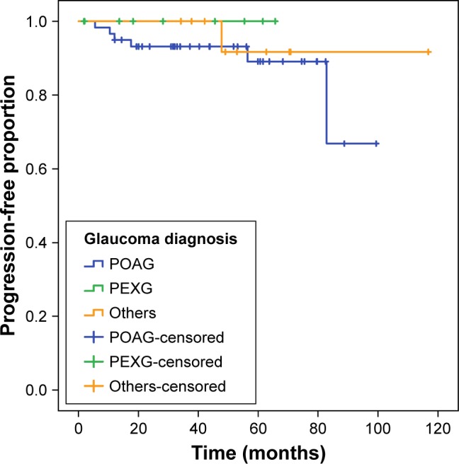
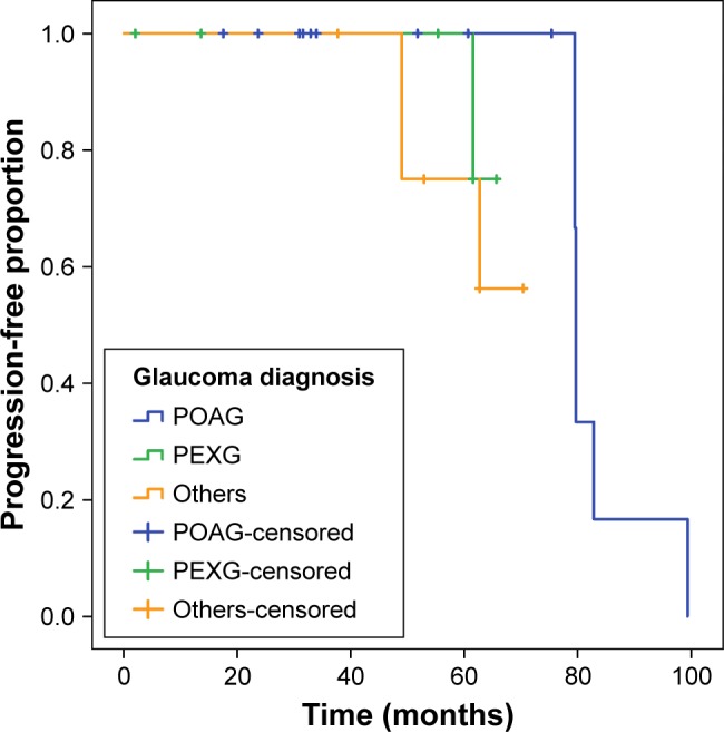
Materials and methods: Two-hundred and forty eyes of 120 glaucoma patients (51% female) with a mean age of 64.5 years were included. Inclusion criteria were at least a visual field (VF) and a 48-hour diurnal phasing of IOP including nocturnal measurement. Glaucoma progression was defined as - if available - confirmed progression of reproducible VF defects in at least three VF examinations or increase of cup area on optic nerve imaging (Heidelberg Retina Tomograph [HRT]) with at least two images after baseline. If results were stable or less than previously mentioned VF or HRT examinations were available, it was classified as "no progression".
Results: Glaucoma progression was seen in seven of 240 eyes in the VF analysis and ten of 240 eyes on HRT. Of all 240 eyes, 92 and 41 eyes fulfilled the criteria to be included for progression evaluation on VF and HRT analysis, respectively. Mean time to progression ± standard error was 3.6±0.2 years on VF and 4.5±0.3 years on HRT. Univariate and multivariate Cox regression analyses revealed short-term IOP fluctuation (P<0.0001) and maximum IOP (P<0.001) as risk factors for glaucoma progression on VF. There was no significant influence of demographic characteristics, ocular or general health on glaucoma progression.
Conclusion: Short-term IOP fluctuation was associated with the progression of glaucoma in this non-selected cohort of glaucoma patients receiving phasing of IOP.
Keywords: glaucoma imaging; glaucoma progression; long-term IOP fluctuation; short-term IOP fluctuation; visual field.
Conflict of interest statement
Disclosure The authors report no conflicts of interest in this work.
Figures
References
-
- Leske MC, Heijl A, Hyman L, Bengtsson B. Early Manifest Glaucoma Trial: design and baseline data. Ophthalmology. 1999;106(11):2144–2153. - PubMed
-
- Oddone F, Centofanti M, Rossetti L, et al. Exploring the Heidelberg Retinal Tomograph 3 diagnostic accuracy across disc sizes and glaucoma stages: a multicenter study. Ophthalmology. 2008;115(8):1358–1365.e1. - PubMed
-
- Wollstein G, Garway-Heath DF, Fontana L, Hitchings RA. Identifying early glaucomatous changes. Comparison between expert clinical assessment of optic disc photographs and confocal scanning ophthalmoscopy. Ophthalmology. 2000;107(12):2272–2277. - PubMed
-
- The Advanced Glaucoma Intervention Study (AGIS): 7 The relationship between control of intraocular pressure and visual field deterioration. The AGIS Investigators. Am J Ophthalmol. 2000;130(4):429–440. - PubMed
LinkOut - more resources
Full Text Sources
