. 2018 Dec 13:12:4253-4260.
doi: 10.2147/DDDT.S188095. eCollection 2018.

MMI-0100 ameliorates lung inflammation in a mouse model of acute respiratory distress syndrome by reducing endothelial expression of ICAM-1

Affiliations

MMI-0100 ameliorates lung inflammation in a mouse model of acute respiratory distress syndrome by reducing endothelial expression of ICAM-1

Binchan He et al. Drug Des Devel Ther. .

Abstract

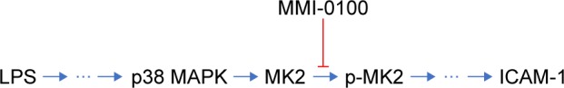
Purpose: ICAM-1 plays a critical role in the development of acute respiratory distress syndrome (ARDS). MK2 regulates the expression of ICAM-1 in human pulmonary microvascular endothelial cells. To explore whether the inhibition of MK2 activation has the same effect in experimental animals, MMI-0100, a peptide-mediated inhibitor of MK2, was used to verify whether MMI-0100 can ameliorate lung inflammation in a mouse model of ARDS by reducing endothelial expression of ICAM-1.

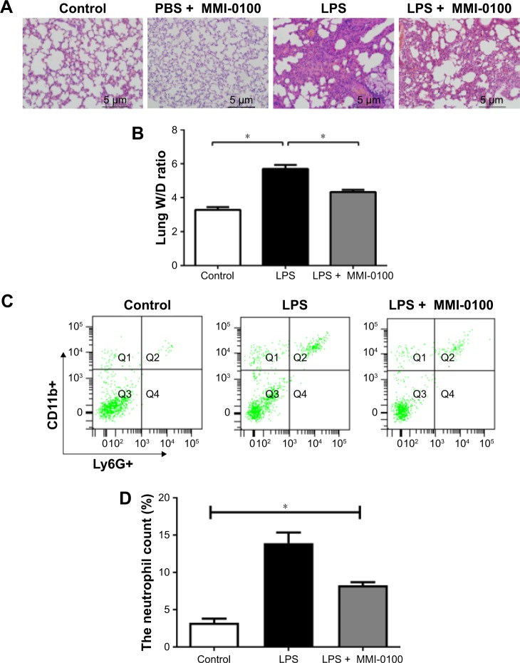
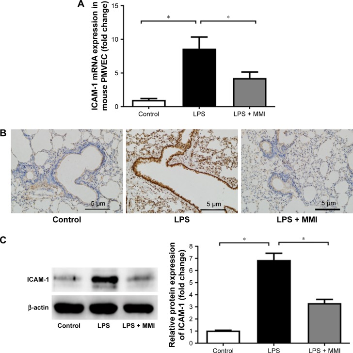
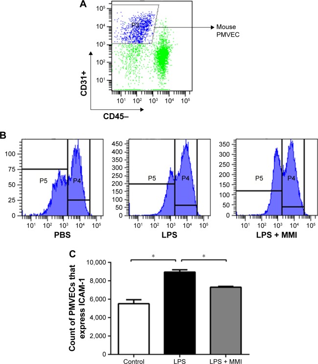
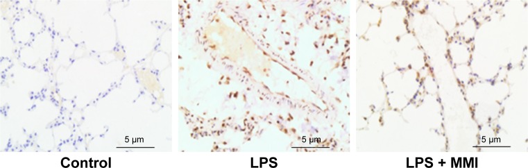
Methods: In this study, C57BL/6 mice were randomly divided into three groups: a control group, an lipopolysaccharides (LPS) group, and an LPS plus MMI-0100 group. Mice were killed 24 hours after the administration of LPS and MMI-0100. The mouse lung tissue histopathology, wet/dry weight ratio (W/D), and the neutrophil count were used to measure the severity of lung inflammation in mice. The pulmonary microvascular endothelial cells (PMVECs) of the mice were isolated. The mRNA expression of ICAM-1 in mouse PMVECs was determined using RT-PCR, and the protein expression of MK2 and ICAM-1 in mouse PMVECs was analyzed using Western blotting and immunohistochemistry.

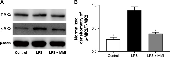
Results: We found that the level of phosphorylated MK2 in the LPS plus MMI-0100 group was reduced. Compared with the LPS group, the LPS plus MMI-0100 group of mice showed less severe inflammation, including a lower W/D and neutrophil count. The mRNA and protein expression of ICAM-1 in the LPS group was significantly higher than in the control group in mouse PMVECs, and the ICAM-1 level was reduced after the administration of MMI-0100.

Conclusion: These data indicate that MMI-0100 ameliorates lung inflammation in a mouse model of ARDS by reducing endothelial expression of ICAM-1.

Keywords: acute respiratory distress syndrome; inflammation; pulmonary microvascular endothelial cell.


Conflict of interest statement

Disclosure The authors report no conflicts of interest in this work.

Figures

Figure 1
Figure 1
The damage caused by LPS in the mouse lungs. Notes: The mice were killed 24 hours after administration of LPS or PBS. (A) The upper lobes of the right lung of each mouse were harvested, and the degree of acute lung injury was assessed by H&E staining (×200). (B) The middle lobes of the right lung were collected, weighed quickly to record the wet weight, and then dried in an oven at 85°C for 24 hours to be able to record the dry weight. The W/Ds were calculated. (C, D) The number of neutrophils in each mouse lung was determined using a flow cytometer counting cells stained with antibodies against CD11b+ and Ly6G+. The values presented are the mean ± SEM (n=15 in each group). Comparisons were made by one-way ANOVA, and the results were representative of three independent experiments. *P<0.05. Abbreviations: LPS, lipopolysaccharides; W/Ds, wet/dry weight ratios.
Figure 2
Figure 2
ICAM-1 expression in mice. Notes: After euthanasia, the lung cells were collected and stained with CD45-PE-labeled and CD31-APC-labeled antibodies for 30 minutes at 25°C. The mouse lung microvascular endothelial cells were collected using CD45- and CD31+ immunomagnetic beads. (A) mRNA expression of ICAM-1 in PMVECs was increased significantly in the LPS group. The values presented are mean ± SEM. (n=15 in each group). Comparisons were made by one-way ANOVA, *P<0.05. (B) Immunohistochemistry (200×) revealed that ICAM-1 protein in LPS-stimulated mouse lung tissues was higher than in the control group or the LPS plus MMI-0100 group. (C) ICAM-1 protein level in mouse PMVECs was measured by Western blotting (left); quantitative densitometry of ICAM-1 protein level in mouse PMVECs (right). The values presented are the mean ± SEM. The results are representative of three independent experiments (*P<0.05). Abbreviations: LPS, lipopolysaccharides; PMVECs, pulmonary microvascular endothelial cells.
Figure 3
Figure 3
The expression of ICAM-1 on the surface of mouse PMVECs. Notes: The mice were euthanized 24 hours after administration of LPS or PBS, and then the lung cells were collected. (A) PMVECs were labeled with the antibodies CD31+ and CD45-, and then the expression was assessed using a flow cytometer. (B) ICAM-1 was labeled with the antibody against CD54+. (C) The expression of ICAM-1 on the membranes of mouse PMVECs was higher in the LPS group than in the other groups. The values presented are the mean ± SEM (n=15 in each group). Comparisons were made by one-way ANOVA, and the results are representative of three independent experiments (*P<0.05). Abbreviations: LPS, lipopolysaccharides; PMVECs, pulmonary microvascular endothelial cells.
Figure 4
Figure 4
Protein expression of MK2 in mice. After euthanasia, the mouse PMVECs were collected. Notes: (A) The levels of protein expression of total MK2 (T-MK2) and phosphorylated MK2 (p-MK2) in mouse PMVECs were measured by Western blotting. (B) Quantitative densitometry of p-MK2/T-MK2. The values presented are mean ± SEM. The results were representative of three independent experiments (*P<0.05). Abbreviations: LPS, lipopolysaccharides; PMVECs, pulmonary microvascular endothelial cells.
Figure 5
Figure 5
Expression of p-MK2 in mouse lung. Notes: Immunohistochemistry of mouse lung sections with anti-p-MK2 antibody revealed less expression of p-MK2 in the LPS + MMI group than in the LPS group. Magnification ×200. Abbreviations: LPS, lipopolysaccharides; p-MK2, phosphorylated MK2.
Figure 6
Figure 6
A simplified model of the molecular pathway. Notes: Upon stimulation by LPS, the p38 MAPK pathway is activated, and this is followed by the activation of MK2. MMI-0100 is able to inhibit the activation of p-MK2 and reduce the expression of ICAM-1. Abbreviations: LPS, lipopolysaccharides; p-MK2, phosphorylated MK2.

Similar articles

Cited by

References

    1. Rubenfeld GD, Caldwell E, Peabody E, et al. Incidence and outcomes of acute lung injury. N Engl J Med. 2005;353(16):1685–1693. - PubMed
    1. Chen W, Chen YY, Tsai CF, et al. Incidence and outcomes of acute respiratory distress syndrome: a nationwide registry-based study in Taiwan, 1997 to 2011. Medicine. 2015;94(43):e1849. - PMC - PubMed
    1. Montefort S, Roche WR, Howarth PH, et al. Intercellular adhesion molecule-1 (ICAM-1) and endothelial leucocyte adhesion molecule-1 (ELAM-1) expression in the bronchial mucosa of normal and asthmatic subjects. Eur Respir J. 1992;5(7):815–823. - PubMed
    1. Sumagin R, Lomakina E, Sarelius IH. Leukocyte-endothelial cell interactions are linked to vascular permeability via ICAM-1-mediated signaling. Am J Physiol Heart Circ Physiol. 2008;295(3):H969–H977. - PMC - PubMed
    1. Bird MD, Morgan MO, Ramirez L, Yong S, Kovacs EJ. Decreased pulmonary inflammation after ethanol exposure and burn injury in intercellular adhesion molecule-1 knockout mice. J Burn Care Res. 2010;31(4):652–660. - PMC - PubMed

MeSH terms

Substances