. 2018 Dec 11:13:8443-8460.
doi: 10.2147/IJN.S179273. eCollection 2018.

Drug delivery to atherosclerotic plaques using superparamagnetic iron oxide nanoparticles

Affiliations

Drug delivery to atherosclerotic plaques using superparamagnetic iron oxide nanoparticles

Jasmin Matuszak et al. Int J Nanomedicine. .

Abstract

Introduction: Magnetic drug targeting utilizes superparamagnetic iron oxide nanoparticles (SPIONs) to accumulate drugs in specified vasculature regions.

Methods: We produced SPIONs conjugated with dexamethasone phosphate (SPION-DEXA). The efficacy of magnetic drug targeting was investigated in a rabbit model of atherosclerosis induced by balloon injury and high cholesterol diet.

Results: In vitro, SPION-DEXA were well-tolerated by endothelial cells. SPION-DEXA were internalized by human peripheral blood mononuclear cells and induced CD163 expression comparable with the free drug. In vivo, magnetic targeting of SPIONs to abdominal aorta was confirmed by histology. Upon vascular injury followed by high-cholesterol diet, early administration of SPION-DEXA enhanced the inflammatory burden in the plaques. Increased macrophage content and larger intima- media thickness were observed in animals treated with SPION-DEXA compared with controls. In advanced atherosclerosis, no beneficial effect of local glucocorticoid therapy was detectable.

Conclusion: Magnetic drug targeting represents an efficient platform to deliver drugs to diseased arteries in vivo. However, targeting of vascular injury in the lipid-rich environment using dexamethasone-conjugated SPIONs may cause accelerated inflammatory response.

Keywords: dexamethasone; macrophage accumulation; magnetic drug targeting; magnetic nanoparticles; rabbit model of atherosclerosis.


Conflict of interest statement

Disclosure The authors report no conflicts of interest in this work.

Figures

Figure 1
Figure 1
The release curve of the DEXA from SPION-DEXA. Notes: Percentage of released DEXA phosphate is shown. Dashed line indicates the amount of drug released after 24 hours. Abbreviations: DEXA, dexamethasone; SPION-DEXA, SPIONs conjugated with dexamethasone phosphate.
Figure 2
Figure 2
Effects of SPION-DEXA on endothelial cell viability and migration. Notes: (A) Real-time cell analysis and (B) live-cell microscopy. HUVECs were treated with DEXA, SPION-DEXA, or SPIONs alone for up to 72 hours. Negative controls represent untreated cells. Cell index is displayed as x-fold of untreated controls. Data are expressed as mean ± SEM; n=3. (C) Endothelial cell migration. HUVECs were pretreated with free DEXA (1 µg/mL), SPION-DEXA (1 µg/mL DEXA, corresponding to 40 µg Fe/mL), or SPIONs (40 µg Fe/mL) overnight. A gap between two cell layers was created using a cell culture insert. After removal of the insert, cell migration was monitored for 24 hours. Analysis was performed with ImageJ. Data are expressed as mean ± SEM; **P<0.01 vs untreated control (one-way ANOVA); n=3. Abbreviations: DEXA, dexamethasone; HUVECs, human umbilical vein endothelial cells; SEM, standard error of the mean; SPION-DEXA, SPIONs conjugated with dexamethasone phosphate.
Figure 3
Figure 3
In vitro effects of SPION-DEXA on monocytic cells. Notes: (A) Flow cytometric analysis of CD163 expression in PBMCs treated with free DEXA (1 µg/mL), SPION-DEXA (1 µg/mL DEXA, corresponding to 40 µg Fe/mL), or control SPIONs for 48 hours. (B) Fluorescence intensity quantification. Data are expressed as mean ± SEM. ***P<0.001 vs untreated control (signed rank test); n=3. (C) SPION uptake by PBMCs. Data are expressed as mean ± SEM. ***P<0.001 vs untreated control (signed rank test); n=3. (D) THP-1 chemotaxis toward MCP-1 was quantified after treatment with DEXA, SPION-DEXA, or SPIONs alone for 2 hours. Nanoparticle-untreated positive control values (with MCP-1) were set to 100%. Data are expressed as mean ± SEM. *P<0.05; ***P<0.001 vs corresponding concentrations of control SPIONs (signed rank test); n=3. Abbreviations: DEXA, dexamethasone; MCP-1, monocyte chemoattractant protein-1; ns, not significant; Neg Co, negative control; PBMCs, peripheral blood mononuclear cells; Pos Co, positive control; SEM, standard error of the mean; SPION-DEXA, SPIONs conjugated with dexamethasone phosphate.
Figure 4
Figure 4
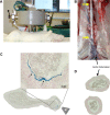
Magnetic targeting setup and efficacy. Notes: SPIONs were magnetically targeted to the injured aortic region directly after the ballooning. (A) Experimental setup and placement of magnetic tip are shown. (B) Following the magnet exposure for 30 minutes, the animals were sacrificed and the excised aorta analyzed histologically. Atherosclerotic plaque is visible as indicated by yellow arrows. (C) The presence of iron in the targeted region was visualized by Prussian blue staining (arrows). Scale bar: 50 µm. (D) No iron accumulation was detected in aortic bifurcation region by histology. The overview images of the artery cross-sections were taken at ×10 objective magnification. M indicates the tip of the magnet. Abbreviations: SPIONs, superparamagnetic iron oxide nanoparticles.
Figure 5
Figure 5
In vivo effects of early MDT with SPION-DEXA. Notes: Following the balloon injury, the animals were intra-arterially administered either control SPION (n=5) or SPION-DEXA (n=4) under external magnetic field. Control group animals (Co) did not receive any treatment with nanoparticles. After 5 weeks of high-cholesterol diet and normal diet for 2 weeks, animals were sacrificed and the excised aortas analyzed histochemically. (A) Normalized plaque area; (B) maximum thickness of intima (measured on Crossman’s trichrome stained sections); and (C) the calculated IMT are shown; (D) Macrophage-positive area (RAM-11 staining) in animals receiving early administration of control SPION or SPION-DEXA. Graphs show median, 25th and 75th percentile. *P=0.025 vs nanoparticle-untreated control; #P=0.0285 (one-tailed t-test), P=0.057 (two-tailed t-test) vs control SPION group. Abbreviations: IMT, intima–media thickness; MDT, magnetic drug targeting; ns, not significant; SPIONs, superparamagnetic iron oxide nanoparticles; SPION-DEXA, SPIONs conjugated with dexamethasone phosphate.
Figure 6
Figure 6
In vivo effects of late MDT with SPION-DEXA. Notes: Following the balloon injury, the animals were fed high-cholesterol diet for 5 weeks and normal diet for 2 weeks. Subsequently, the animals received intra-arterial administration of either control SPION or SPION-DEXA under external magnetic field and additional 4 weeks of normal diet. Control group animals (Co) did not receive any treatment with nanoparticles. (A) Normalized plaque area; (B) maximum thickness of intima; and (C) the calculated IMT are shown. (D) Macrophage-positive area (RAM-11 staining) in animals receiving early administration of control SPION or SPION-DEXA. Graphs show median, 25th and 75th percentile. Abbreviations: IMT, intima–media thickness; MDT, magnetic drug targeting; ns, not significant; SPION, superparamagnetic iron oxide nanoparticles; SPION-DEXA, SPIONs conjugated with dexamethasone phosphate.

Similar articles

Cited by

References

    1. Lozano R, Naghavi M, Foreman K, et al. Global and regional mortality from 235 causes of death xfor 20 age groups in 1990 and 2010: a systematic analysis for the Global Burden of Disease Study 2010. Lancet. 2012;380(9859):2095–2128. - PMC - PubMed
    1. Libby P, Theroux P. Pathophysiology of coronary artery disease. Circulation. 2005;111(25):3481–3488. - PubMed
    1. Cicha I, Garlichs CD, Alexiou C. Cardiovascular therapy through nanotechnology – how far are we still from bedside? Eur J Nanomed. 2014;6(2):63–87.
    1. Thorat ND, Bohara RA, Yadav HM, Tofail SAM. Multi-modal MR imaging and magnetic hyperthermia study of Gd doped Fe3O4 nanoparticles for integrative cancer therapy. RSC Adv. 2016;6(97):94967–94975.
    1. Lyer S, Tietze R, Jurgons R, et al. Visualisation of tumour regression after local chemotherapy with magnetic nanoparticles – a pilot study. Anticancer Res. 2010;30(5):1553–1557. - PubMed

MeSH terms