

Folic acid-nanoscale gadolinium-porphyrin metal-organic frameworks: fluorescence and magnetic resonance dual-modality imaging and photodynamic therapy in hepatocellular carcinoma
- PMID: 30587985
- PMCID: PMC6304077
- DOI: 10.2147/IJN.S177880
Folic acid-nanoscale gadolinium-porphyrin metal-organic frameworks: fluorescence and magnetic resonance dual-modality imaging and photodynamic therapy in hepatocellular carcinoma
Abstract
Background: Hepatocellular carcinoma (HCC) is the most common primary liver cancer and severely threatens human health. Since the prognosis of advanced HCC remains poor, there is an urgent need to develop new therapeutic approaches. Porphyrin metal-organic frameworks are a class of porous organic-inorganic hybrid functional materials with good biocompatibility.
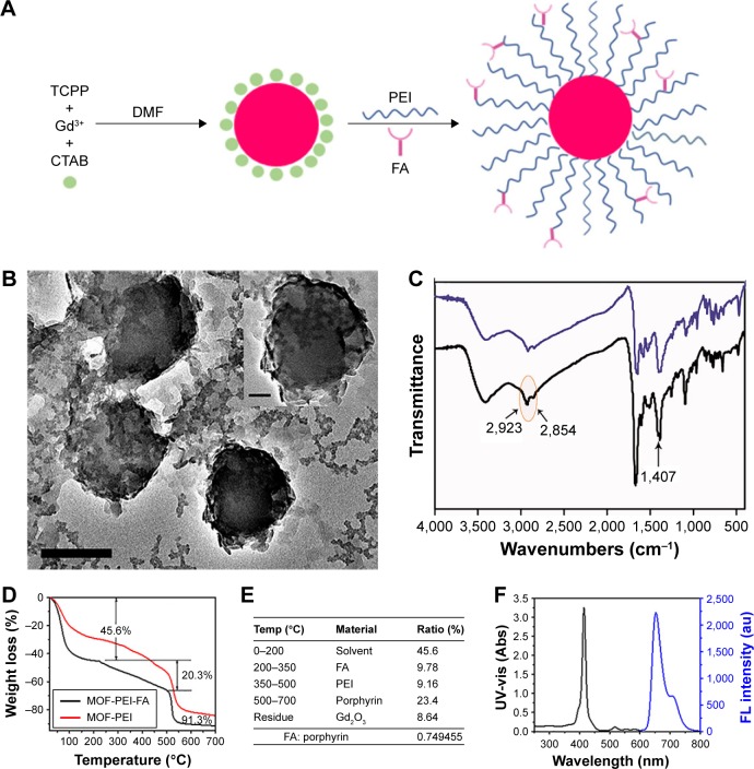
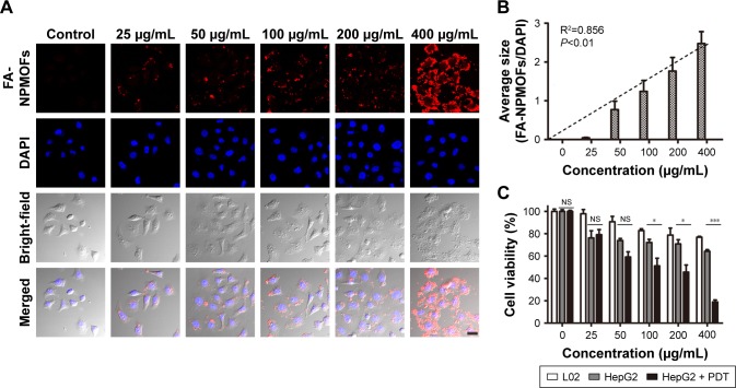
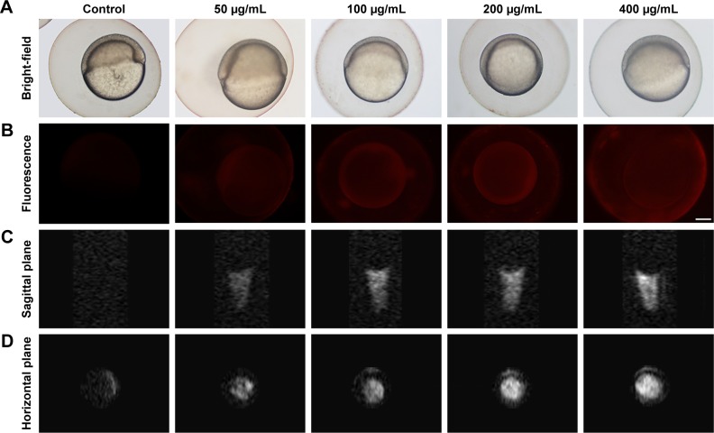
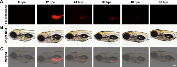
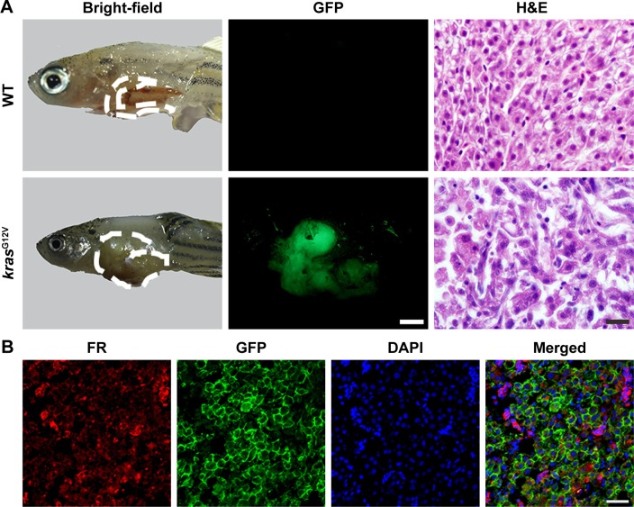
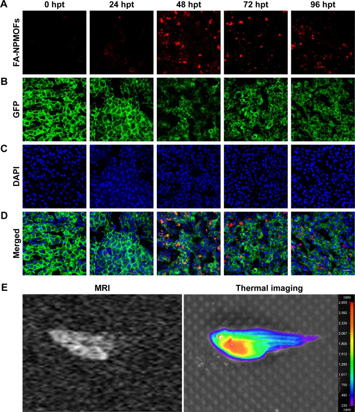
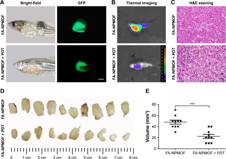
Methods: Gadolinium-porphyrin metal-organic frameworks were used as a skeleton for folic acid (FA) conjugation to synthesize a novel type of nanoparticle, denoted as folic acid-nanoscale gadolinium-porphyrin metal-organic frameworks (FA-NPMOFs). The FA-NPMOFs were characterized using transmission electron microscopy, Fourier transform infrared spectroscopy and thermogravimetric-differential thermal analysis. The biotoxicity and imaging capability of the FA-NPMOFs were determined using HepG2 cells and embryonic and larval zebrafish. The delivery and photodynamic therapeutic effect of FA-NPMOFs were explored in transgenic zebrafish with doxycycline-induced HCC.
Results: FA-NPMOFs were spherical in structure with good dispersion and water solubility. They showed low biotoxicity, emitted bright red fluorescence, and exhibited an excellent magnetic resonance imaging capability, both in vitro and in vivo. Meanwhile, the FA-NPMOFs exhibited a strong affinity for folate receptor (FR)-expressing cells and were delivered to the tumor site in a targeted manner. Moreover, HCC tumor cells were eliminated following laser irradiation.
Conclusion: FA-NPMOFs can be used for dual-modality imaging and photodynamic therapy in HCC and show promise for use as a carrier in new therapies for HCC and other FR-positive tumors.
Keywords: dual-modality imaging; folic acid; hepatocellular carcinoma; nanoscale gadolinium-porphyrin metal-organic frameworks; photodynamic therapy.
Conflict of interest statement
Disclosure The authors report no conflicts of interest in this work.
Figures
Similar articles
-
Fluorescent Imaging-Guided Chemo- and Photodynamic Therapy of Hepatocellular Carcinoma with HCPT@NMOFs-RGD Nanocomposites.Int J Nanomedicine. 2022 Mar 25;17:1381-1395. doi: 10.2147/IJN.S353803. eCollection 2022. Int J Nanomedicine. 2022. PMID: 35369034 Free PMC article.
-
Rapid fabrication of carbon quantum dots as multifunctional nanovehicles for dual-modal targeted imaging and chemotherapy.Acta Biomater. 2016 Dec;46:151-164. doi: 10.1016/j.actbio.2016.09.027. Epub 2016 Sep 20. Acta Biomater. 2016. PMID: 27662808
-
Fluorescent Imaging-Guided Chemotherapy-and-Photodynamic Dual Therapy with Nanoscale Porphyrin Metal-Organic Framework.Small. 2017 May;13(17). doi: 10.1002/smll.201603459. Epub 2017 Feb 28. Small. 2017. PMID: 28244202
-
Porphyrin-Based Metal-Organic Framework Compounds as Promising Nanomedicines in Photodynamic Therapy.ChemMedChem. 2020 Oct 5;15(19):1766-1775. doi: 10.1002/cmdc.202000353. Epub 2020 Aug 31. ChemMedChem. 2020. PMID: 32715651 Review.
-
Recent Advances in Glutathione Depletion-Enhanced Porphyrin-Based nMOFs for Photodynamic Therapy.Pharmaceutics. 2025 Feb 12;17(2):244. doi: 10.3390/pharmaceutics17020244. Pharmaceutics. 2025. PMID: 40006611 Free PMC article. Review.
Cited by
-
Metal-organic frameworks for hepatocellular carcinoma therapy and mechanism.Front Pharmacol. 2022 Sep 26;13:1025780. doi: 10.3389/fphar.2022.1025780. eCollection 2022. Front Pharmacol. 2022. PMID: 36225574 Free PMC article.
-
BioMOF-Based Anti-Cancer Drug Delivery Systems.Nanomaterials (Basel). 2023 Mar 6;13(5):953. doi: 10.3390/nano13050953. Nanomaterials (Basel). 2023. PMID: 36903831 Free PMC article. Review.
-
Porphyrin NanoMetal-Organic Frameworks as Cancer Theranostic Agents.Molecules. 2022 May 12;27(10):3111. doi: 10.3390/molecules27103111. Molecules. 2022. PMID: 35630585 Free PMC article. Review.
-
Development of folic acid modified reduction-responsive micelles for the targeted release of sorafenib in liver cancer.Ther Deliv. 2025 Jul;16(7):637-649. doi: 10.1080/20415990.2025.2513223. Epub 2025 May 30. Ther Deliv. 2025. PMID: 40448268
-
Nanomedicine in Hepatocellular Carcinoma: A New Frontier in Targeted Cancer Treatment.Pharmaceutics. 2021 Dec 25;14(1):41. doi: 10.3390/pharmaceutics14010041. Pharmaceutics. 2021. PMID: 35056937 Free PMC article. Review.
References
-
- Chen JG, Zhang SW. Liver cancer epidemic in China: past, present and future. Semin Cancer Biol. 2011;21(1):59–69. - PubMed
MeSH terms
Substances
LinkOut - more resources
Full Text Sources
Medical
Molecular Biology Databases
