. 2018 Dec 19:6:e5976.
doi: 10.7717/peerj.5976. eCollection 2018.

The oldest ceratosaurian (Dinosauria: Theropoda), from the Lower Jurassic of Italy, sheds light on the evolution of the three-fingered hand of birds

Affiliations

The oldest ceratosaurian (Dinosauria: Theropoda), from the Lower Jurassic of Italy, sheds light on the evolution of the three-fingered hand of birds

Cristiano Dal Sasso et al. PeerJ. .

Abstract

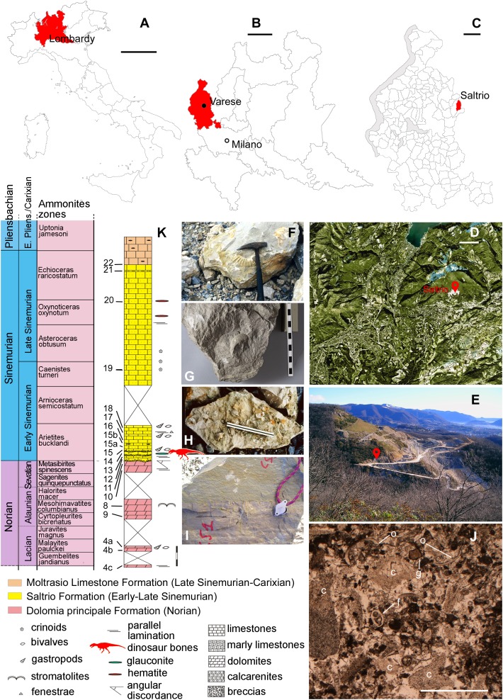
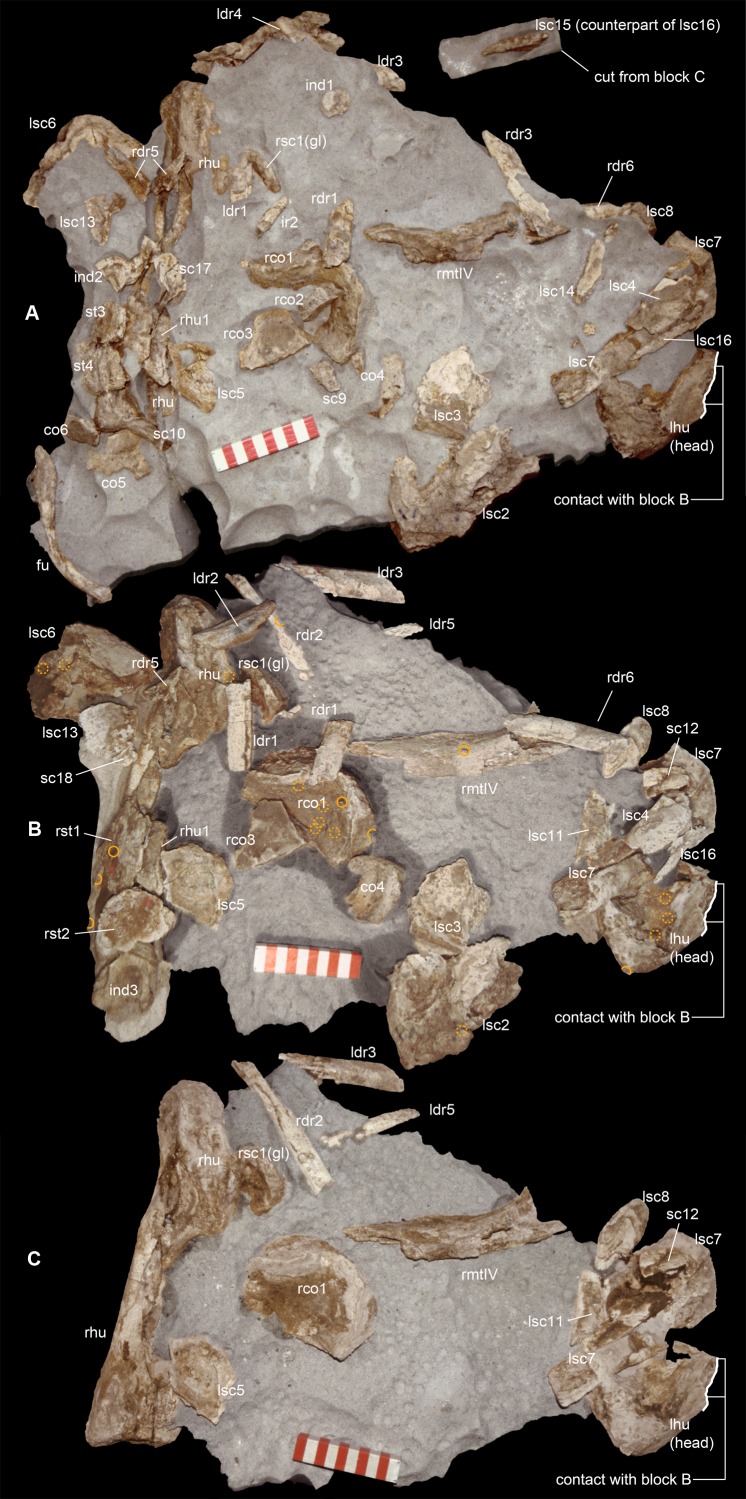
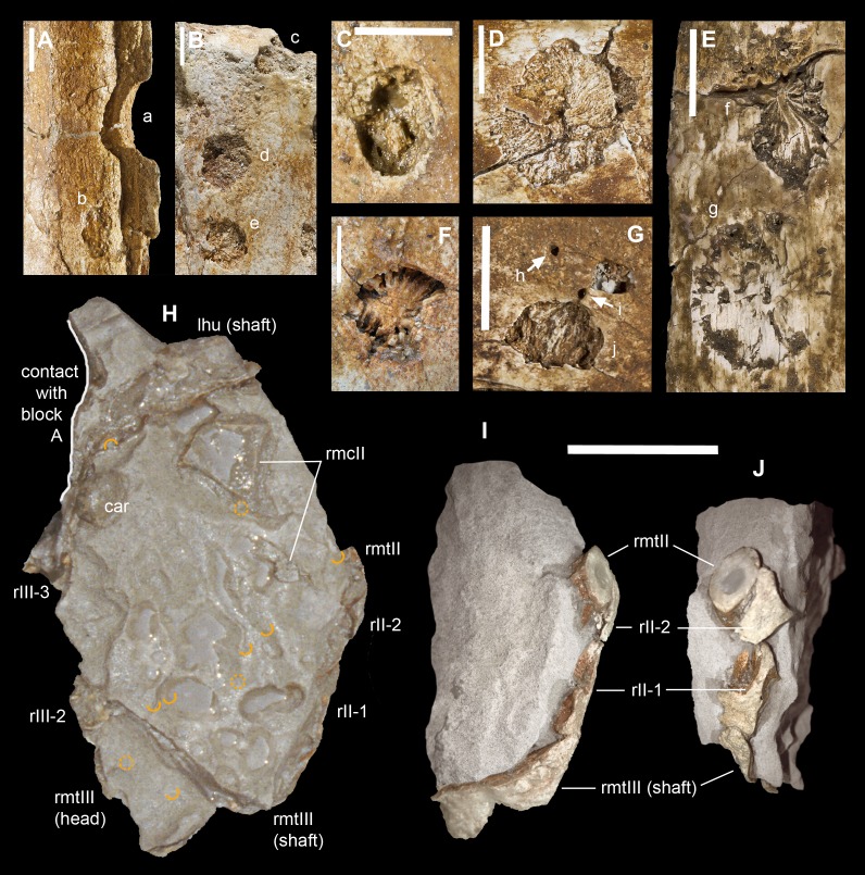
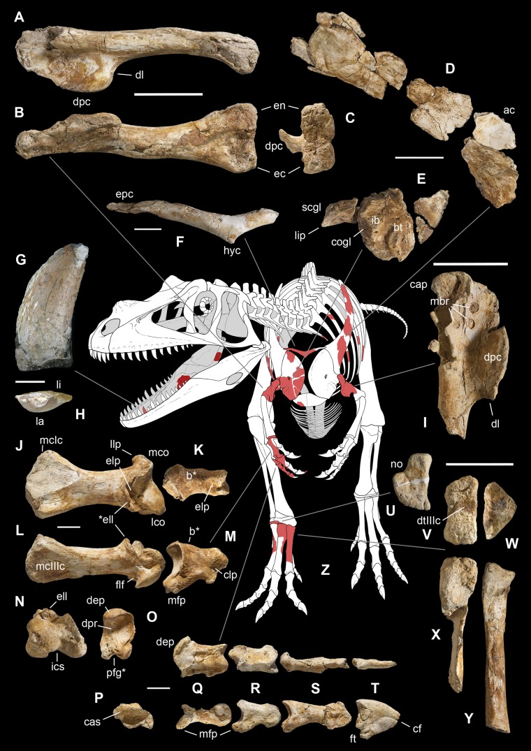
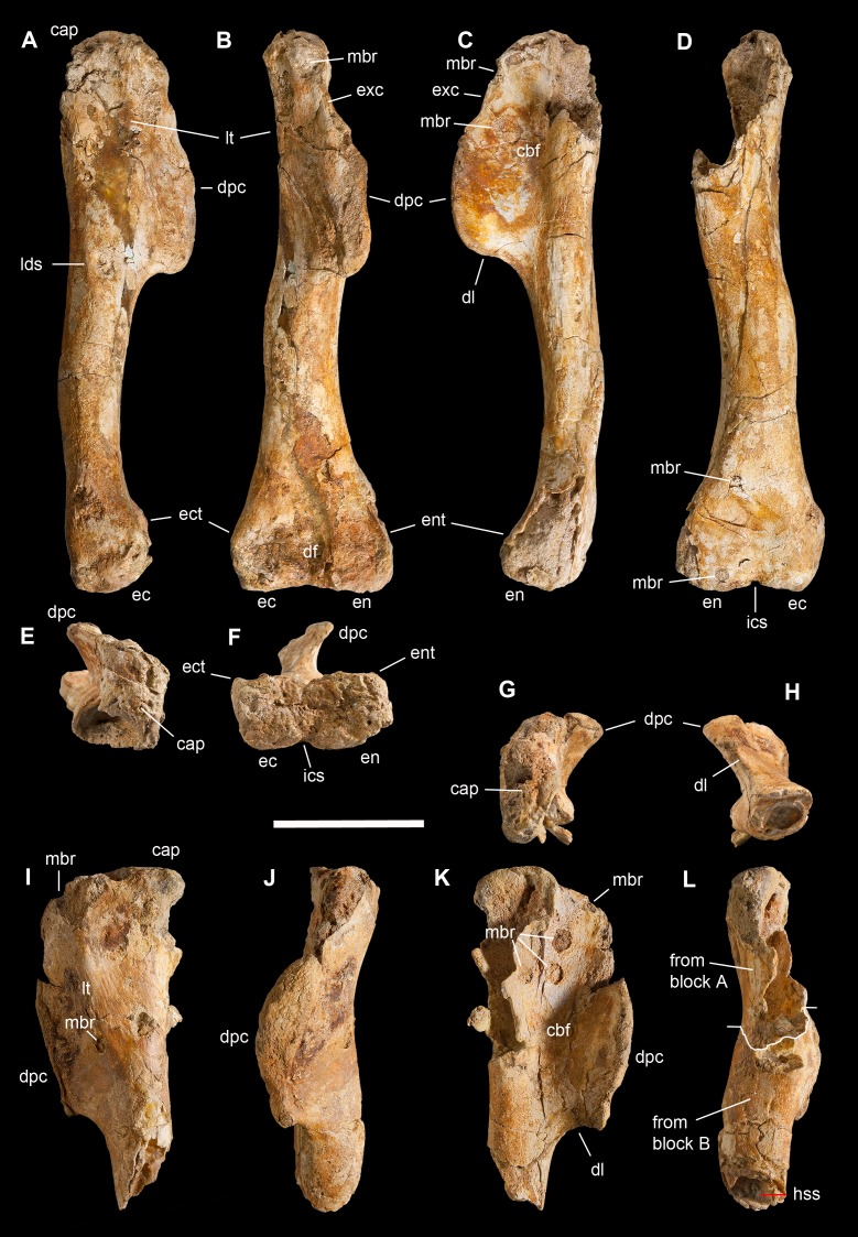
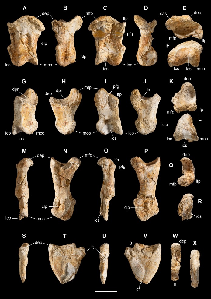
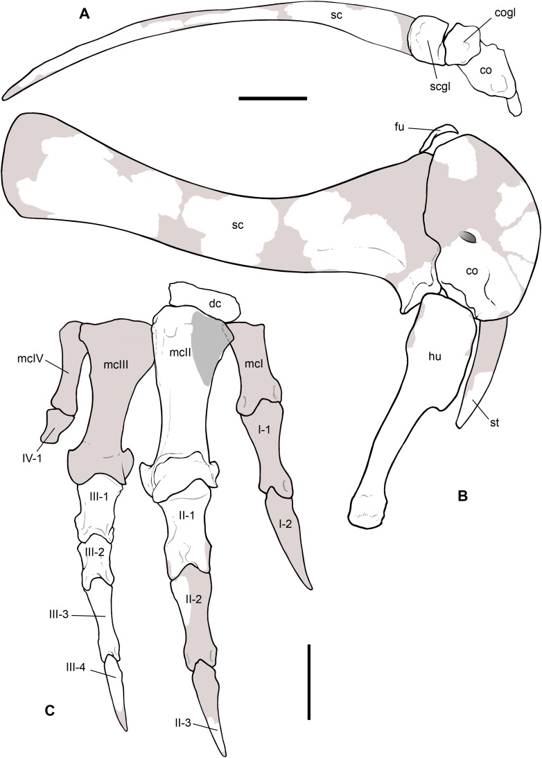
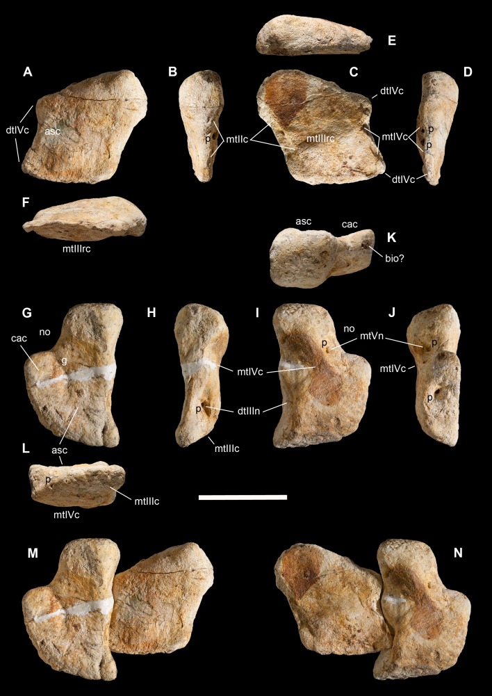
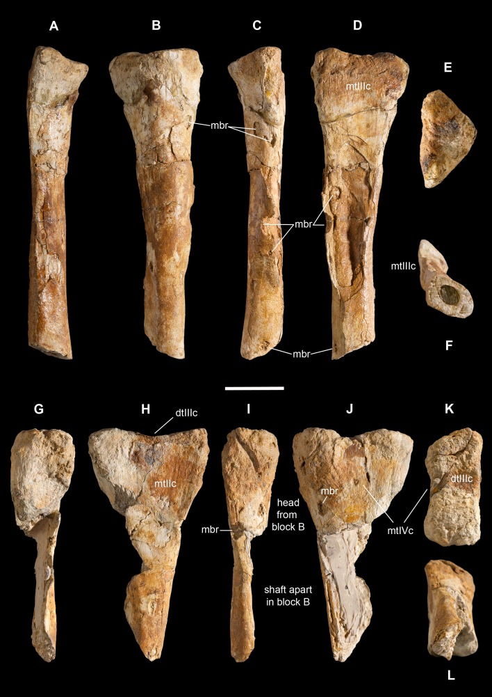
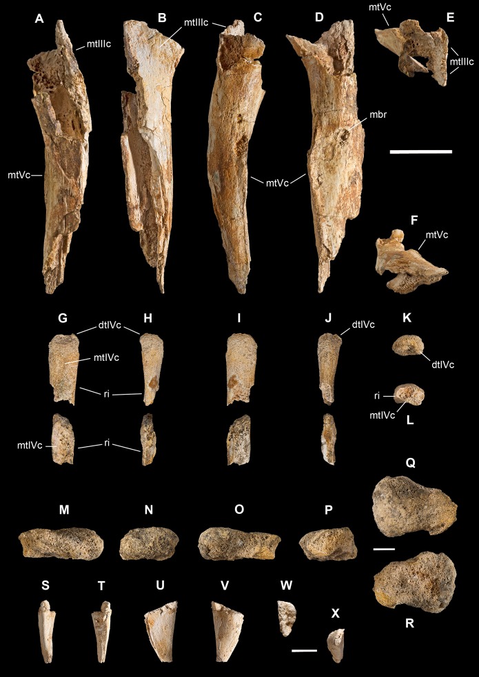
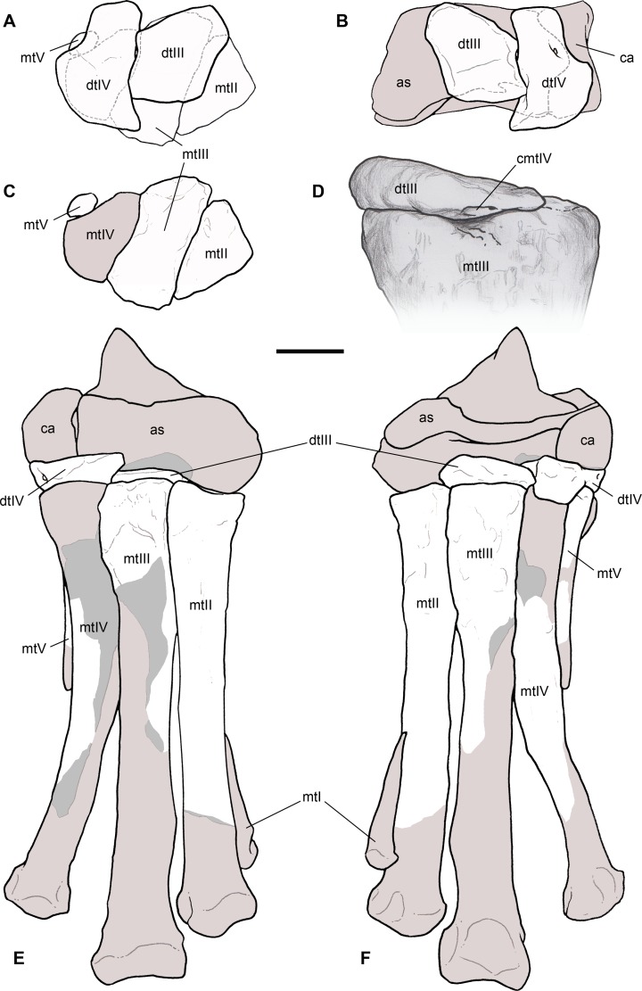
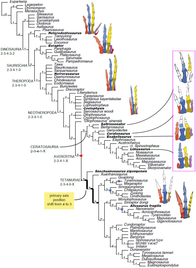
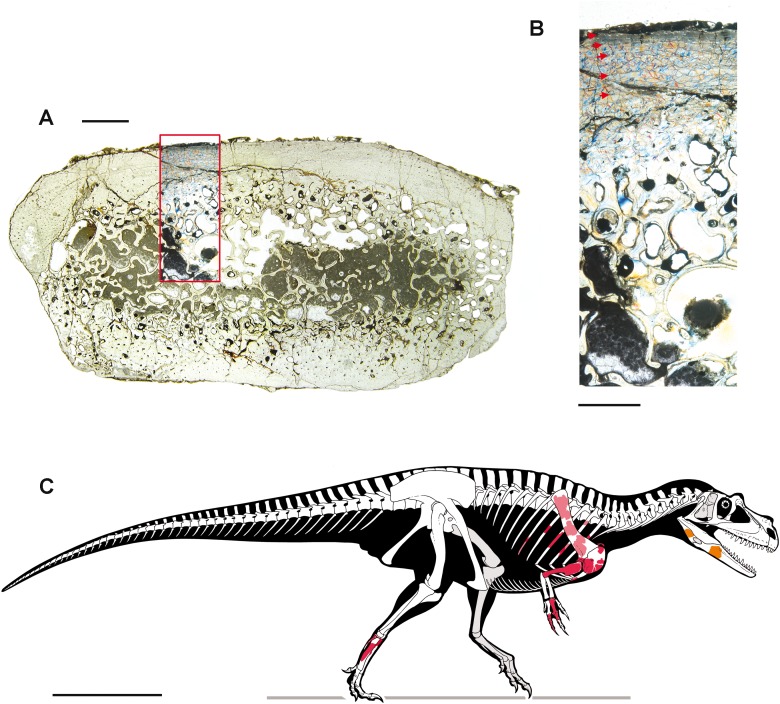
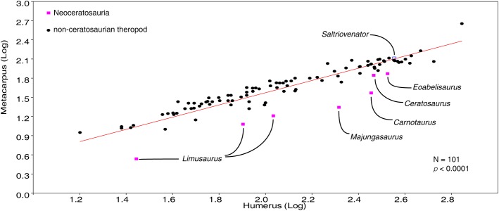
The homology of the tridactyl hand of birds is a still debated subject, with both paleontological and developmental evidence used in support of alternative identity patterns in the avian fingers. With its simplified phalangeal morphology, the Late Jurassic ceratosaurian Limusaurus has been argued to support a II-III-IV digital identity in birds and a complex pattern of homeotic transformations in three-fingered (tetanuran) theropods. We report a new large-bodied theropod, Saltriovenator zanellai gen. et sp. nov., based on a partial skeleton from the marine Saltrio Formation (Sinemurian, lowermost Jurassic) of Lombardy (Northern Italy). Taphonomical analyses show bone bioerosion by marine invertebrates (first record for dinosaurian remains) and suggest a complex history for the carcass before being deposited on a well-oxygenated and well-illuminated sea bottom. Saltriovenator shows a mosaic of features seen in four-fingered theropods and in basal tetanurans. Phylogenetic analysis supports sister taxon relationships between the new Italian theropod and the younger Early Jurassic Berberosaurus from Morocco, in a lineage which is the basalmost of Ceratosauria. Compared to the atrophied hand of later members of Ceratosauria, Saltriovenator demonstrates that a fully functional hand, well-adapted for struggling and grasping, was primitively present in ceratosaurians. Ancestral state reconstruction along the avian stem supports 2-3-4-1-X and 2-3-4-0-X as the manual phalangeal formulae at the roots of Ceratosauria and Tetanurae, confirming the I-II-III pattern in the homology of the avian fingers. Accordingly, the peculiar hand of Limusaurus represents a derived condition restricted to late-diverging ceratosaurians and cannot help in elucidating the origin of the three-fingered condition of tetanurans. The evolution of the tridactyl hand of birds is explained by step-wise lateral simplification among non-tetanuran theropod dinosaurs, followed by a single primary axis shift from digit position 4 to 3 at the root of Tetanurae once the fourth finger was completely lost, which allowed independent losses of the vestigial fourth metacarpal among allosaurians, tyrannosauroids, and maniraptoromorphs. With an estimated body length of 7.5 m, Saltriovenator is the largest and most robust theropod from the Early Jurassic, pre-dating the occurrence in theropods of a body mass approaching 1,000 Kg by over 25 My. The radiation of larger and relatively stockier averostran theropods earlier than previously known may represent one of the factors that ignited the trend toward gigantism in Early Jurassic sauropods.

Keywords: Aves; Ceratosauria; Dinosauria; Hand evolution; Italy; Lower Jurassic; Osteology; Phylogeny; Taphonomy; Theropoda.


Conflict of interest statement

Cristiano Dal Sasso is an employee of the Museo di Storia Naturale di Milano, Italy.

Figures

Figure 1
Figure 1. Fossil location and geological setting.
(A–C) Outline maps of Italy, Lombardy, Varese Province, and Saltrio Municipality; (D) satellite view of the Saltrio area, with map marker indicating the Saltrio quarry; (E) map marker indicating the stratigraphic log in the Saltrio quarry; (F) the ammonite Paracoroniceras cf. gmuendense and (G) the nautiloid Cenoceras striatum, both found associated in the layer containing the dinosaur bones; (H) glauconite present as accessory mineral in block C (counterpart of block A of Fig. 2); (I) the discordance between the Dolomia Principale Fm. and the Saltrio Fm.; (J) thin sections of the layer embedding the dinosaur bones; (K) stratigraphic log of the Saltrio quarry, based on Croce (2005), with geological time scale and ammonites zones based on Sacchi Vialli (1964) and Ogg & Hinnov (2012). Abbreviations: c, crinoids; f, foraminifers; g, gastropods; o, ostracods. Scale bars equal 200 km in (A), 30 km in (B), six km in (C), one km in (D), one mm in (K), and 150 cm in (L). Photos by F. Berra, G. Bindellini, M. Croce, and G. Pasini; drawings by M. Croce and S. Maganuco.
Figure 2
Figure 2. Taphonomy of the Saltrio theropod (block A).
Bones of Saltriovenator mapped in temporal sequence (A–C), gradually emerging from the embedding rock during acid preparation of block A. Numbers refer to each fragment, not to a specific anatomical position. The latter is reported in other figures, for fragments that were later reconnected into more complete bones. Abbreviations as in text, and as follows: ind, indeterminate bone; ir, indeterminate rib; l (left) and r (right) are specified for fragments of paired bones certainly (appendicular elements) or tentatively (ribs) positioned in the skeleton. Macroborings facing front, side and back are mapped respectively with yellow circles, semicircles, and hatched circles. Scale bars equal 10 cm. Photos by G. Bindellini and C. Dal Sasso.
Figure 3
Figure 3. Macroborings, and taphonomy of the Saltrio theropod (block B).
Selected macroborings (A–G) on bones from blocks A and B, and bones of Saltriovenator mapped in temporal sequence (H–J), gradually emerging from block B during acid preparation. (A) Semicircular feeding mark (a) splitted after diagenetic crushing of the bone (right metatarsal II); elliptical and flat-bottomed boring (b). (B) Feeding mark trenching the left humeral head (c); subcircular boring (d) and feebly septate circular boring (e). (C) Elliptical boring with deepening edges and a central peak on the shaft of the left humerus. (D) Circular boring with radial waves on the right humerus. (E) Dorsal rib with wavy, markedly septate elliptical boring (f) and feebly septate circular mark with zigzagging margin (g). (F) Asterisk-like septate boring on the shaft of right metatarsal II. (G) Tiny burrows penetrating the cortex of the right coracoid (h, i), and an enigmatic boring with irregular bottom and margins (j). Abbreviations as in text, symbols as in Fig. 2. Scale bars equal 10 mm in (A)–(E) and (G), five mm in (F), and 10 cm in (1)–(3). Photos by G. Bindellini, C. Dal Sasso, and M. Zilioli.
Figure 4
Figure 4. Selected elements used in the diagnosis of Saltriovenator zanellai n. gen. n. sp.
Right humerus in medial (A), frontal (B) and distal (C) views; (D) left scapula, medial view; (E) right scapular glenoid and coracoid, lateral view; (F) furcula, ventral view; tooth, labial (G) and apical (H) views; (I) left humerus, medial view; right second metacarpal in dorsal (J), lateral (L) and distal (N) views; first phalanx of the right second digit in dorsal (K), lateral (M) and proximal (O) views; (P–T) right third digit in proximal, dorsal and lateral views; (U) right distal tarsal IV, proximal view; third right metatarsal in proximal (V) and frontal (X) views; second right metatarsal, proximal (W) and frontal (Y) views; (Z) reconstructed skeleton showing identified elements (red). Abbreviations as in text, asterisks mark autapomorphic traits. Scale bars: 10 cm in (A)–(E), (I), and (U)–(Y); two cm in (F), and (J)–(T); one cm in (G). Photos by G. Bindellini, C. Dal Sasso and M. Zilioli; drawing by M. Auditore.
Figure 5
Figure 5. Cranio-mandibular fragments, tooth, and ribs of Saltriovenator zanellai.
Indeterminate cranial fragment (A–B); right splenial in lateral (C), rostral (D) and ventral (E) views; right prearticular in lateral (F) and rostral views (G); sketch of the right prearticular of MOR 693 (Allosaurus fragilis) with virtual cross-section (H) diagnostic for G, also confirmed by CT slicing of the left side element of MOR 693 (I); splenial and prearticular in medial view, positioned in a reconstructed right lower jawof Saltriovenator (J). Maxillary or dentary tooth in labial (K) and apical (L) views; close-up of the distal carina and denticles in lingual (M) and distal (N) views. Left cervical rib (O) in craniolateral view; fragmentary right (P) and left (Q) dorsal ribs in craniolateral view. Abbreviations as in text, ribs labeled as in Fig. 2 maps and caption. Scale bars equal two cm in (A)–(I), five cm in (J), one cm in (K), five mm in (L), one mm in (M)–(N), five cm in (O)–(Q). Photos by G. Bindellini, C. Dal Sasso, and M. Zilioli; drawing by C. Dal Sasso.
Figure 6
Figure 6. Pectoral girdle of Saltriovenator zanellai.
Left scapula in lateral (A), caudal (B), medial (C), and cranial (D) views; right scapular glenoid and coracoid in medial (E), caudal (F), lateral (G), and cranial (H) views; right coracoid in ventral (I) and dorsal (J) views; furcula in cranial (K), caudal (L), right lateral (M), ventral (N, with selected craniocaudal cross-sections), and dorsal (O) views; caudolateral portion of the right sternal plate in dorsal (P), lateral (Q), and ventral (R) views. Each bone fragment is labeled on the side cropping out in Fig. 2. Abbreviations as in text. The position of co4 and co6 is hypothetical. Scale bars equal 10 cm in (A)–(J), five cm in (K)–(R). Photos by G. Bindellini and C. Dal Sasso; drawings by M. Auditore.
Figure 7
Figure 7. Humeri of Saltriovenator zanellai.
Right humerus in (A) lateral, (B) cranial, (C) medial, (D) caudal, (E) proximal, and (F) distal views; left humerus in (G) proximal, (H) distal, (I) lateral, (J) cranial, (K) medial, and (L) caudal views. Abbreviations as in text. Scale bar equals 10 cm. Photos by G. Bindellini.
Figure 8
Figure 8. Manus of Saltriovenator zanellai, second metacarpal and digit.
Right second metacarpal (II) in (A) dorsal, (B) medial, (C) palmar, (D) lateral, (E) proximal, and (F) distal views; first phalanx of the right second digit (II-1) in (G) proximal, (H) distal, (I) dorsal, (J) medial, (K) palmar, and (L) lateral views; second phalanx of the right second digit (II-2) in (M) dorsal, (N) medial, (O) palmar, (P) lateral, (Q) proximal, and (R) distal views;. Abbreviations as in text. Scale bar equals five cm. Photos by G. Bindellini.
Figure 9
Figure 9. Manus of Saltriovenator zanellai, third digit.
Right first phalanx (III-1) in (A) dorsal, (B) medial, (C) palmar, (D) lateral, (E) proximal, and (F) distal views; right second phalanx (III-2) in (G) dorsal, (H) medial, (I) palmar, (J) lateral, (K) proximal, and (L) distal views; right third phalanx (III-3) in (M) dorsal, (N) medial, (O) palmar, (P) lateral, (Q) proximal, and (R) distal views; right fourth (ungual) phalanx (III-4) in (S) dorsal, (T) medial, (U) palmar, (V) lateral, (W) proximal, and (X) distal views. Abbreviations as in text. Scale bar equals two cm. Photos by G. Bindellini.
Figure 10
Figure 10. Reconstruction of the pectoral girdle and forelimb of Saltriovenator zanellai.
Composite right scapula and coracoid in ventral view (A), and composite right pectoral girdle, humerus and manus in lateral view (B–C). The scapular body and blade, and the proximal portion of the humerus are reversed left elements. Preserved elements in white, reconstructed bone in light gray, exposed inner bone in gray, hidden bone in dotted lines. Abbreviations as in text. Scale bar equals 10 cm in (A) and (B), five cm in (C). Drawings by M. Auditore.
Figure 11
Figure 11. Distal tarsals of Saltriovenator zanellai.
Right distal tarsal III in (A) proximal, (B) medial, (C) distal, (D) lateral, (E) caudal, (F) cranial views; right distal tarsal IV in (G) proximal, (H) medial, (I) distal, (J) lateral, (K) caudal, (L) cranial views; articulated right distal tarsals III + IV in proximal (M) and distal (N) views. Abbreviations as in text. Scale bar equals five cm. Photos by G. Bindellini.
Figure 12
Figure 12. Metatarsals II and III of Saltriovenator zanellai.
Right metatarsal II in (A) cranial, (B) medial, (C) caudal), (D) lateral, (E) proximal, and (F) distal views; right metatarsal III in (G) cranial, (H) medial, (I) caudal), (J) lateral, (K) proximal, and (L) distal views. Abbreviations as in text. Scale bar equals five cm. Photos by G. Bindellini.
Figure 13
Figure 13. Metatarsals IV and V, carpal and ungual of Saltriovenator zanellai.
Right metatarsal IV in (A) cranial, (B) medial, (C) caudal), (D) lateral, (E) proximal, and (F) distal views; right metatarsal IV in (G) cranial, (H) medial, (I) caudal), (J) lateral, (K) proximal, and (L) distal views. Carpal bone in (M) ?cranial, (N) ?medial, (O) ?caudal), (P) ?lateral, (Q) ?proximal, and (R) ?distal views; distal portion of manual ungual phalanx (?II-3) in (S) dorsal, (T) palmar, (U) ?medial, (V) ?lateral, (W) proximal, and (X) distal views. Abbreviations as in text. Scale bar equals five cm. Photos by G. Bindellini.
Figure 14
Figure 14. Reconstruction of the right ankle and foot of Saltriovenator zanellai.
Distal tarsals superimposed and articulated to metatarsals in proximal view (A), and to astragalus and calcaneum in distal view (B); metatarsals II–V in proximal view (C); close-up of the perfect contact between distal tarsal III and metatarsal III in lateral view (D); tarsal and metatarsal elements fully reconstructed and articulated in cranial (E), and caudal (F) view. Abbreviations as in text, preserved elements in white (except in D), reconstructed bone in light gray, exposed inner bone in gray, hidden bone in dotted lines. Scale bar equals five cm. Scale bar equals five cm in (A)–(F), three cm in (D). Drawings by M. Auditore.
Figure 15
Figure 15. Phylogenetic affinities of Saltriovenator and evolution of the hand in Theropoda.
Reduced strict consensus of the shortest trees found by the phylogenetic analysis after pruning of Lewisuchus and Teleocrater. Numbers at nodes indicate decay index. Inferred manual phalangeal formula for selected nodes indicated below clade names. Hands of representative members of the avian stem (bold names) in extensor view (Herrerasaurus in flexor view), medial side at left, missing elements in white based on ancestral states inferred at least inclusive node containing the taxon. Red star indicates loss of metacarpal V, blue stars indicate multiple independent losses of metacarpal IV among tetanurans. Drawings by A. Cau.
Figure 16
Figure 16. Bone microstructure (A–B) and skeletal reconstruction (C) of Saltriovenator zanellai.
Mid-shaft thin section of a right dorsal rib. (A) Overall view; (B) close-up of the Haversian system in the compacta and of the microstructure of the outermost cortex showing the presence of an incipient EFS, interpreted on the basis of reduced vascular canals, presence of lamellar bone, and presence of closely spaced LAGs. The outer surface of the bone is at the top, red arrows point to the LAGs. Colors are emphasized due to photography under gypsum plate. (C) Skeletal reconstruction of Saltriovenator zanellai in right lateral view, realized using comparative anatomy and character state inference to predict a plausible range for size and proportions of the missing elements; known elements are mapped on the skeleton in different colors: right bones in red; counterlateral copies of the left bones in light red; bones from the medial side of the lower jaw in orange. Scale bars equal two mm in (A), one mm in (B), and one m in (C). Photos by M. Zilioli; drawing by M. Auditore.
Figure 17
Figure 17. Plot of metacarpus vs humerus length in Theropoda.
The metacarpus-humerus ratio in Saltriovenator perfectly fits the general theropod pattern, whereas more advanced ceratosaurians have the shortest metacarpi compared to the humerus among theropods. This suggests that the acquisition of a stout and robust metacarpus in Ceratosauria (present in Saltriovenator) preceded the relative size reduction of the metapodium (present in neoceratosaurs and extreme in abelisauroids). Data from Dececchi & Larsson (2011) and Wang et al. (2017). Diagram by A. Cau.

References

    1. Allain R. The postcranial anatomy of the megalosaur Dubreuillosaurus valesdunensis (Dinosauria Theropoda) from the Middle Jurassic of Normandy, France. Journal of Vertebrate Paleontology. 2005;25(4):850–858. doi: 10.1671/0272-4634(2005)025[0850:TPAOTM]2.0.CO;2. - DOI
    1. Allain R, Chure DJ. Poekilopleuron bucklandii, the theropod dinosaur from the Middle Jurassic (Bathonian) of Normandy. Palaeontology. 2002;45(6):1107–1121. doi: 10.1111/1475-4983.00277. - DOI
    1. Allain R, Tykoski R, Aquesbi N, Jalil N-E, Monbaron M, Russell D, Taquet P. An abelisauroid (Dinosauria: Theropoda) from the Early Jurassic of the High Atlas Mountains, Morocco, and the radiation of ceratosaurs. Journal of Vertebrate Paleontology. 2007;27(3):610–624. doi: 10.1671/0272-4634(2007)27[610:aadtft]2.0.co;2. - DOI
    1. Amorosi A. Detecting compositional, spatial, and temporal attributes of glaucony: a tool for provenance research. Sedimentary Geology. 1997;109(1–2):135–153. doi: 10.1016/s0037-0738(96)00042-5. - DOI
    1. Andrews CW. On some remains of a theropodous dinosaur from the Lower Lias of Barrow-on-Soar. Annals and Magazine of Natural History, Series. 1921;9(8):570–576.

LinkOut - more resources