

Morphological characterization and staging of bumble bee pupae
- PMID: 30588402
- PMCID: PMC6302898
- DOI: 10.7717/peerj.6089
Morphological characterization and staging of bumble bee pupae
Abstract
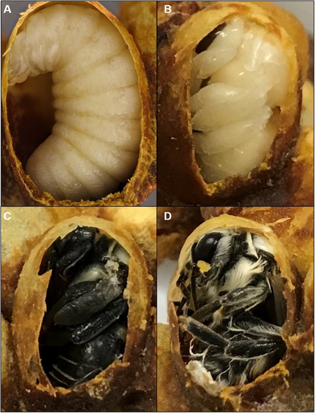
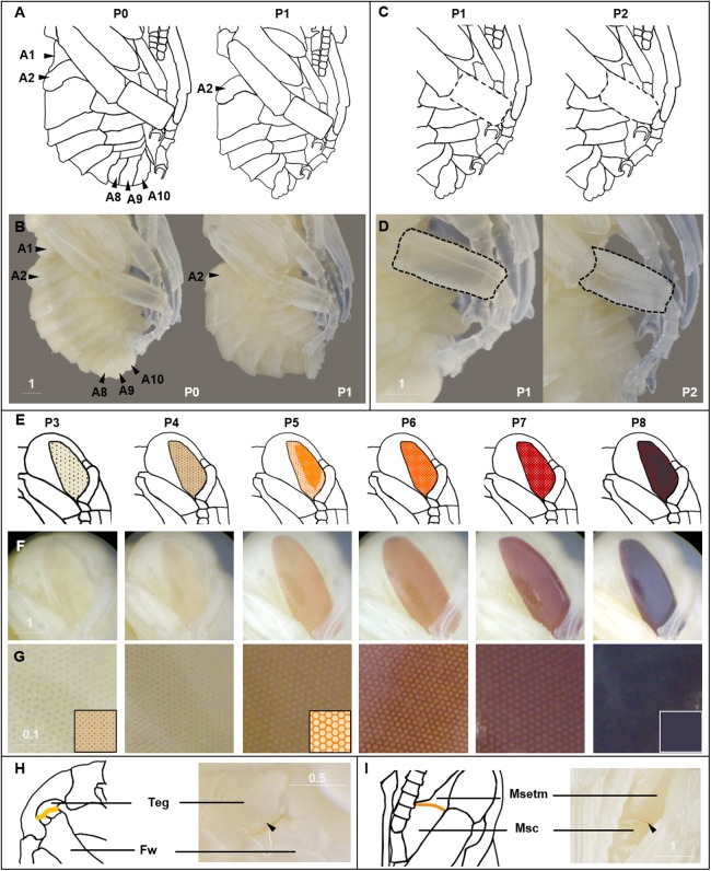
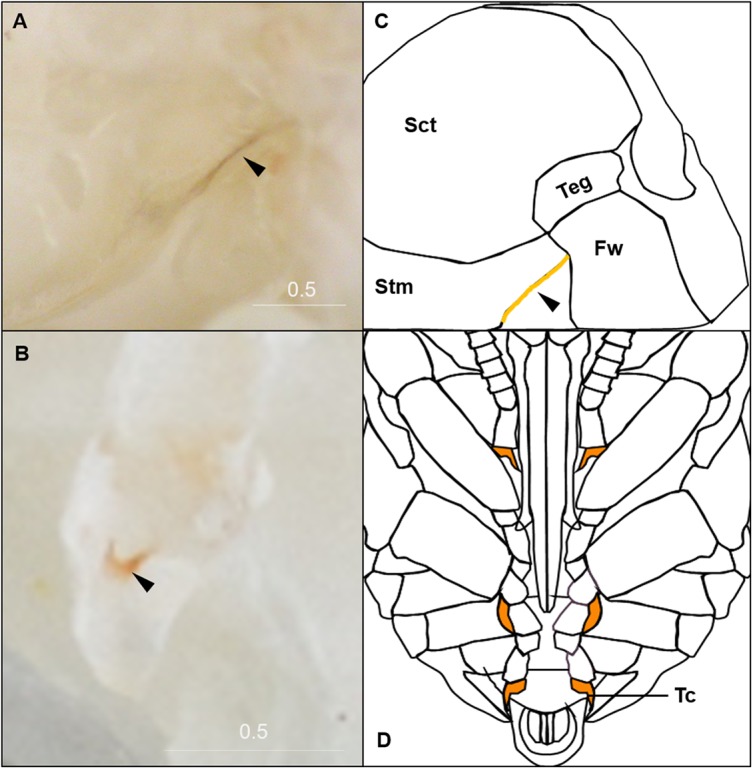
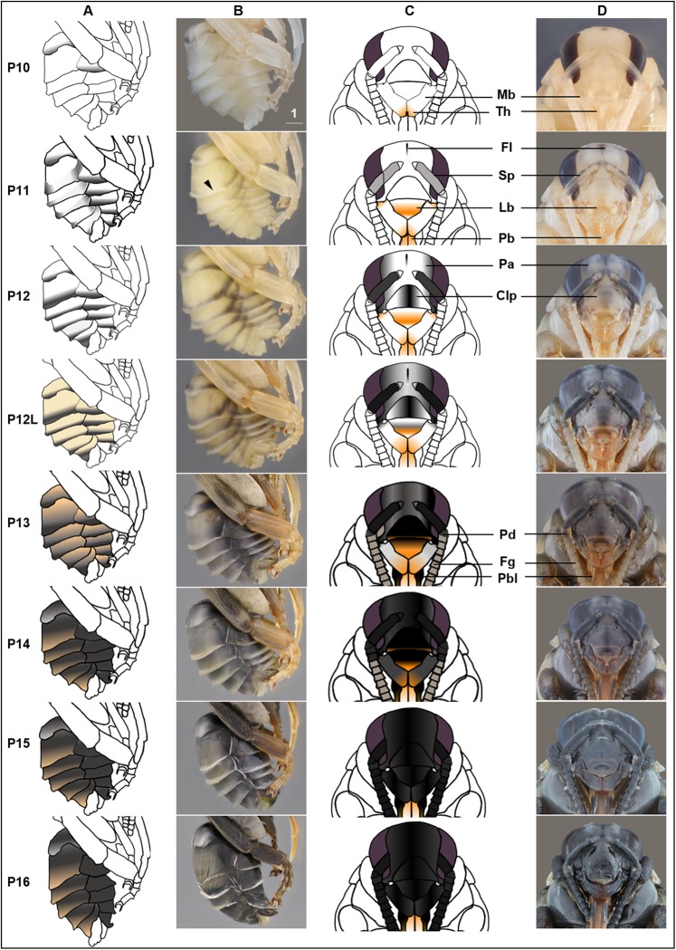
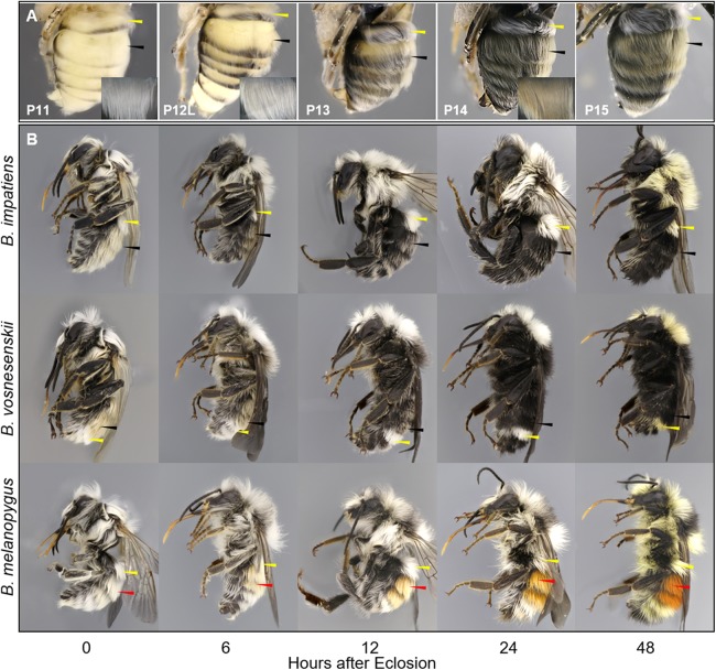
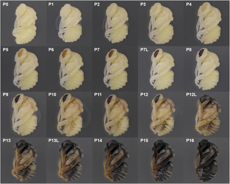
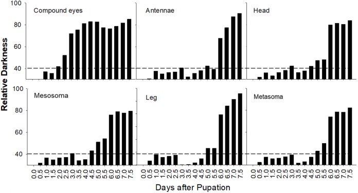
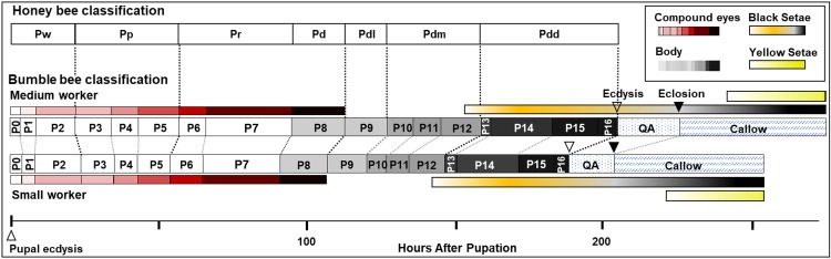
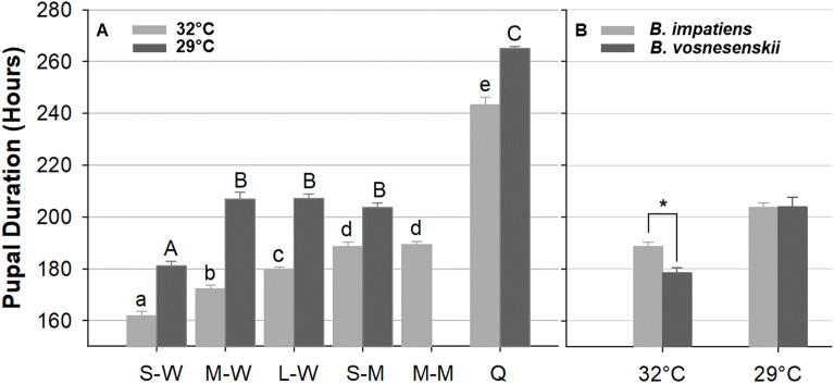
Bumble bees (Hymenoptera: Apidae, Bombus) are important pollinators and models for studying mechanisms underlying developmental plasticity, such as factors influencing size, immunity, and social behaviors. Research on such processes, as well as expanding use of gene-manipulation and gene expression technologies, requires a detailed understanding of how these bees develop. Developmental research often uses time-staging of pupae, however dramatic size differences in these bees can generate variation in developmental timing. To study developmental mechanisms in bumble bees, appropriate staging of developing bees using morphology is necessary. In this study, we describe morphological changes across development in several bumble bee species and use this to establish morphology-based staging criteria, establishing 20 distinct illustrated stages. These criteria, defined largely by eye and cuticle pigmentation patterns, are generalizable across members of the subgenus Pyrobombus, and can be used as a framework for study of other bumble bee subgenera. We examine the effects of temperature, caste, size, and species on pupal development, revealing that pupal duration shifts with each of these factors, confirming the importance of staging pupae based on morphology rather than age and the need for standardizing sampling.
Keywords: Bumble bee; Development; Pupa; Pupal duration; Staging.
Conflict of interest statement
The authors declare that they have no competing interests.
Figures
Similar articles
-
Why Are Queens Broodless? Failed Nest Initiation Not Linked to Parasites, Mating Status, or Ovary Development in Two Bumble Bee Species of Pyrobombus (Hymenoptera: Apidae: Bombus).J Econ Entomol. 2020 Apr 6;113(2):575-581. doi: 10.1093/jee/toz330. J Econ Entomol. 2020. PMID: 31814010
-
USBombus, a database of contemporary survey data for North American Bumble Bees (Hymenoptera, Apidae, Bombus) distributed in the United States.Biodivers Data J. 2015 Dec 30;(3):e6833. doi: 10.3897/BDJ.3.e6833. eCollection 2015. Biodivers Data J. 2015. PMID: 26751762 Free PMC article.
-
Ecological Drivers and Consequences of Bumble Bee Body Size Variation.Environ Entomol. 2022 Dec 16;51(6):1055-1068. doi: 10.1093/ee/nvac093. Environ Entomol. 2022. PMID: 36373400 Review.
-
Novel Characteristics of Immune Responsive Protein IRP30 in the Bumble Bee Bombus lantschouensis (Hymenoptera: Apidae).J Insect Sci. 2020 Jan 1;20(2):11. doi: 10.1093/jisesa/ieaa017. J Insect Sci. 2020. PMID: 32219449 Free PMC article.
-
The Importance of Males to Bumble Bee (Bombus Species) Nest Development and Colony Viability.Insects. 2020 Aug 5;11(8):506. doi: 10.3390/insects11080506. Insects. 2020. PMID: 32764336 Free PMC article. Review.
Cited by
-
Strain-level analysis reveals the vertical microbial transmission during the life cycle of bumblebee.Microbiome. 2021 Nov 4;9(1):216. doi: 10.1186/s40168-021-01163-1. Microbiome. 2021. PMID: 34732245 Free PMC article.
-
Evidence for and against deformed wing virus spillover from honey bees to bumble bees: a reverse genetic analysis.Sci Rep. 2020 Oct 8;10(1):16847. doi: 10.1038/s41598-020-73809-3. Sci Rep. 2020. PMID: 33033296 Free PMC article.
-
Developmental transcriptomes predict adult social behaviours in the socially flexible sweat bee, Lasioglossum baleicum.Mol Ecol. 2025 Aug;34(15):e17244. doi: 10.1111/mec.17244. Epub 2023 Dec 18. Mol Ecol. 2025. PMID: 38108560 Free PMC article.
-
A homeotic shift late in development drives mimetic color variation in a bumble bee.Proc Natl Acad Sci U S A. 2019 Jun 11;116(24):11857-11865. doi: 10.1073/pnas.1900365116. Epub 2019 May 1. Proc Natl Acad Sci U S A. 2019. PMID: 31043564 Free PMC article.
-
Genetic Modification of a Hox Locus Drives Mimetic Color Pattern Variation in a Highly Polymorphic Bumble Bee.Mol Biol Evol. 2023 Dec 1;40(12):msad261. doi: 10.1093/molbev/msad261. Mol Biol Evol. 2023. PMID: 38039153 Free PMC article.
References
-
- Baker JR, Kuhn ED, Bambara SB. Nests and immature stages of leafcutter bees (Hymenoptera: Megachilidae) Journal of the Kansas Entomological Society. 1985;58:290–313.
-
- Bortolotti L, Duchateau MJ, Sbrenna G. Effect of juvenile hormone on caste determination and colony processes in the bumble bee Bombus terrestris. Entomologia Experimentalis et Applicata. 2001;101(2):143–158. doi: 10.1046/j.1570-7458.2001.00899.x. - DOI
LinkOut - more resources
Full Text Sources
