

Ocular surface heat effects on ocular hemodynamics detected by real-time measuring device
- PMID: 30588420
- PMCID: PMC6288544
- DOI: 10.18240/ijo.2018.12.04
Ocular surface heat effects on ocular hemodynamics detected by real-time measuring device
Abstract
Aim: To investigate the ocular hemodynamic effects of applying a hot compress to the eye.
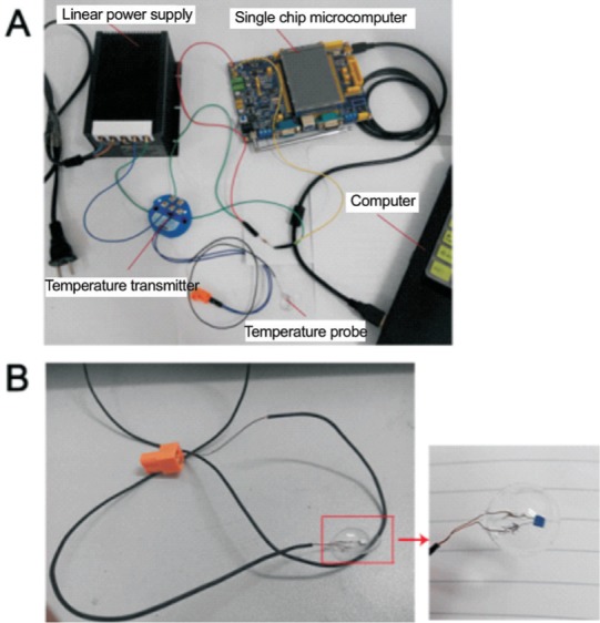
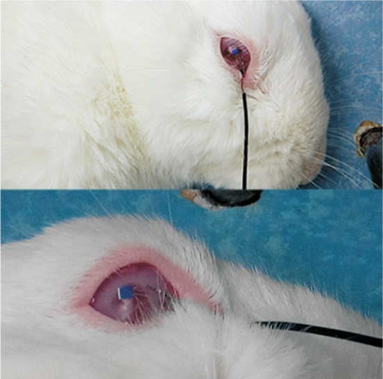
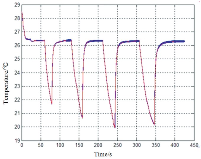
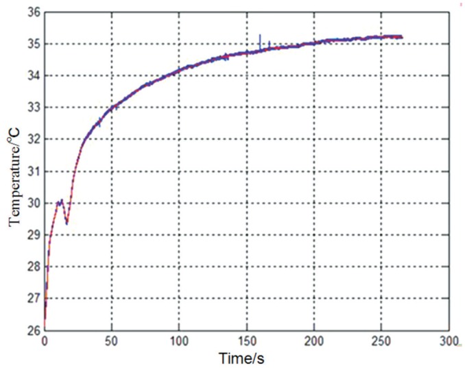
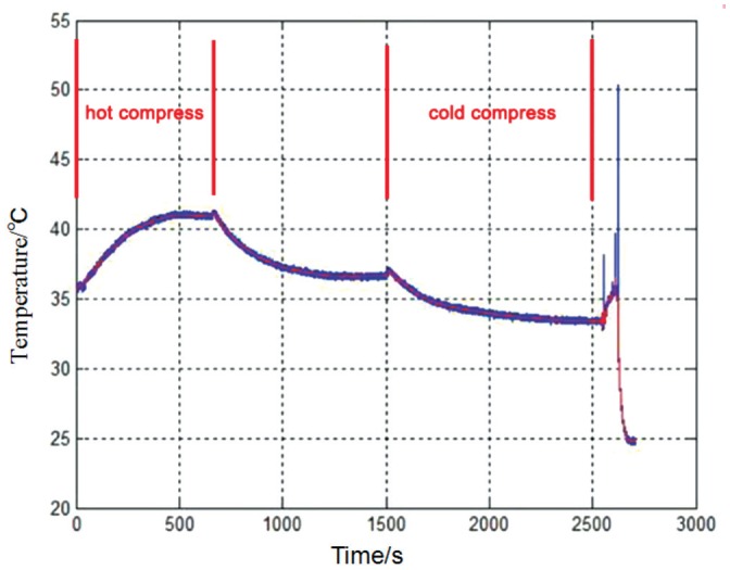
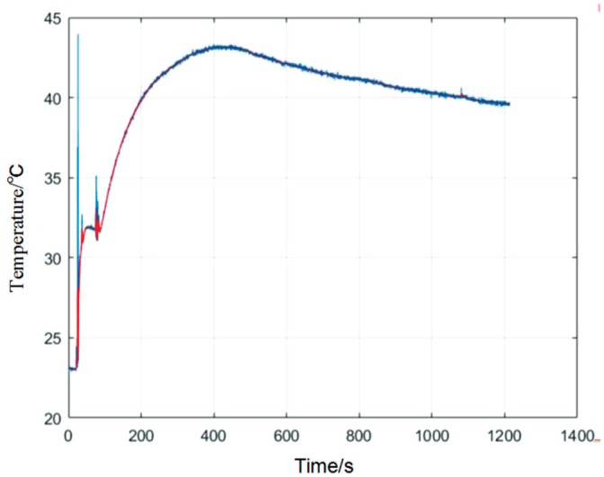
Methods: The right eyes of five New Zealand white rabbits, both male and female, were hot-compressed for 18min. An independently designed novel ocular contact-type temperature measuring device was used to measure the ocular surface temperature before and after the heating. Relevant retrobulbar hemodynamic parameters such as peak systolic velocity (PSV), end diastolic velocity (EDV), and resistance index (RI) of each of the central retinal artery (CRA), long posterior ciliary artery (LPCA), and ophthalmic artery (OA), as well as the mean velocity (Vm) of the central retinal vein (CRV), were measured using a color Doppler flow imaging (CDFI) technique and expressed as mean values with standard deviation (mean±SD). A statistical analysis was conducted based on a paired t-test and the Wilcoxon signed-rank test.
Results: The employed real-time temperature measuring device was able to accurately measure ocular surface temperature during the hot-compress process. The temperature increased after the hot compress was applied. Analysis showed that the PSV and EDV values of the CRA and LPCA significantly increased after the application of the hot compress, as did the Vm of the CRV. There were no significant changes in the EDV of the OA nor the RI of each artery.
Conclusion: This experiment, which is the first of its kind, confirms that the retrobulbar blood flow velocities can increase upon heating the ocular surface. This simple method may be useful in the future.
Keywords: color Doppler flow imaging; ocular hemodynamics; ocular surface heating; temperature detection device.
Figures

Similar articles
-
Doppler ultrasonographic measurement of short-term effects of valsalva maneuver on retrobulbar blood flow.J Clin Ultrasound. 2017 Nov 12;45(9):551-555. doi: 10.1002/jcu.22487. Epub 2017 Apr 25. J Clin Ultrasound. 2017. PMID: 28440860
-
Color doppler imaging of ocular hemodynamic changes in Behçet's disease.Jpn J Ophthalmol. 2004 Mar-Apr;48(2):101-5. doi: 10.1007/s10384-003-0024-0. Jpn J Ophthalmol. 2004. PMID: 15060789
-
Color Doppler evaluation of the ocular arterial flow changes in chronic obstructive pulmonary disease.Eur J Radiol. 2006 Jan;57(1):63-8. doi: 10.1016/j.ejrad.2005.06.010. Epub 2005 Jul 26. Eur J Radiol. 2006. PMID: 16051458
-
Blood-flow velocities in the extraocular vessels in normal volunteers.Am J Ophthalmol. 1996 Sep;122(3):364-70. doi: 10.1016/s0002-9394(14)72063-x. Am J Ophthalmol. 1996. PMID: 8794709
-
Color Doppler Imaging Analysis of Ocular Blood Flow Velocities in Normal Tension Glaucoma Patients: A Meta-Analysis.J Ophthalmol. 2015;2015:919610. doi: 10.1155/2015/919610. Epub 2015 Oct 29. J Ophthalmol. 2015. PMID: 26634152 Free PMC article. Review.
Cited by
-
Measurement of Retrobulbar Blood Flow and Vascular Reactivity-Relevance for Ocular and Cardiovascular Diseases.Diagnostics (Basel). 2023 Nov 23;13(23):3514. doi: 10.3390/diagnostics13233514. Diagnostics (Basel). 2023. PMID: 38066755 Free PMC article. Review.
References
-
- Abegão Pinto L, Vandewalle E, de Clerck E, Marques-Neves C, Stalmans I. Ophthalmic artery doppler waveform changes associated with increased damage in glaucoma patients. Invest Ophthalmol Vis Sci. 2012;53(4):2448–2453. - PubMed
-
- Schumann J, Orgül S, Gugleta K, Dubler B, Flammer J. Interocular difference in progression of glaucoma correlates with interocular differences in retrobulbar circulation. Am J Ophthalmol. 2000;129(6):728–733. - PubMed
-
- Yamazaki Y, Drance SM. The relationship between progression of visual field defects and retrobulbar circulation in patients with glaucoma. Am J Ophthalmol. 1997;124(3):287–295. - PubMed
LinkOut - more resources
Full Text Sources