

Evaluation of Virtual Reality for Detection of Lung Nodules on Computed Tomography
- PMID: 30588506
- PMCID: PMC6299745
- DOI: 10.18383/j.tom.2018.00053
Evaluation of Virtual Reality for Detection of Lung Nodules on Computed Tomography
Abstract
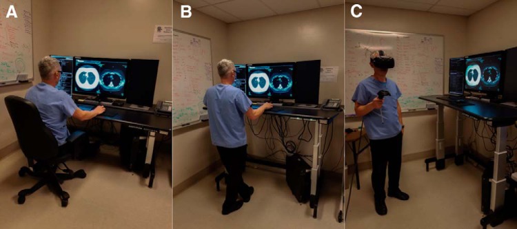
Virtual reality (VR) systems can offer benefits of improved ergonomics, but their resolution may currently be limited for the detection of small features. For detection of lung nodules, we compared the performance of VR versus standard picture archiving and communication system (PACS) monitor. Four radiologists and 1 novice radiologist reviewed axial computed tomography (CTs) of the thorax using standard PACS monitors (SM) and a VR system (HTC Vive, HTC). In this study, 3 radiologists evaluated axial lung-window CT images of a Lungman phantom. One radiologist and the novice radiologist reviewed axial lung-window patient CT thoracic images (32 patients). This HIPAA-compliant study was approved by the institutional review board. Detection of 227 lung nodules on patient CTs did not result in different sensitivity with SM compared with VR. Detection of 23 simulated Lungman phantom lung nodules on CT with SM resulted in statistically greater sensitivity (78.3%) than with VR (52.2%, P = .041) for 1 of 3 radiologists. The trend was similar but not significant for the other radiologists. There was no significant difference in the time spent by readers reviewing CT images with VR versus SM. These findings indicate that performance of a commercially available VR system for detection of lung nodules may be similar to traditional radiology monitors for assessment of small lung nodules on CTs of the thorax for most radiologists. These results, along with the potential of improving ergonomics for radiologists, are promising for the future development of VR in diagnostic radiology.
Keywords: Computed tomography; Cost saving; Lung nodule; Virtual CT; Virtual reality.
Conflict of interest statement
Conflict of Interest: The authors have no conflict of interest to declare.
Figures
References
-
- Faisal A. Computer science: Visionary of virtual reality. Nature. 2017;551:298.
-
- Edwards PW. Stereoscopic and postural radiology of the chest. Tubercle. 1931;12:529–532.
-
- Grier GW. Stereoscopy of the accessory sinuses. Am J Roentgenol Radium Ther. 1923;10:501–502.
-
- Thomson E. Stereoscopic Roentgen pictures. Electr Eng. 1896;2:256.
-
- Robinett W, Rolland JP. A computational model for the stereoscopic optics of a head-mounted display. Presence-Teleop Virt. 1992;1:45–62.
LinkOut - more resources
Full Text Sources
Research Materials
